

t(X;14)(p11;q32) in MALT lymphoma involving GPR34 reveals a role for GPR34 in tumor cell growth
- PMID: 22966169
- PMCID: PMC3496954
- DOI: 10.1182/blood-2011-11-389908
t(X;14)(p11;q32) in MALT lymphoma involving GPR34 reveals a role for GPR34 in tumor cell growth
Abstract
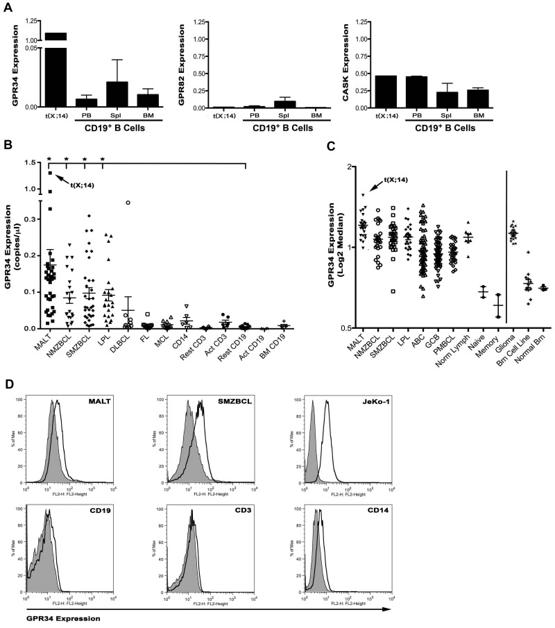
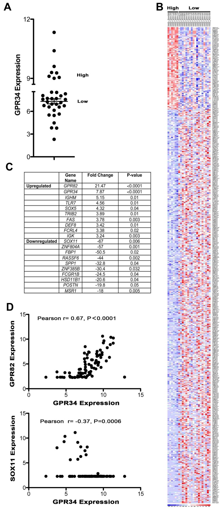
Genetic aberrations, including trisomies 3 and 18, and well-defined IGH translocations, have been described in marginal zone lymphomas (MZLs); however, these known genetic events are present in only a subset of cases. Here, we report the cloning of an IGH translocation partner on chromosome X, t(X;14)(p11.4;q32) that deregulates expression of an poorly characterized orphan G-protein-coupled receptor, GPR34. Elevated GPR34 gene expression was detected independent of the translocation in multiple subtypes of non-Hodgkin lymphoma and distinguished a unique molecular subtype of MZL. Increased expression of GPR34 was also detected in tissue from brain tumors and surface expression of GPR34 was detected on human MZL tumor cells and normal immune cells. Overexpression of GPR34 in lymphoma and HeLa cells resulted in phosphorylation of ERK, PKC, and CREB; induced CRE, AP1, and NF-κB-mediated gene transcription; and increased cell proliferation. In summary, these results are the first to identify a role for a GPR34 in lymphoma cell growth, provide insight into GPR34-mediated signaling, identify a genetically unique subset of MZLs that express high levels of GPR34, and suggest that MEK inhibitors may be useful for treatment of GPR34-expressing tumors.
Figures


Similar articles
-
t(X;14)(p11.4;q32.33) is recurrent in marginal zone lymphoma and up-regulates GPR34.Haematologica. 2012 Feb;97(2):184-8. doi: 10.3324/haematol.2011.052639. Epub 2011 Nov 4. Haematologica. 2012. PMID: 22058210 Free PMC article.
-
Clinical, histopathological, and molecular features of mucosa-associated lymphoid tissue (MALT) lymphoma carrying the t(X;14) (p11;q32)/GPR34-immunoglobulin heavy chain gene.Leuk Lymphoma. 2017 Sep;58(9):1-4. doi: 10.1080/10428194.2017.1289525. Epub 2017 Feb 28. Leuk Lymphoma. 2017. PMID: 28278700 No abstract available.
-
The t(14;18)(q32;q21)/IGH-MALT1 translocation in MALT lymphomas contains templated nucleotide insertions and a major breakpoint region similar to follicular and mantle cell lymphoma.Blood. 2010 Mar 18;115(11):2214-9. doi: 10.1182/blood-2009-08-236265. Epub 2009 Nov 25. Blood. 2010. PMID: 19965626
-
Molecular pathogenesis of mucosa-associated lymphoid tissue lymphoma.J Clin Oncol. 2005 Sep 10;23(26):6370-8. doi: 10.1200/JCO.2005.05.011. J Clin Oncol. 2005. PMID: 16155022 Review.
-
MALT lymphoma: many roads lead to nuclear factor-κb activation.Histopathology. 2011 Jan;58(1):26-38. doi: 10.1111/j.1365-2559.2010.03699.x. Histopathology. 2011. PMID: 21261681 Review.
Cited by
-
Novel lysophosphoplipid receptors: their structure and function.J Lipid Res. 2014 Oct;55(10):1986-95. doi: 10.1194/jlr.R046920. Epub 2014 Jun 2. J Lipid Res. 2014. PMID: 24891334 Free PMC article. Review.
-
Novel GPR34 and CCR6 mutation and distinct genetic profiles in MALT lymphomas of different sites.Haematologica. 2018 Aug;103(8):1329-1336. doi: 10.3324/haematol.2018.191601. Epub 2018 Apr 19. Haematologica. 2018. PMID: 29674500 Free PMC article.
-
Molecular mechanisms of target recognition by lipid GPCRs: relevance for cancer.Oncogene. 2016 Aug 4;35(31):4021-35. doi: 10.1038/onc.2015.467. Epub 2015 Dec 7. Oncogene. 2016. PMID: 26640151 Review.
-
The role of orphan G protein-coupled receptors in pain.Heliyon. 2024 Mar 26;10(7):e28818. doi: 10.1016/j.heliyon.2024.e28818. eCollection 2024 Apr 15. Heliyon. 2024. PMID: 38590871 Free PMC article. Review.
-
Molecular classification and identification of an aggressive signature in low-grade B-cell lymphomas.Hematol Oncol. 2023 Oct;41(4):644-654. doi: 10.1002/hon.3187. Epub 2023 May 30. Hematol Oncol. 2023. PMID: 37254453 Free PMC article.
References
-
- Willis TG, Dyer MJ. The role of immunoglobulin translocations in the pathogenesis of B-cell malignancies. Blood. 2000;96(3):808–822. - PubMed
-
- Ott G, Katzenberger T, Greiner A, et al. The t(11;18)(q21;q21) chromosome translocation is a frequent and specific aberration in low-grade but not high-grade malignant non-Hodgkin's lymphomas of the mucosa-associated lymphoid tissue (MALT-) type. Cancer Res. 1997;57(18):3944–3948. - PubMed
-
- Auer IA, Gascoyne RD, Connors JM, et al. t(11;18)(q21;q21) is the most common translocation in MALT lymphomas. Ann Oncol. 1997;8(10):979–985. - PubMed
-
- Levine EG, Arthur DC, Machnicki J, et al. Four new recurring translocations in non-Hodgkin lymphoma. Blood. 1989;74(5):1796–1800. - PubMed
-
- Willis TG, Jadayel DM, Du MQ, et al. Bcl10 is involved in t(1;14)(p22;q32) of MALT B cell lymphoma and mutated in multiple tumor types. Cell. 1999;96(1):35–45. - PubMed
Publication types
MeSH terms
Substances
Grants and funding
LinkOut - more resources
Full Text Sources
Miscellaneous
