. 2012 Dec 6;120(24):4869-72.
doi: 10.1182/blood-2012-06-438598. Epub 2012 Sep 10.

Platelets increase the proliferation of ovarian cancer cells

Affiliations

Platelets increase the proliferation of ovarian cancer cells

Min Soon Cho et al. Blood. .

Abstract

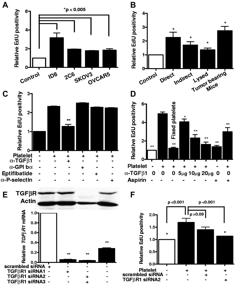
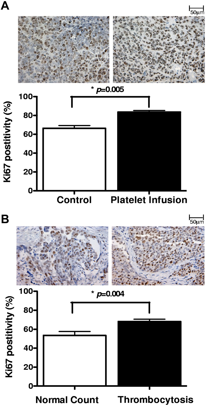
Platelets promote metastasis and angiogenesis, but their effect on tumor cell growth is uncertain. Here we report a direct proliferative effect of platelets on cancer cells both in vitro and in vivo. Incubation of platelets with ovarian cancer cells from murine (ID8 and 2C6) or human (SKOV3 and OVCAR5) origin increased cell proliferation. The proliferative effect of platelets was not dependent on direct contact with cancer cells, and preincubation of platelets with blocking antibodies against platelet adhesion molecules did not alter their effect on cancer cells. The proliferative effect of platelets was reduced by fixing platelets with paraformaldehyde, preincubating platelets with a TGF-β1-blocking antibody, or reducing expression of TGF-βR1 receptor on cancer cells with siRNA. Infusing platelets into mice with orthotopic ovarian tumors significantly increased the proliferation indices in these tumors. Ovarian cancer patients with thrombocytosis had higher tumor proliferation indices compared with patients with normal platelet counts.


Figures

Figure 1
Figure 1
Proliferative effect of platelet on ovarian cancer cells in vitro. (A) After incubation with platelets, the proliferation rate of murine (ID8 and 2C6) and human (SKOV3 and OVCAR5) ovarian cancer cells was assessed by measuring Edu incorporation. Results are normalized to Edu positivity among buffer-exposed cells. (B) Effect of manipulating platelets on proliferative response of cancer cells was investigated by either directly incubating platelets on ID8 cells (direct) or separating them using a porous membrane (indirect). Lysed platelets were prepared by repeated applications of freeze and thaw and sonication. Platelets from tumor-bearing mice were isolated from moribund C57BL/6 female mice (n = 3) by IVC venipuncture 3-4 weeks after inoculation of ID8 cells. (C) Blocking antibodies against GPIbα (5 μg/mL), P-selectin (4 μg/mL), TGF-β1 (5 μg/mL), or GPIIb-IIIa blocking agent Eptifibatide (0.5 μM) or aspirin (15 μg/mL) was added to platelets before incubation with cancer cells, and after 24 hours Edu incorporation was measured. Results were normalized to cancer cells incubated with buffer alone. (D) Increasing concentrations of TGF-β1 blocking antibody and aspirin were used for blocking platelet-induced cancer cell proliferation. All results are compared with the second bar. (E) Knockdown of TGF-βR1 gene expression after transfection of SKOV3 human ovarian cancer cells with siRNA/liposome mixture (2 μg siRNA and 3 μL lipofectamine) was investigated by measuring protein and mRNA products, using Western blotting with anti–TGF-βR1 antibody (above) and real-time RT-PCR (below), respectively. (F) ID8 cells were transfected with either TGF-βR1 siRNA or scrambled siRNA before incubation with platelets. All results are compared with the second bar. The reduction in the proliferative response induced by scrambled siRNA was not statistically significant (P > .09). Each experiment was repeated at least 3 times in triplicates. The cumulative results are summarized as bar graphs. *P < .05 (t test). **P < .01 (t test).
Figure 2
Figure 2
Proliferative effect of platelet on ovarian cancer cells in vivo. The proliferation index was measured in (A) tumors resected from tumor-bearing mice infused with buffer (control; n = 10) or platelets (n = 10) and (B) surgical specimens from ovarian cancer patients with normal platelet counts (n = 10) or thrombocytosis (n = 10), by Ki67 immunostaining. From each tissue block 5 sections were prepared, and from each section 5 fields were counted at original magnification ×200 of a light microscope by 2 different observers as independent blinded assessments. The ratio of positive Ki67 nuclear staining to total number of nuclei was measured for each field. The cumulative results are summarized as bar graphs, and a representative Ki67 staining is shown above each column.

Comment in

Similar articles

Cited by

References

    1. Nash GF, Turner LF, Scully MF, Kakkar AK. Platelets and cancer. Lancet Oncol. 2002;3(7):425–430. - PubMed
    1. Jain S, Harris J, Ware J. Platelets: linking hemostasis and cancer. Arterioscler Thromb Vasc Biol. 2010;30(12):2362–2367. - PMC - PubMed
    1. Egan K, Crowley D, Smyth P, et al. Platelet adhesion and degranulation induce pro-survival and pro-angiogenic signalling in ovarian cancer cells. PLoS One. 2011;6(10):e26125. - PMC - PubMed
    1. Nieswandt B, Hafner M, Echtenacher B, Mannel DN. Lysis of tumor cells by natural killer cells in mice is impeded by platelets. Cancer Res. 1999;59(6):1295–1300. - PubMed
    1. Karpatkin S, Pearlstein E, Ambrogio C, Coller BS. Role of adhesive proteins in platelet tumor interaction in vitro and metastasis formation in vivo. J Clin Invest. 1988;81(4):1012–1019. - PMC - PubMed

Publication types

MeSH terms

Substances