. 2012 Nov;32(22):4510-22.
doi: 10.1128/MCB.00463-12. Epub 2012 Sep 10.

Internal amino acids promote Gap1 permease ubiquitylation via TORC1/Npr1/14-3-3-dependent control of the Bul arrestin-like adaptors

Affiliations

Internal amino acids promote Gap1 permease ubiquitylation via TORC1/Npr1/14-3-3-dependent control of the Bul arrestin-like adaptors

Ahmad Merhi et al. Mol Cell Biol. 2012 Nov.

Abstract

Ubiquitylation of many plasma membrane proteins promotes their endocytosis followed by degradation in the lysosome. The yeast general amino acid permease, Gap1, is ubiquitylated and downregulated when a good nitrogen source like ammonium is provided to cells growing on a poor nitrogen source. This ubiquitylation requires the Rsp5 ubiquitin ligase and the redundant arrestin-like Bul1 and Bul2 adaptors. Previous studies have shown that Gap1 ubiquitylation involves the TORC1 kinase complex, which inhibits the Sit4 phosphatase. This causes inactivation of the protein kinase Npr1, which protects Gap1 against ubiquitylation. However, the mechanisms inducing Gap1 ubiquitylation after Npr1 inactivation remain unknown. We here show that on a poor nitrogen source, the Bul adaptors are phosphorylated in an Npr1-dependent manner and bound to 14-3-3 proteins that protect Gap1 against downregulation. After ammonium is added and converted to amino acids, the Bul proteins are dephosphorylated, dissociate from the 14-3-3 proteins, and undergo ubiquitylation. Furthermore, dephosphorylation of Bul requires the Sit4 phosphatase, which is essential to Gap1 downregulation. The data support the emerging concept that permease ubiquitylation results from activation of the arrestin-like adaptors of the Rsp5 ubiquitin ligase, this coinciding with their dephosphorylation, dissociation from the inhibitory 14-3-3 proteins, and ubiquitylation.


Figures
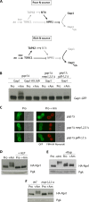
Fig 1
Fig 1
Ammonium must be converted to amino acids to induce Gap1 ubiquitylation and downregulation. (A) Model of control of Gap1 ubiquitylation by the nitrogen supply conditions. When highly active, factors and their stimulatory (arrows) or inhibitory (T bars) effects are in black, and when poorly active, they are in gray (see the text). (B) Strains EK008 (gap1Δ ura3), MA001 (mep1Δ mep2Δ mep3Δ gap1Δ ura3), and MA005 (gdh1Δ gdh2Δ gap1Δ ura3) transformed with the pJOD10 (GAL-GAP1-GFP) or pCJ038 (GAL-Gap1K9R,K16R-GFP) plasmids were grown on raffinose Pro medium. Gap1 was induced with galactose for 2 h, and glucose was then added for 1 h to repress synthesis of Gap1. Am (20 mM) was then added for 15 min, and crude cell extracts were prepared and immunoblotted with anti-GFP antibody. (C) Strains and growth conditions before Am addition were the same as those in panel B. Cells were examined by fluorescence microscopy before and 2 h after addition of Am (20 mM). Cells were also stained with the lipophilic dye FM4-64 to visualize the vacuolar membrane. (D) Wild-type strain 23344c (ura3) transformed with the pAS103 (HA-Npr1) plasmid was grown on glucose Pro medium. Cells were filtered before and 30 min after addition of Am (50 mM). Cell extracts were treated or not with ALP and immunoblotted with anti-HA and anti-Pgk antibodies. (E) Same as in panel D, except that cells were filtered before and 30 min after addition of Am (50 mM) and/or rapamycin (Rap). (F) Same as in panel D except that strains were EK008 (gap1Δ ura3) and MA001 (mep1Δ mep2Δ mep3Δ gap1Δ ura3) and Am was added for 30 min.
Fig 2
Fig 2
The Bul proteins contain a highly conserved PY-containing sequence and arrestin-related motifs. Schematic representation of the yeast Bul1 and Bul2 proteins as defined by the Pfam database (http://pfam.sanger.ac.uk/). The Bul1-N Pfam motif (PF04425, residues 108 to 543 in Bul1) is one of the six protein domains of the “arrestin N-like” Pfam motif clan (CL0135). The upper alignment shows the sequences of Bul1 and Bul2 that contain the PY motif; this sequence is specifically conserved among proteins of the Bul1_N Pfam family. The lower alignments correspond to two of the most highly conserved amino acid regions of the “arrestin N-like” clan. The sequences were aligned using ClustalW. Vertical arrows mark the amino acid residues mutated in Bul1.
Fig 3
Fig 3
The PPSY and arrestin motifs of Bul1 are essential to Am-induced Gap1 downregulation. (A) Cells were grown on solid medium containing Pro or Am as sole N source and with d-histidine (4 mM) when indicated. In the top six series of growth tests, the strains were EK008 (gap1Δ ura3), MA013 (gap1Δ bul1Δ ura3), MA014 (gap1Δ bul2Δ ura3), and JA479 (gap1Δ bul1Δ bul2Δ ura3) transformed with pFL38 (empty vector), pJOD10 (GAL-GAP1-GFP), or pCJ038 (GAL-Gap1K9R,K16R-GFP). In the bottom five series of growth tests, the strain was MA052 (gap1Δ bul1Δ bul2Δ ura3 leu2) cotransformed with pMA114 (GAL-GAP1-GFP LEU2) and a plasmid expressing the indicated Bul1 mutant, native Bul1, or no Bul1. (B) Strain EK008 (gap1Δ ura3) transformed with pJOD10 (GAL-GAP1-GFP) plasmid and strain MA052 (gap1Δ bul1Δ bul2Δ ura3 leu2) cotransformed with pMA114 (GAL-GAP1-GFP LEU2) and a second plasmid expressing the indicated Bul1 mutant, native Bul1, or no Bul1 were grown on galactose Pro medium. Glucose was added for 2 h to stop Gap1 synthesis, and Am (20 mM) was then added for 30 min. Crude cell extracts were prepared and immunoblotted with anti-GFP antibodies. (C) Strains and growth conditions before addition of Am were the same as those in panel B. Cells were examined by fluorescence microscopy before and 2 h after addition of Am (20 mM). (D) Strain OS27-1 (bul1Δ bul2Δ ura3) transformed with the empty vector pFL38 (−), pMA121 (Bul1-FLAG), or pMA136 (Bul1-PPSY>AASY-FLAG) was grown on glucose Pro medium. Crude cell extracts were prepared before and 2 h after addition of Am and immunoblotted with anti-Gap1 or anti-Pma1 antibodies.
Fig 4
Fig 4
A Bul1-Gap1 chimera is constitutively targeted to the vacuole. (A) Schematic representation of the Gap1-GFP protein fused to the PY-containing Bul1 sequence. (B) Strain EK008 (gap1Δ ura3) transformed with pJOD10 (Gap1-GFP), pMA155 (PPSY-Gap1-GFP), or pMA160 (AASY-Gap1-GFP) and strain CJ005 (gap1Δ rsp5/npi1 ura3) transformed with pJOD10 (Gap1-GFP) or pMA155 (PPSY-Gap1-GFP) were grown on solid galactose Pro medium with or without d-histidine. (C) Strains EK008 (gap1Δ ura3) and JA479 (gap1Δ bul1Δ bul2Δ ura3) transformed with the indicated plasmid were grown on galactose Pro medium. Glucose was added for 2 h to repress Gap1 synthesis, and when indicated, Am (20 mM) was added. Gap1-GFP localization was examined by florescence microscopy. (D) Same as panel C except that strains were EK008 (gap1Δ ura3) and EN121 (gap1Δ end3Δ ura3) transformed with the indicated plasmid. (E) Same as panel C except that the strain was CJ005 (gap1Δ rsp5/npi1 ura3) transformed with the indicated plasmid.
Fig 5
Fig 5
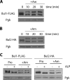
Ammonium-induced downregulation of Gap1 is associated with Bul1 dephosphorylation. (A and B) Strains MA025 (gap1Δ bul2Δ BUL1-FLAG ura3) and MA032 (gap1Δ BUL2-HA ura3) transformed with a GAP1-expressing plasmid were grown on minimal Pro medium, and Am (50 mM) was added to the culture at time zero. Crude cell extracts were immunoblotted with anti-FLAG (A) and anti-HA (B) antibodies. (C) Strains and conditions were the same as those in panels A and B except that cells were collected 30 min after Am addition and cell extracts were treated or not with alkaline phosphatase (ALP).
Fig 6
Fig 6
Ammonium-induced downregulation of Gap1 is associated with Bul1 ubiquitylation. (A) Strains 23344c (ura3) and 27038a (npi1/rsp5 ura3) transformed with pMA121 (Bul1-FLAG) plasmid were grown on Pro medium. Cells were collected before and 30 min after addition of Am. Crude cell extracts were immunoblotted with anti-FLAG or anti-Pgk antibodies. (B) Strain OS27-1 (bul1Δ bul2Δ ura3) transformed with pMA121 (Bul1-FLAG) or the empty plasmid was grown on Pro medium, and Am (50 mM) was added to part of the culture for 30 min. Crude cell extracts were immunoprecipitated with anti-FLAG antibody under denaturing conditions before immunoblotting with anti-Ub, anti-FLAG, or anti-Pgk antibodies. (C) Same as in panel A except that the strain was OS27-1 (bul1Δ bul2Δ ura3) transformed with pMA121 (Bul1-FLAG) or pMA136 (Bul1PPSY/AASY-FLAG). (D) Same as in panel A except that the strain was OS27-1 (bul1Δ bul2Δ) transformed with pMA121 (Bul1-FLAG), pMA126 (Bul1Y281A,G284A,I287A,G289A), or pMA130 (Bul1Y378A,F382A,F384A).
Fig 7
Fig 7
Inactivation of Npr1 kinase is sufficient to trigger dephosphorylation and ubiquitylation of Bul1. (A) Strains 23344c (ura3), 30788a (npr1Δ ura3), and 30788d (npr1Δ rsp5/npi1 ura3) transformed with pMA121 (Bul1-FLAG) were grown on Pro medium. Cells were collected before and 30 min after addition of Am. Crude cell extracts were immunoblotted with anti-FLAG or anti-Pgk antibodies. (B) Strains 23344c (ura3), 30788a (npr1Δ ura3), and JA413 (npr1Δ bul1Δ bul2Δ ura3) transformed with pEL003 (Gap1-GFP) were grown on Pro medium. When indicated, Am (20 mM) was added for 2 h. Gap1-GFP localization was determined by fluorescence microscopy. (C) Strains 23344c (ura3) (wild type) and 30035b [npr1(ts) ura3] transformed with pMA121 (Bul1-FLAG) were grown at 25°C on Pro medium. Cultures were then transferred to 35°C for 30 min. Crude cell extracts were immunoblotted with anti-FLAG antibody. The ubiquitylated form of Bul1 is marked with a filled circle. (D) Strain EK008 (gap1Δ ura3) transformed with pJOD10 (GAL-GAP1-GFP) plasmid and strain MA003 [npr1(ts) gap1Δ ura3] transformed with pJOD10 (GAL-GAP1-GFP) or pCJ038 (GAL-Gap1K9R,K16R-GFP) plasmid were grown at 25°C on galactose Pro medium. The cells were incubated at 35°C for 2 h, and Gap1-GFP localization was determined by fluorescence microscopy.
Fig 8
Fig 8
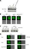
Sit4 phosphatase is required for Bul1 dephosphorylation and Gap1 endocytosis. (A) Strains 23344c (ura3) and 35064c (sit4Δ ura3) transformed with pAS103 (HA-Npr1) were grown on Pro medium. When indicated, Am was added for 30 min. Crude cell extracts were immunoblotted with anti-HA antibody. (B) Strains 23344c (ura3), 30788a (npr1Δ ura3), and 35064c (sit4Δ ura3) transformed with pEL003 (Gap1-GFP) were grown on Pro medium. Gap1-GFP localization was determined by fluorescence microscopy. (C) (Top) Strains 23344c (ura3) and 35064c (sit4Δ ura3) transformed with pMA121 (Bul1-FLAG) were grown on Pro medium. Cells were collected before and 30 min after Am addition, and crude cell extracts were immunoblotted with anti-FLAG antibody. (Bottom) Strain 35064c (sit4Δ ura3) transformed with pMA121 (Bul1-FLAG) was grown on Pro medium. Am was added for 30 min, and crude cell extracts were treated or not with ALP. (D) Strains 23344c (ura3) and 35064c (sit4Δ ura3) transformed with pEL003 (Gap1-GFP) were grown on minimal proline medium. Gap1-GFP localization was determined before and 2 h after addition of Am (20 mM).
Fig 9
Fig 9
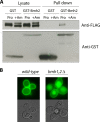
14-3-3 proteins bind to phosphorylated Bul1 and protect Gap1 against downregulation. (A) Strain MA025 (Bul1-FLAG) transformed with pRS426-GST-BMH2 or pRS426-GST was grown on Pro medium, and cells were collected before and 30 min after Am addition. Cells were lysed, and GST was pulled down as described in Materials and Methods. Lysates and pulled-down fractions were immunoblotted with anti-GST or anti-FLAG antibodies. (B) Strain 10560-6b (his3 leu2 trp1 ura3-52) cotransformed with pCJ366 (HIS3 LEU2 TRP1) and pEL003 (Gap1-GFP) and strain RRY1217 (bmh1Δ::HIS3 bmh2Δ::HIS3 his3 leu2 trp1 ura3-52) cotransformed with pCJ362 (LEU2 TRP1) and pEL003 (Gap1-GFP) were grown on Pro medium. Gap1-GFP localization was determined by fluorescence microscopy.
Fig 10
Fig 10
A model for the nitrogen control of Gap1 ubiquitylation. (Left) For the meaning of the black and gray shading, see the Fig. 1A legend. On a poor N source such as proline or urea, TORC1 phosphorylates neither Tap42 nor Npr1. Sit4 phosphatase is active and acts on Npr1 to maintain it in a hypophosphorylated and active state. Active Npr1 phosphorylates the Bul adaptors, which are bound to the 14-3-3 proteins and thus inhibited. Hence, Gap1 is not ubiquitylated and is stable and active at the plasma membrane. When a good nitrogen source such as Am is provided to cells, TORC1 is activated by internal amino acids via the EGO complex and phosphorylates Npr1, which is thus inactive. TORC1 also phosphorylates Tap42, which associates with Sit4, thus possibly redirecting the phosphatase to the Bul adaptors. The Bul adaptors undergo dephosphorylation, dissociate from the 14-3-3 factors, and can thus recruit the Rsp5 Ub ligase to the Gap1 permease, which is ubiquitylated and downregulated. Npr1 inactivation also induces Rsp5-dependent ubiquitylation of the Bul adaptors. Dephosphorylation and ubiquitylation of Bul proteins appear independent from each other, as lack of either modification does not impair the other.

Comment in

Similar articles

Cited by

References

    1. Béchet J, Grenson M, Wiame JM. 1970. Mutations affecting the repressibility of arginine biosynthetic enzymes in Saccharomyces cerevisiae. Eur. J. Biochem. 12:31–39 - PubMed
    1. Beck T, Schmidt A, Hall MN. 1999. Starvation induces vacuolar targeting and degradation of the tryptophan permease in yeast. J. Cell Biol. 146:1227–1238 - PMC - PubMed
    1. Becuwe M, et al. 2012. A molecular switch on an arrestin-like protein relays glucose signaling to transporter endocytosis. J. Cell Biol. 196:247–259 - PMC - PubMed
    1. Binda M, et al. 2009. The Vam6 GEF controls TORC1 by activating the EGO complex. Mol. Cell 35:563–573 - PubMed
    1. Boeckstaens M, Andre B, Marini AM. 2007. The yeast ammonium transport protein Mep2 and its positive regulator, the Npr1 kinase, play an important role in normal and pseudohyphal growth on various nitrogen media through retrieval of excreted ammonium. Mol. Microbiol. 64:534–546 - PubMed

Publication types

MeSH terms