. 2012 Nov;32(22):4595-610.
doi: 10.1128/MCB.00724-12. Epub 2012 Sep 10.

Upstream stimulatory factor 2 and hypoxia-inducible factor 2α (HIF2α) cooperatively activate HIF2 target genes during hypoxia

Affiliations

Upstream stimulatory factor 2 and hypoxia-inducible factor 2α (HIF2α) cooperatively activate HIF2 target genes during hypoxia

Matthew R Pawlus et al. Mol Cell Biol. 2012 Nov.

Abstract

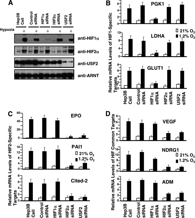
While the functions of hypoxia-inducible factor 1α (HIF1α)/aryl hydrocarbon receptor nuclear translocator (ARNT) and HIF2α/ARNT (HIF2) proteins in activating hypoxia-inducible genes are well established, the role of other transcription factors in the hypoxic transcriptional response is less clear. We report here for the first time that the basic helix-loop-helix-leucine-zip transcription factor upstream stimulatory factor 2 (USF2) is required for the hypoxic transcriptional response, specifically, for hypoxic activation of HIF2 target genes. We show that inhibiting USF2 activity greatly reduces hypoxic induction of HIF2 target genes in cell lines that have USF2 activity, while inducing USF2 activity in cells lacking USF2 activity restores hypoxic induction of HIF2 target genes. Mechanistically, USF2 activates HIF2 target genes by binding to HIF2 target gene promoters, interacting with HIF2α protein, and recruiting coactivators CBP and p300 to form enhanceosome complexes that contain HIF2α, USF2, CBP, p300, and RNA polymerase II on HIF2 target gene promoters. Functionally, the effect of USF2 knockdown on proliferation, motility, and clonogenic survival of HIF2-dependent tumor cells in vitro is phenocopied by HIF2α knockdown, indicating that USF2 works with HIF2 to activate HIF2 target genes and to drive HIF2-depedent tumorigenesis.


Figures

Fig 1
Fig 1
Some reported HIF/USF common target genes are indeed activated by USF2 or HIF2αTM in Hep3B cells. qPCR detection of reported HIF/USF common target gene expression levels in normoxic Hep3B cells or Hep3B cells transfected with 3 μg of HIF1αTM, HIF2αTM, or USF2 plasmid or with 1.5 μg USF2 plus 1.5 μg of HIF1αTM (or HIF2αTM).
Fig 2
Fig 2
USF2 siRNA reduces hypoxic induction of several known HIF2 but not HIF1 target genes in Hep3B cells. (A) Western blot analysis of HIF1α, HIF2α, USF2, and ARNT proteins in normoxic and hypoxic Hep3B cells or Hep3B cells transfected with control siRNA or siRNA against HIF1α, HIF2α, or USF2. (B to D) qPCR analysis of known HIF1-specific target genes (B), HIF2-specific target genes (C), and HIF1/HIF2 common targets (D) in normoxic and hypoxic Hep3B cells or Hep3B cells targeted with the indicated siRNAs. Results are presented relative to those for WT Hep3B cells cultured under normoxia. qPCR results were normalized to 18S rRNA expression; error bars are ±1 SD from three independent experiments in this and the other figures.
Fig 3
Fig 3
USF2 siRNA reduces HIF2 but not HIF1 target gene expression in normoxic RCC4 cells. (A) qPCR analysis of HIF1α, HIF2α, and USF2 mRNA in normoxic RCC4 cells or RCC4 cells transfected with control, HIF1α, HIF2α, or USF2 siRNA. (B to D) qPCR analysis of known HIF1-specific (B), HIF2-specific (C), and HIF1/HIF2 common (D) target genes in normoxic RCC4 cells or RCC4 cells transfected with control, HIF1α, HIF2α, or USF2 siRNA.
Fig 4
Fig 4
Increased FL USF2 expression in Hif1α−/− mouse ES cells restores hypoxic induction of HIF2 target genes. (A) Western blot analysis of USF2 protein in HIF2 functional (PRC3, PRC3/pVHL, RCC4, RCC4/pVHL, and Hep3B) and not functional (mouse ES) cells. Full-length USF2 and the splicing variant of USF2 are indicated. (B) Western blot of USF2 in mouse ES, Hep3B, 293T, or 293T cells transfected with USF2ΔE4 expression vectors. The Western blot gel was run longer to separate a nonspecific band from USF2ΔE4. (C) Western blot of endogenous mouse USF2 and Flag-tagged human USF2 proteins in Hif1α−/− or Hif1α−/−/USF2 shRNA mES cells or Hif1α−/−/USF2 shRNA/hUSF2-Flag ES cells, using anti-USF2 or anti-Flag antibodies. NS, nonspecific band. (D) Western blot of endogenous HIF2α and HIF1α proteins in normoxic and hypoxic (Hx) ES cells. (E and F) qPCR analysis of endogenous known HIF1/HIF2 target genes (E) and HIF1-specific target genes (F) in normoxic and hypoxic WT, Hif1α−/−, Hif1α−/−/USF2 shRNA/hUSF2-Flag, or Hif1α−/−/USF2 shRNA/hUSF2-Flag/HIF2α siRNA cells.
Fig 5
Fig 5
USF2 binds to the human PAI1 promoter at multiple sites. (A) Cloned human PAI1 promoter/Luc reporter, showing the locations of consensus USF/HIF common (CACGTG), HIF (ACGTG), or USF2 (CANNTG, where the NN nucleotides are CG or GC) binding sites. The previously reported HIF or USF binding sites are represented by squares with bold borders, and the additional potential HIF or USF binding sites are represented by white squares. (B) Relative luciferase activity of PAI1/Luc reporter in response to activation by indicated plasmids in nanograms. (C) Gel shift assay of USF2 binding to potential USF2 binding sites on the PAI1 promoter using 30-mer double-stranded oligonucleotides centered at the indicated number and nuclear extracts prepared from USF2-transfected HEK293T cells. A 200-fold excess of cold WT competitor oligonucleotide or cold USF2-binding-site-mutated oligonucleotide (Mut) was added in competition assays.
Fig 6
Fig 6
USF2 binding to the PAI1 promoter is important to maintain basal activity of the PAI1 promoter and important for HIF2 to activate the PAI1 promoter. (A) Schematic representation of PAI1/Luc reporters in which USF binding sites were sequentially deleted. In addition, all the deletion mutants were based on PAI1 MUSF2 +5/Luc, in which the USF2 binding site at position +5 was mutated. (B) Activation of the PAI1 MUSF2 +5/Luc deletion reporters by 400 ng of empty vector as a control (activation of the vector is 100%), HIF2αTM, or USF2 or 200 ng each of HIF2αTM and USF2 expression plasmids in HEK293T cells. The relative basal activity of these deletion reporters to the full-length PAI1/Luc reporter is also indicated. (C) Gel shift assay of USF2 binding to additional USF2 binding sites on the PAI1 promoter using 30-mer double-stranded oligonucleotides centered at the indicated number. (D) Activation of single or combined USF2/HIF-binding-site-mutated PAI1/Luc reporters by the same activators used for panel B in HEK293T cells. The basal activity of these reporters relative to that of the WT PAI1/Luc reporter is also indicated.
Fig 7
Fig 7
USF2 is critical for CBP and p300 protein recruitment to HIF2 target genes (PAI1 and EPO) in vivo. ChIP analysis of Pol II, CBP, p300, USF2, and HIF2α binding to the HIF2 target gene PAI1 promoter at the region close to the transcription start site and HRE (PAI1 from positions −248 to −128) (A), the PAI promoter away from the transcription start site and HRE (PAI1 from positions −808 to −608) (B), the HIF2 target gene EPO promoter close to the EPO transcription start site (EPO from positions −128 to −23) (C), and the EPO enhancer that is close to the validated HRE (EPO from positions +2970 to +3058) (D) in normoxic (Nx) or hypoxic (Hx) parental Hep3B, Hep3B/ARNT shRNA, or Hep3B/USF2 shRNA cells. No Chro., no chromatin.
Fig 8
Fig 8
HIF2α, USF2, and p300 physically interact in vitro and in vivo. (A) Western blot analysis of p300 protein coprecipitated by HIF2α or USF2 protein (top), p300 protein in lysates (middle), and HIF2α/USF2 protein (bottom) in lysates of HEK293T cells or HEK293T cells transfected with HIF2αTM-Flag or USF2-Flag. (B) Western blot detection of HIF1α (top), HIF2α (middle), and USF2 protein (bottom) in hypoxic Hep3B cell lysates or coprecipitated with beads or the USF2, HIF1α, or HIF2α protein. (C) The PAI1 promoter indicating the locations of ChIP qPCR primers in relation to the positions of validated USF/HIF binding sites. (D) ChIP and re-ChIP were conducted in hypoxic Hep3B cells, and precipitated DNA was analyzed for the indicated regions of the PAI1 promoter. The antibodies listed first were used in the first precipitation, while the antibodies listed second were used to precipitate the DNA-protein complex immunoprecipitated by the first antibody. PAI intron 4 served as a negative control.
Fig 9
Fig 9
USF2 is required for HIF2α-dependent tumorigenic properties in RCC4 cells. (A) Western blot analysis of HIF2α and USF2 proteins in normoxic RCC4, RCC4/USF2 shRNA, or RCC4/HIF2α shRNA cells. (B) Cell number of normoxic RCC4, RCC4/USF2 shRNA, or RCC4/HIF2α shRNA cells on different days. Assays were performed in triplicate, and results are presented as averages from three independent experiments. (C) USF2 or HIF2α shRNA similarly reduced RCC4 cell motility in scratch assays. Assays were performed in duplicate, and the results are graphed as the average percent scratch closure at each time point from three experiments. (D) Clonogenic survival assays demonstrated that USF2 and HIF2 shRNA decreased colony formation. Assays were conducted in duplicate with plating of 500, 1,000, or 1,500 cells. Representative plates from 1,000 cells are shown. (E) The average number of CFU from 1,000 cells was graphed. The raw numbers are also shown.
Fig 10
Fig 10
USF2 is required for HIF2-dependent tumorigenicity in PRC3 cells in vitro. PRC3 cells lack functional pVHL and HIF1α gene expression. HIF2α protein and HIF2 target genes are constitutively expressed. (A) The efficiency of USF2 and HIF2α protein knockdown in PRC3 cells by USF2 or HIF2 shRNA is demonstrated by Western blotting. (B) In vitro cellular motility was measured by scratch assay and found to be decreased in both PRC3/USF2 shRNA and PRC3/HIF2α shRNA cells. (C) Colony formation in a clonogenic survival assay where 1,000 cells were plated was likewise decreased by USF2 or HIF2 shRNA in comparison to that for wild-type controls. (D) Average colony counts from 1,000 cells are quantified in PRC3, PRC3/USF2 shRNA, and PRC3/USF2 shRNA cells. The raw clone number is given at the top of each bar.

Similar articles

Cited by

References

    1. Alfranca A, et al. 2002. c-Jun and hypoxia-inducible factor 1 functionally cooperate in hypoxia-induced gene transcription. Mol. Cell. Biol. 22:12–22 - PMC - PubMed
    1. Allen RR, Qi L, Higgins PJ. 2005. Upstream stimulatory factor regulates E box-dependent PAI-1 transcription in human epidermal keratinocytes. J. Cell. Physiol. 203:156–165 - PubMed
    1. Anderson CJ, Hoare SF, Ashcroft M, Bilsland AE, Keith WN. 2006. Hypoxic regulation of telomerase gene expression by transcriptional and post-transcriptional mechanisms. Oncogene 25:61–69 - PubMed
    1. Aprelikova O, Wood M, Tackett S, Chandramouli GV, Barrett JC. 2006. Role of ETS transcription factors in the hypoxia-inducible factor-2 target gene selection. Cancer Res. 66:5641–5647 - PubMed
    1. Arany Z, et al. 1996. An essential role for p300/CBP in the cellular response to hypoxia. Proc. Natl. Acad. Sci. U. S. A. 93:12969–12973 - PMC - PubMed

Publication types

MeSH terms

Substances

LinkOut - more resources