. 2012:2012:215810.
doi: 10.1155/2012/215810. Epub 2012 Jun 28.

Imaging features of superficial and deep fibromatoses in the adult population

Affiliations

Imaging features of superficial and deep fibromatoses in the adult population

Eric A Walker et al. Sarcoma. 2012.

Abstract

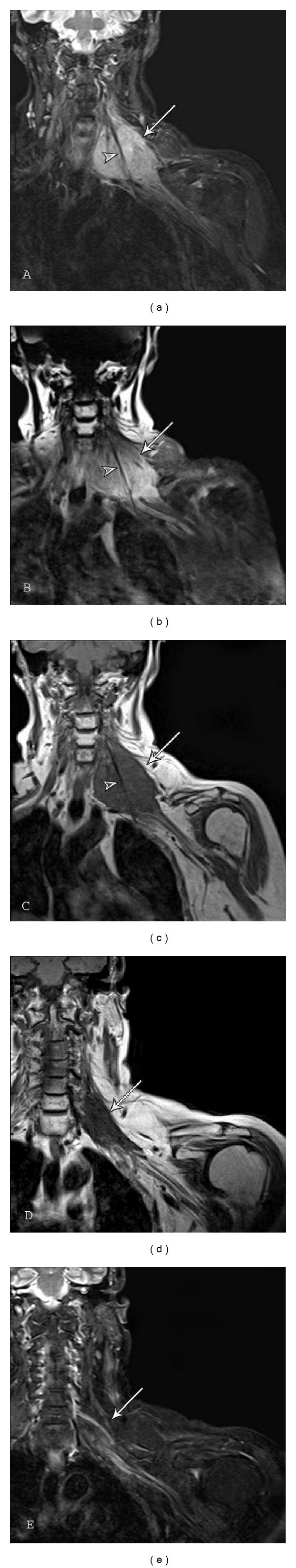
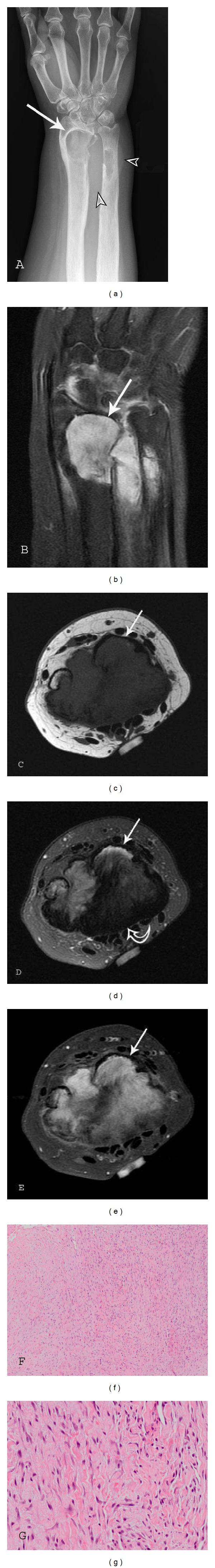
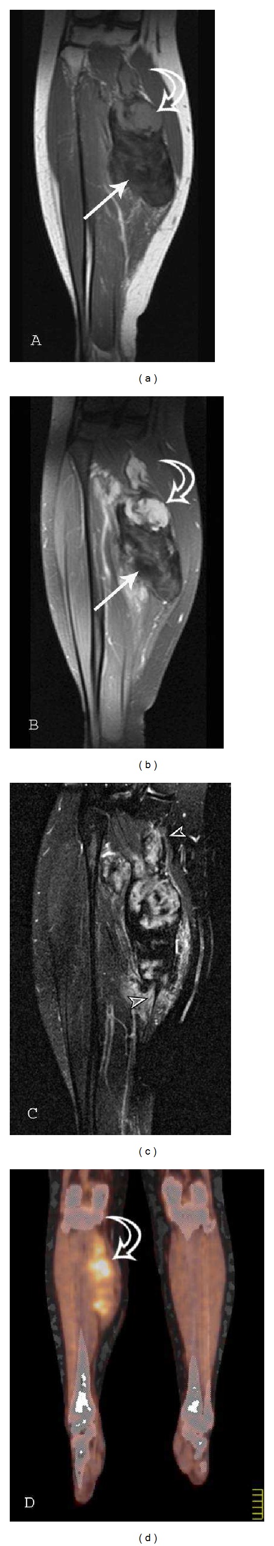
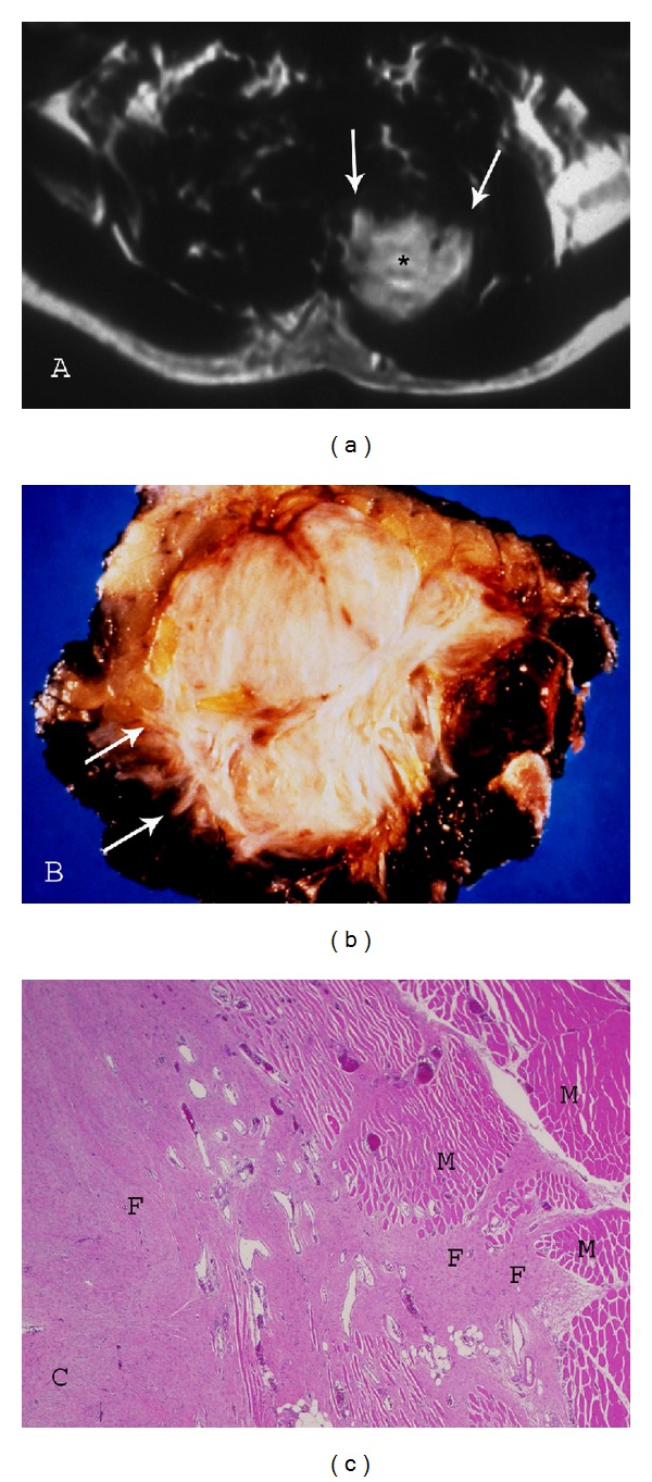
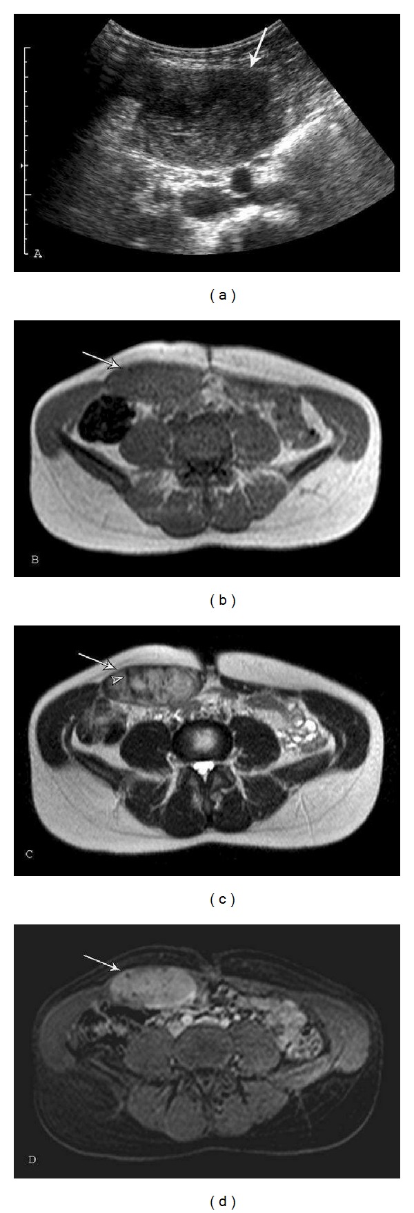
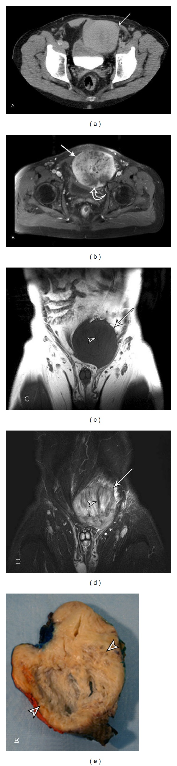
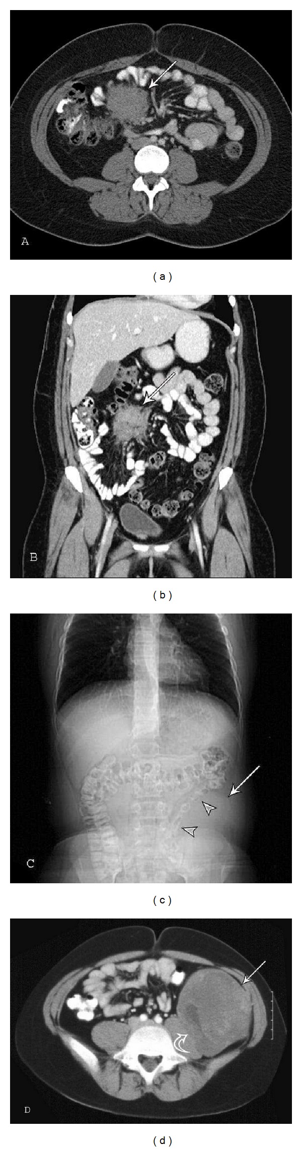
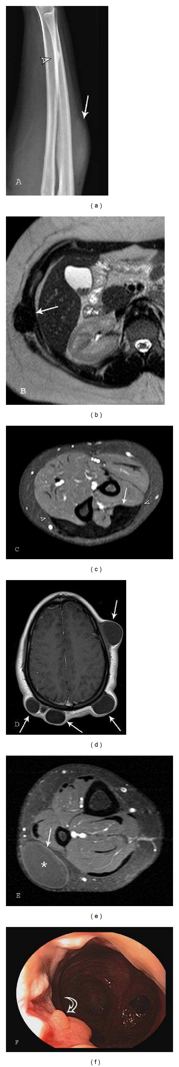
The fibromatoses are a group of benign fibroblastic proliferations that vary from benign to intermediate in biological behavior. This article will discuss imaging characteristics and patient demographics of the adult type superficial (fascial) and deep (musculoaponeurotic) fibromatoses. The imaging appearance of these lesions can be characteristic (particularly when using magnetic resonance imaging). Palmar fibromatosis demonstrates multiple nodular or band-like soft tissue masses arising from the proximal palmar aponeurosis and extending along the subcutaneous tissues of the finger in parallel to the flexor tendons. T1 and T2-weighted signal intensity can vary from low (higher collagen) to intermediate (higher cellularity), similar to the other fibromatoses. Plantar fibromatosis manifests as superficial lesions along the deep plantar aponeurosis, which typically blend with the adjacent plantar musculature. Linear tails of extension ("fascial tail sign") along the aponeurosis are frequent. Extraabdominal and abdominal wall fibromatosis often appear as a heterogeneous lesion with low signal intensity bands on all pulse sequences and linear fascial extensions ("fascial tail" sign) with MR imaging. Mesenteric fibromatosis usually demonstrates a soft tissue density on CT with radiating strands projecting into the adjacent mesenteric fat. When imaging is combined with patient demographics, a diagnosis can frequently be obtained.


Figures

Figure 1
Figure 1
Palmar Fibromatosis. Lateral radiograph (a) of the right hand 5th finger in a 48-year-old man shows a flexion (Dupuytren) contracture of the proximal interphalangeal (PIP) joint. MR images of a 73-year-old man with pathologically proven palmar fibromatosis. (b) Axial T1-weighted (TR 500/TE 21) and (c) axial proton-density-weighted (TR 1500/TE 35) fat suppressed images of the hand at the level of the metacarpal bones show nodular areas of low-signal intensity in the volar subcutaneous fat (arrows) located superficial to the flexor tendons of the fourth and fifth fingers. Increased signal surrounds the nodules (curved arrow) on fluid sensitive sequence. (d) Axial T1-weighted (TR 500/TE 21) fat-suppressed image after administration of gadolinium contrast demonstrates moderate and diffuse enhancement (curved arrow) surrounding the nodules.
Figure 2
Figure 2
Plantar fibromatosis: a 54-year-old male who presents with left foot pain for one year. A longitudinal ultrasound color Doppler image (a) demonstrates a soft tissue mass with heterogeneous echotexture and internal color Doppler flow (arrow). (b) Sagittal T1-weighted (TR539.4/TE15) fat saturation postcontrast sequence demonstrates a fusiform, enhancing lesion with linear extension (fascial tail sign) along the plantar aponeurosis (arrow). Short axis MR images ((c)–(e)) demonstrate a well-defined mass (arrows) in the medial aspect of the plantar aponeurosis (c) Short axis T1-weighted (TR568/TE15) sequence reveals lesion signal intensity similar to skeletal muscle. There is heterogeneity with several foci of low signal (curved arrows) within the lesion. (d) Short axis T2-weighted (TR2693/TE60) with fat suppression reveals intermediate-to-high heterogeneous signal (arrow) and (e) T1-weighted (TR638.7/TE15) postcontrast fat saturation sequences demonstrate marked heterogeneous enhancement (arrow). Curved arrows indicate band-like areas of higher collagen content and low cellularity. Note the lower T1 and T2 signal intensity and lack of enhancement in these foci.
Figure 3
Figure 3
Extraabdominal fibromatosis of the popliteus and soleus muscles after pregnancy: a 27-year-old female with growth of a calf desmoid tumor noted during and after pregnancy. (a) Transverse sonography of the lesion (arrow) during a needle biopsy demonstrates a well-defined, heterogeneous hypoechoic mass. (b) T1-weighted (TR450/TE24) image reveals a heterogeneous lesion (arrow) measuring 6.6 centimeters (cm) × 2.9 cm in the coronal plane with signal similar to skeletal muscle. (c) Coronal T2- (TR3000/TE90) and (d) T1-weighted (TR450/TE24) postcontrast sequences with fat suppression shows a heterogeneous lesion (arrow) with central enhancement and significant band-like low signal component (arrowheads) predominantly at the periphery. (e) Coronal T1-weighted (TR484.913/TE7) postcontrast with fat saturation was obtained one year and nine months following the other MR images ((b)–(d)) and five months after partum. Both the size (9.8 cm × 3.3 cm in the coronal plane) of the desmoid tumor (arrow) and relative proportion of enhancing cellular tissue have increased under hormonal stimulation.
Figure 4
Figure 4
Multicentric extraabdominal fibromatosis of the right ankle: a 22-year-old male with ankle pain with prolonged standing. (a) AP radiograph of the right ankle demonstrates soft tissues masses (arrows) and mature periosteal reaction (curved arrow) of the fibula adjacent to the proximal lesion. (b) Coronal T1-weighted (TR420/TE10) sequence reveals predominantly intermediate signal lesions (arrows) of the subcutaneous tissues of the lateral ankle. (c) Coronal T2-weighted (TR4730.91/TE70) fat saturation image shows heterogeneous lesions (arrows) with moderate to marked enhancement on the (d) coronal T1-weighted (TR420/TE10) postcontrast fat suppression image. Low-signal-intensity bands (small arrowheads) of mature collagenized tissue within this desmoid tumor are best appreciated on the T2FS sequence. Photograph (e) of resected gross specimen demonstrates infiltrative borders (arrow) and a coarsely trabeculated surface.
Figure 5
Figure 5
Extraabdominal fibromatosis of the left brachial plexus: a 64-year-old female presented with left shoulder and upper arm pain. The lesion was not amenable to surgical resection because of the intimate relationship with neurovascular structures of the left brachial plexus. (a) Coronal T2-weighted (TR4130/TE30) fat saturation and (b) coronal T1-weighted (TR576/TE11) postcontrast fat suppression sequences reveal a heterogeneous intermediate-to-high signal lesion with moderate and diffuse enhancement (arrows). Note the low-intensity band (arrowheads) corresponding to an acellular, collagen rich area interspersed between the highly vascularized fascicles of spindle cells. (c) Coronal T1-weighted (TR560/TE11) image obtained at presentation is compared to (d) coronal T1-weighted (TR572/TE14) and (e) STIR (TR5560/TE34) images obtained two years and four months after the previous images and status after completing radiotherapy (50.4 gray in 28 fractions). The lesion (arrows) reveals decrease in size and lower T1 and STIR signal indicating mature collagenized tissue after treatment.
Figure 6
Figure 6
Extraabdominal fibromatosis of the right forearm with osseous involvement: a 47-year-old female with recurrent right distal forearm desmoid. (a) AP radiograph and (b) coronal T1-weighted (TR452/TE6.24) fat saturation (FS) postcontrast image demonstrate a distal forearm soft tissue mass (arrows) with involvement of the distal radius and ulna. Note the soft tissue density (arrowheads) on the radiograph. Axial (c) T1-weighted (TR428/TE9.5), (d) T2FS (TR3263/TE68.3), and (e) T1FS postcontrast sequences reveal a volar soft tissue mass deep to the flexor tendons with deep invasion and marrow replacement of the distal radius (arrows) and ulna. Low-signal, predominantly collagenous component (curved arrow) is best appreciated on the T2 fat suppression image. Photomicrographs (f) low- and (g) high-power hematoxylin-eosin (H-E) stain reveal spindled or stellate cells with bland nuclear features in a background of thick collagenous bands.
Figure 7
Figure 7
Extraabdominal fibromatosis of the medial soleus muscle: a 22-year-old female presents with a painless soft tissue mass of the right calf. (a) Coronal T1-weighted (TR550/TE12) and (b) coronal T1-weighted (TR552/TE12) postcontrast fat suppression sequences demonstrate a heterogeneous predominately low T1-weighted signal lesion of the medial soleus muscle (arrow). (c) Coronal T2-weighted (TR3000/TE70) image with fat saturation reveals the lesion growing along the fascia (fascial tail sign) (arrowheads) at the proximal and distal aspects of the lesion. (d) Coronal PET-CT fusion image reveals heterogeneous FDG uptake, which is the most common reported pattern of deep fibromatosis. Note the intermediate T1-weighted signal with marked enhancement corresponding to an area of high cellularity (curved arrows on (a) and (b)) at the proximal aspect of the lesion. This immature area demonstrates higher FDG uptake (curved arrow on (c)).
Figure 8
Figure 8
Paraspinal fibromatosis with infiltrative borders. (a) Axial postcontrast T1-weighted (TR500/TE20) sequence demonstrates fibromatosis of the paraspinal muscles with prominent enhancement (asterisk) and infiltrative margin (arrows). (b) Photograph of gross specimen reveals multiple collagenized bands and irregular, spiculated margin (arrows). (c) Photomicrograph (original magnification, ×100; H-E stain) also illustrates the marginal invasion of muscle (M) by the collagenized fibromatosis lesion (F).
Figure 9
Figure 9
Abdominal fibromatosis of the right rectus abdominis muscle: a 35-year-old female with right rectus abdominis fibromatosis. The lesion continued to grow despite discontinuing oral contraceptive pills and taking ibuprofen. (a) Transverse ultrasound image demonstrates a well-defined, heterogeneous hypoechoic mass. (b) Axial T1- (TR148/TE4.6) and T2-weighted (TR441.2/TE100) sequences reveal a heterogeneous lesion (arrows) with T1 signal similar to skeletal muscle and intermediate to high T2 signal. Low-signal bands (arrowhead) are best evaluated on the (c) T2-weighted image. (d) Axial dynamic thrive (TR4.27/TE2.06) postcontrast fat suppression sequence reveals moderate and diffuse enhancement (arrow) of this lesion with high cellularity and scattered nonenhancing foci corresponding to the collagenized bands.
Figure 10
Figure 10
Abdominal fibromatosis: a 65-year-old male with a slowly growing painless suprapubic mass. (a) Axial CT and T1 (TE13/TR643) fat saturation image postcontrast reveal a well-defined soft-tissue mass (arrows) in the lower left rectus abdominis that displaces the bladder to the right. Lesion attenuation on CT is higher than skeletal muscle reflecting higher collagen component. An area of necrosis (curved arrow in (b)) is noted in the posterior aspect of the lesion. Lesion margins and heterogeneous enhancement are better appreciated on MR. (c) Coronal T1-weighted (TE11/TR427) and (d) STIR (TE78/TR4810) sequences show the lesion (arrows) with mildly ill-defined borders, mild peripheral edema, and band-like areas (arrowheads) of low signal within the lesion. (e) Photograph of the sectioned gross specimen, resected after radiation therapy, reveals a large area of central necrosis (arrowheads).
Figure 11
Figure 11
Intraabdominal fibromatosis. Mesenteric fibromatosis: a 29-year-old female with right-sided flank pain. (a) Axial and (b) coronal CT images demonstrate a mass (arrows) in the small bowel mesentery with irregular margins and attenuation similar to skeletal muscle. The mesenteric fat surrounds the lesion outlining the extent. Retroperitoneal fibromatosis: a 12-year-old male with a mass palpated in the left lower quadrant on routine physical exam. (c) CT topogram reveals an intraabdominal soft tissue mass (arrow) displacing the descending colon (arrowheads). (d) Axial CT shows a large retroperitoneal heterogeneous lesion (arrow) causing mass effect on the left psoas muscle (curved arrow) with scattered areas of mild-to-moderate enhancement.
Figure 12
Figure 12
Imaging of gardner syndrome: a teenage female with Gardner syndrome. Images were obtained from age 13 to 16 years-of-age. (a) Lateral radiograph of the forearm demonstrates an osteoma (arrowhead) of the distal radial diaphysis. Also note the soft tissue mass (arrow) corresponding to extraabdominal desmoid imaged with MR in 11c. (b) Axial T2 (TR544.326/TE100) sequence through the upper abdomen demonstrates fibromatosis (arrow) involving the left intercostal muscles. (c) Axial T1 (TR539.09/TE15) fat saturation postcontrast of the upper forearm reveals a mature (collagenized) extraabdominal fibromatosis (arrow) along the dorsal superficial fascia with no significant enhancement. Note the fascial tail sign (arrowheads). (d) Axial T1 (TR491/TE11) postcontrast of the scalp and (e) axial T1 (TR667/TE10) fat suppression postcontrast of the calf reveal multiple epidermal inclusion cysts (arrows) of the subcutaneous tissues with mild peripheral enhancement. Follow-up endoscopy (f) of the patient status post colectomy for multiple tubular adenomas demonstrates development of an adenomatous polyp within the distal rectum.

Similar articles

Cited by

References

    1. Weiss SW, Goldblum JR, Enzinger FM. Fibromatoses. In: Weiss SW, Goldblum JR, editors. Enzinger and Weiss' Soft Tissue Tumors. Philadelphia, Pa, USA: Mosby Elsevier; 2008. pp. 227–228.
    1. Fletcher CDM, Krishnan Unni K, Mertens F, editors. Pathology and Genetics of Tumours of Soft Tissue and Bone (IARC WHO Classification of Tumours) 3rd edition 2006.
    1. Murphey MD, Ruble CM, Tyszko SM, Zbojniewicz AM, Potter BK, Miettinen M. From the archives of the AFIP: musculoskeletal fibromatoses: radiologic-pathologic correlation. Radiographics. 2009;29(7):2143–2176. - PubMed
    1. Yost J, Winters T, Fett HC. Dupuytren’s contracture: a statistical study. The American Journal of Surgery. 1955;90(4):568–571. - PubMed
    1. Kempson RL, Armed Forces Institute of Pathology (U.S.), Universities Associated for Research and Education in Pathology . Tumors of the Soft Tissues. Atlas of Tumor Pathology. 3rd edition. Washington, DC, USA: Armed Forces Institute of Pathology; 2001.