

Gallic Acid Enriched Fraction of Phyllanthus emblica Potentiates Indomethacin-Induced Gastric Ulcer Healing via e-NOS-Dependent Pathway
- PMID: 22966242
- PMCID: PMC3433150
- DOI: 10.1155/2012/487380
Gallic Acid Enriched Fraction of Phyllanthus emblica Potentiates Indomethacin-Induced Gastric Ulcer Healing via e-NOS-Dependent Pathway
Abstract
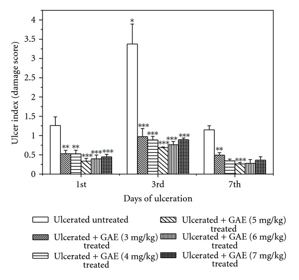
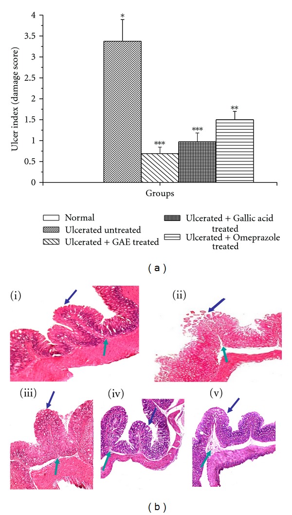
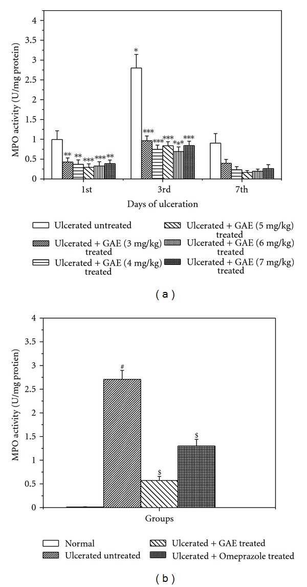
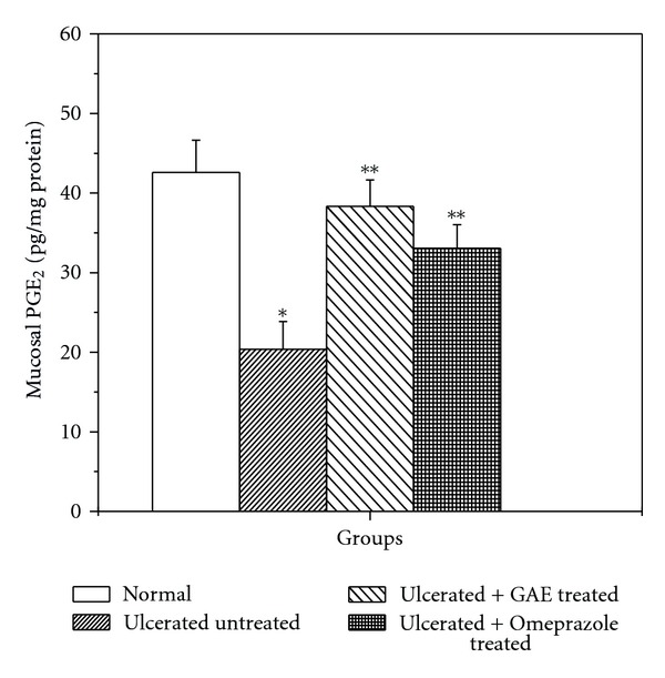
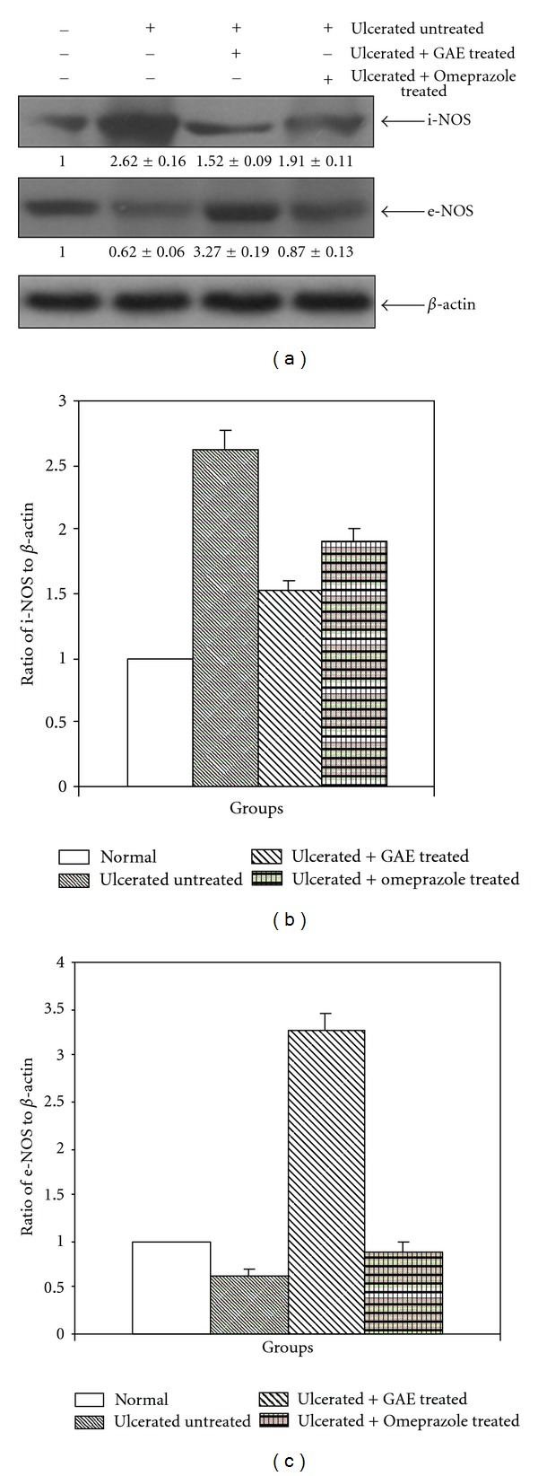
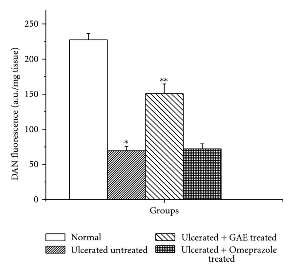
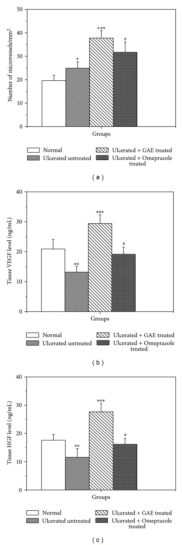
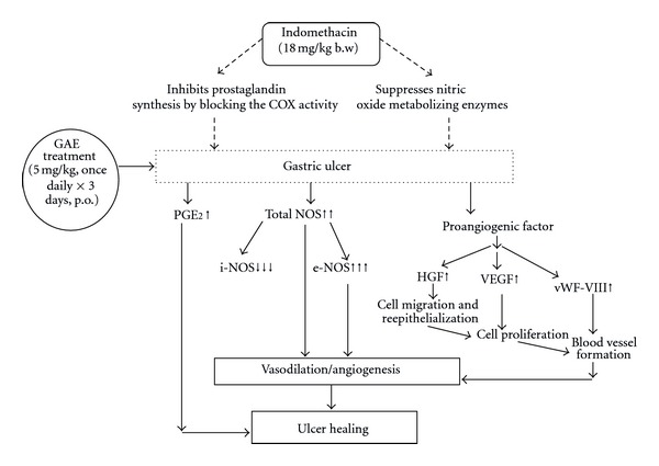
The healing activity of gallic acid enriched ethanolic extract (GAE) of Phyllanthus emblica fruits (amla) against the indomethacin-induced gastric ulceration in mice was investigated. The activity was correlated with the ability of GAE to alter the cyclooxygenase- (COX-) dependent healing pathways. Histology of the stomach tissues revealed maximum ulceration on the 3rd day after indomethacin (18 mg/kg, single dose) administration that was associated with significant increase in inflammatory factors, namely, mucosal myeloperoxidase (MPO) activity and inducible nitric oxide synthase (i-NOS) expression. Proangiogenic parameters such as the levels of prostaglandin (PG) E(2), vascular endothelial growth factor (VEGF), hepatocyte growth factor (HGF), von Willebrand Factor VIII, and endothelial NOS (e-NOS) were downregulated by indomethacin. Treatment with GAE (5 mg/kg/day) and omeprazole (3 mg/kg/day) for 3 days led to effective healing of the acute ulceration, while GAE could reverse the indomethacin-induced proinflammatory changes of the designated biochemical parameters. The ulcer healing activity of GAE was, however, compromised by coadministration of the nonspecific NOS inhibitor, N-nitro-L-arginine methyl ester (L-NAME), but not the i-NOS-specific inhibitor, L-N6-(1-iminoethyl) lysine hydrochloride (L-NIL). Taken together, these results suggested that the GAE treatment accelerates ulcer healing by inducing PGE(2) synthesis and augmenting e-NOS/i-NOS ratio.
Figures
Similar articles
-
Improved antiulcer and anticancer properties of a trans-resveratrol analog in mice.J Pharmacol Exp Ther. 2009 Mar;328(3):829-38. doi: 10.1124/jpet.108.145334. Epub 2008 Dec 9. J Pharmacol Exp Ther. 2009. PMID: 19066340
-
Ellagic acid facilitates indomethacin-induced gastric ulcer healing via COX-2 up-regulation.Acta Biochim Biophys Sin (Shanghai). 2012 Jul;44(7):565-76. doi: 10.1093/abbs/gms034. Epub 2012 May 24. Acta Biochim Biophys Sin (Shanghai). 2012. PMID: 22626975
-
dl-trans-3,4-Dihydroxy-1-selenolane (DHSred) heals indomethacin-mediated gastric ulcer in mice by modulating arginine metabolism.Biochim Biophys Acta. 2014 Dec;1840(12):3385-92. doi: 10.1016/j.bbagen.2014.09.004. Epub 2014 Sep 16. Biochim Biophys Acta. 2014. PMID: 25218693
-
Healing of duodenal ulcers is not impaired by indomethacin or rofecoxib, the selective COX-2 inhibitor, in rats.Digestion. 2002;66(3):145-53. doi: 10.1159/000066759. Digestion. 2002. PMID: 12481160
-
Nitric oxide and prostaglandin systems in the stimulation of hypothalamic-pituitary-adrenal axis by neurotransmitters and neurohormones.J Physiol Pharmacol. 2004 Dec;55(4):679-703. J Physiol Pharmacol. 2004. PMID: 15613736 Review.
Cited by
-
Clinical Potential of Himalayan Herb Bergenia ligulata: An Evidence-Based Study.Molecules. 2022 Oct 18;27(20):7039. doi: 10.3390/molecules27207039. Molecules. 2022. PMID: 36296631 Free PMC article. Review.
-
Role of dietary polyphenols in the management of peptic ulcer.World J Gastroenterol. 2015 Jun 7;21(21):6499-517. doi: 10.3748/wjg.v21.i21.6499. World J Gastroenterol. 2015. PMID: 26074689 Free PMC article. Review.
-
Recent Insights into the Morphological, Nutritional and Phytochemical Properties of Indian Gooseberry (Phyllanthus emblica) for the Development of Functional Foods.Plants (Basel). 2024 Feb 20;13(5):574. doi: 10.3390/plants13050574. Plants (Basel). 2024. PMID: 38475421 Free PMC article. Review.
-
Polysaccharide-rich fraction of Termitomyces eurhizus accelerate healing of indomethacin induced gastric ulcer in mice.Glycoconj J. 2013 Nov;30(8):759-68. doi: 10.1007/s10719-013-9479-5. Epub 2013 May 30. Glycoconj J. 2013. PMID: 23715800
-
Ameliorative effect of Phytocee™ Cool against carbon tetrachloride-induced oxidative stress.Pharmacognosy Res. 2014 Oct;6(4):320-5. doi: 10.4103/0974-8490.138284. Pharmacognosy Res. 2014. PMID: 25276070 Free PMC article.
References
-
- Khan KH. Roles of emblica officinalis in medicine—a review. Botany Research International. 2009;2(4):218–228.
-
- Anonymous. Case study on Amla-related patent, technology information. Forecasting & Assessment Council (TIFAC) Bulletin. 2001;7:6–6.
-
- Soong Y-Y, Barlow PJ. Quantification of gallic acid and ellagic acid from longan (Dimocarpus longan Lour.) seed and mango (Mangifera indica L.) kernel and their effects on antioxidant activity. Food Chemistry. 2006;97(3):524–530.
-
- Gentile JM, Rahimi S, Zwiesler J, Gentile GJ, Ferguson LR. Effect of selected antimutagens on the genotoxicity of antitumor agents. Mutation Research. 1998;402(1-2):289–298. - PubMed
-
- Indap MA, Radhika S, Motiwale L, Rao KVK. Anticancer activity of phenolic antioxidants against breast cancer cells and a spontaneous mammary tumor. Indian Journal of Pharmaceutical Sciences. 2006;68(4):470–474.
LinkOut - more resources
Full Text Sources
Research Materials
Miscellaneous
