

Unique intense uptake demonstrated by (18)F-FDG positron emission tomography/computed tomography in primary pancreatic lymphoma: A case report
- PMID: 22966351
- PMCID: PMC3436258
- DOI: 10.3892/ol_00000107
Unique intense uptake demonstrated by (18)F-FDG positron emission tomography/computed tomography in primary pancreatic lymphoma: A case report
Abstract
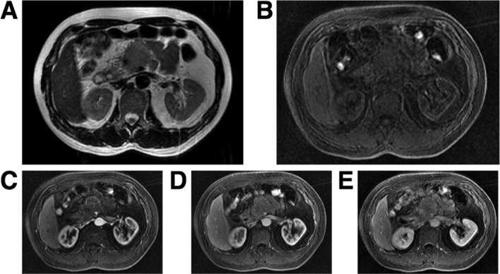
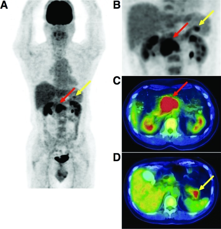
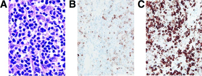
Patients with primary pancreatic lymphoma (PPL), which is rare, require a different therapeutic approach and have a better prognosis than those with pancreatic cancer. However, conventional imaging modalities alone are not able to differentiate between pancreatic cancer and other rare tumors such as PPL, although the accurate diagnosis of PPL is crucial. The development of new modalities such as F-18 2'-deoxy-2fluoro-D-glucose (FDG) positron emission tomography combined with computed tomography (PET/CT) contributes to the evaluation of lymphoma staging. However, few reports are currently available regarding PET/CT findings in PPL. In this study, a 56-year old man with PPL was examined using FDG PET/CT imaging, which showed the unique intense uptake of FDG in the pancreas with atypical findings of malignancy in the CT scan and magnetic resonance images.
Figures
Similar articles
-
18F-FDG PET/CT assists the diagnosis of primary pancreatic lymphoma: Two case reports and literature review.Front Med (Lausanne). 2024 Feb 23;11:1370762. doi: 10.3389/fmed.2024.1370762. eCollection 2024. Front Med (Lausanne). 2024. PMID: 38463493 Free PMC article.
-
Usefulness of (18)F-FDG positron emission tomography/computed tomography for the diagnosis of pyothorax-associated lymphoma: A report of three cases.Oncol Lett. 2010 Sep;1(5):833-836. doi: 10.3892/ol_00000146. Epub 2010 Sep 1. Oncol Lett. 2010. PMID: 22966389 Free PMC article.
-
More advantages in detecting bone and soft tissue metastases from prostate cancer using 18F-PSMA PET/CT.Hell J Nucl Med. 2019 Jan-Apr;22(1):6-9. doi: 10.1967/s002449910952. Epub 2019 Mar 7. Hell J Nucl Med. 2019. PMID: 30843003
-
Hyperaccumulation of (18)F-FDG in order to differentiate solid pseudopapillary tumors from adenocarcinomas and from neuroendocrine pancreatic tumors and review of the literature.Hell J Nucl Med. 2013 May-Aug;16(2):97-102. doi: 10.1967/s002449910084. Epub 2013 May 20. Hell J Nucl Med. 2013. PMID: 23687644 Review.
-
Clinical applications of (18)F-FDG PET in the management of hepatobiliary and pancreatic tumors.Abdom Imaging. 2012 Dec;37(6):983-1003. doi: 10.1007/s00261-012-9845-y. Abdom Imaging. 2012. PMID: 22527152 Review.
Cited by
-
Primary pancreatic lymphoma: two case reports and a literature review.Onco Targets Ther. 2017 Mar 20;10:1687-1694. doi: 10.2147/OTT.S121521. eCollection 2017. Onco Targets Ther. 2017. PMID: 28356755 Free PMC article.
-
Primary Pancreatic Lymphoma: Recommendations for Diagnosis and Management.J Blood Med. 2021 May 5;12:257-267. doi: 10.2147/JBM.S273095. eCollection 2021. J Blood Med. 2021. PMID: 33981170 Free PMC article. Review.
-
18F-FDG PET/CT assists the diagnosis of primary pancreatic lymphoma: Two case reports and literature review.Front Med (Lausanne). 2024 Feb 23;11:1370762. doi: 10.3389/fmed.2024.1370762. eCollection 2024. Front Med (Lausanne). 2024. PMID: 38463493 Free PMC article.
-
Usefulness of (18)F-FDG positron emission tomography/computed tomography for the diagnosis of pyothorax-associated lymphoma: A report of three cases.Oncol Lett. 2010 Sep;1(5):833-836. doi: 10.3892/ol_00000146. Epub 2010 Sep 1. Oncol Lett. 2010. PMID: 22966389 Free PMC article.
-
A case of early stage lung cancer detected by repeated cancer screening with positron emission tomography.Oncol Lett. 2012 Feb;3(2):297-299. doi: 10.3892/ol.2011.492. Epub 2011 Nov 22. Oncol Lett. 2012. PMID: 22740898 Free PMC article.
References
-
- Behrns KE, Sarr MG, Strickler JG. Pancreatic lymphoma: is it a surgical disease? Pancreas. 1994;9:662–667. - PubMed
-
- Freeman C, Berg JW, Cutler SJ. Occurrence and prognosis of extranodal lymphomas. Cancer. 1972;29:252–260. - PubMed
-
- Reed K, Vose PC, Jarstfer BS. Pancreatic cancer: 30 year review (1947–1977) Am J Surg. 1979;138:929–933. - PubMed
-
- Weiler-Sagie M, Bushelev O, Epelbaum R, Dann EJ, Haim N, Avivi I, Ben-Barak A, Ben-Arie Y, Bar-Shalom R, Israel O. (18)F-FDG avidity in lymphoma readdressed: a study of 766 patients. J Nucl Med. 2010;51:25–30. - PubMed
-
- Cronin CG, Swords R, Truong MT, Viswanathan C, Rohren E, Giles FJ, O’Dwyer M, Bruzzi JF. Clinical utility of PET/CT in lymphoma. AJR Am J Roentgenol. 2010;194:W91–W103. - PubMed
LinkOut - more resources
Full Text Sources