Guideline
. 2012 Apr;8(4):445-544.
doi: 10.4161/auto.19496.

Guidelines for the use and interpretation of assays for monitoring autophagy

Daniel J Klionsky  1 Fabio C AbdallaHagai AbeliovichRobert T AbrahamAbraham Acevedo-ArozenaKhosrow AdeliLotta AgholmeMaria AgnelloPatrizia AgostinisJulio A Aguirre-GhisoHyung Jun AhnOuardia Ait-MohamedSlimane Ait-Si-AliTakahiko AkematsuShizuo AkiraHesham M Al-YounesMunir A Al-ZeerMatthew L AlbertRoger L AlbinJavier Alegre-AbarrateguiMaria Francesca AleoMehrdad AlirezaeiAlexandru AlmasanMaylin Almonte-BecerrilAtsuo AmanoRavi AmaravadiShoba AmarnathAmal O AmerNathalie Andrieu-AbadieVellareddy AnantharamDavid K AnnShailendra Anoopkumar-DukieHiroshi AokiNadezda ApostolovaGiuseppe AranciaJohn P ArisKatsuhiko AsanumaNana Y O AsareHisashi AshidaValerie AskanasDavid S AskewPatrick AubergerMisuzu BabaSteven K BackuesEric H BaehreckeBen A BahrXue-Yuan BaiYannick BaillyRobert BaiocchiGiulia BaldiniWalter BalduiniAndrea BallabioBruce A BamberEdward T W BamptonGábor BánhegyiClinton R BartholomewDiane C BasshamRobert C Bast JrHenri BatokoBoon-Huat BayIsabelle BeauDaniel M BéchetThomas J BegleyChristian BehlChristian BehrendsSoumeya BekriBryan BellaireLinda J BendallLuca BenettiLaura BerliocchiHenri BernardiFrancesca BernassolaSébastien BesteiroIngrid Bhatia-KissovaXiaoning BiMartine Biard-PiechaczykJanice S BlumLawrence H BoisePaolo BonaldoDavid L BooneBeat C BornhauserKarina R BortoluciIoannis BossisFrédéric BostJean-Pierre BourquinPatricia BoyaMichaël Boyer-GuittautPeter V BozhkovNathan R BradyClaudio BrancoliniAndreas BrechJay E BrenmanAna BrennandEmery H BresnickPatrick BrestDave BridgesMolly L BristolPaul S BrookesEric J BrownJohn H BrumellNicola Brunetti-PierriUlf T BrunkDennis E BulmanScott J BultmanGeert BultynckLena F BurbullaWilfried BurschJonathan P ButcharWanda BuzgariuSergio P BydlowskiKen CadwellMonika CahováDongsheng CaiJiyang CaiQian CaiBruno CalabrettaJavier Calvo-GarridoNadine CamougrandMichelangelo CampanellaJenny Campos-SalinasEleonora CandiLizhi CaoAllan B CaplanSimon R CardingSandra M CardosoJennifer S CarewCathleen R CarlinVirginie CarmignacLeticia A M CarneiroSerena CarraRosario A CarusoGiorgio CasariCaty CasasRoberta CastinoEduardo CebolleroFrancesco CecconiJean CelliHassan ChaachouayHan-Jung ChaeChee-Yin ChaiDavid C ChanEdmond Y ChanRaymond Chuen-Chung ChangChi-Ming CheChing-Chow ChenGuang-Chao ChenGuo-Qiang ChenMin ChenQuan ChenSteve S-L ChenWenLi ChenXi ChenXiangmei ChenXiequn ChenYe-Guang ChenYingyu ChenYongqiang ChenYu-Jen ChenZhixiang ChenAlan ChengChristopher H K ChengYan ChengHeesun CheongJae-Ho CheongSara CherryRuss Chess-WilliamsZelda H CheungEric ChevetHui-Ling ChiangRoberto ChiarelliTomoki ChibaLih-Shen ChinShih-Hwa ChiouFrancis V ChisariChi Hin ChoDong-Hyung ChoAugustine M K ChoiDooSeok ChoiKyeong Sook ChoiMary E ChoiSalem ChouaibDivaker ChoubeyVinay ChoubeyCharleen T ChuTsung-Hsien ChuangSheau-Huei ChuehTaehoon ChunYong-Joon ChwaeMee-Len ChyeRoberto CiarciaMaria R CirioloMichael J ClagueRobert S B ClarkPeter G H ClarkeRobert ClarkePatrice CodognoHilary A CollerMaría I ColomboSergio CominciniMaria CondelloFabrizio CondorelliMark R CooksonGraham H CoombsIsabelle CoppensRamon CorbalanPascale CossartPaola CostelliSafia CostesAna Coto-MontesEduardo CouveFraser P CoxonJames M CreggJosé L CrespoMarianne J CronjéAna Maria CuervoJoseph J CullenMark J CzajaMarcello D'AmelioArlette Darfeuille-MichaudLester M DavidsFaith E DaviesMassimo De FeliciJohn F de GrootCornelis A M de HaanLuisa De MartinoAngelo De MilitoVincenzo De TataJayanta DebnathAlexei DegterevBenjamin DehayLea M D DelbridgeFrancesca DemarchiYi Zhen DengJörn DengjelPaul DentDonna DentonVojo DereticShyamal D DesaiRodney J DevenishMario Di GioacchinoGilbert Di PaoloChiara Di PietroGuillermo Díaz-ArayaInés Díaz-LaviadaMaria T Diaz-MecoJavier Diaz-NidoIvan DikicSavithramma P Dinesh-KumarWen-Xing DingClark W DistelhorstAbhinav DiwanMojgan Djavaheri-MergnySvetlana DokudovskayaZheng DongFrank C DorseyVictor DosenkoJames J DowlingStephen DoxseyMarlène DreuxMark E DrewQiuhong DuanMichel A DuchosalKaren DuffIsabelle DugailMadeleine DurbeejMichael DuszenkoCharles L EdelsteinAimee L EdingerGustavo EgeaLudwig EichingerN Tony EissaSuhendan EkmekciogluWafik S El-DeiryZvulun ElazarMohamed ElgendyLisa M EllerbyKai Er EngAnna-Mart EngelbrechtSimone EngelenderJekaterina ErenpreisaRicardo EscalanteAudrey EsclatineEeva-Liisa EskelinenLucile EspertVirginia EspinaHuizhou FanJia FanQi-Wen FanZhen FanShengyun FangYongqi FangManolis FantoAlessandro FanzaniThomas FarkasJean-Claude FarréMathias FaureMarcus FechheimerCarl G FengJian FengQili FengYouji FengLászló FésüsRalph FeuerMaria E Figueiredo-PereiraGian Maria FimiaDiane C FingarSteven FinkbeinerToren FinkelKim D FinleyFilomena FioritoEdward A FisherPaul B FisherMarc FlajoletMaria L Florez-McClureSalvatore FlorioEdward A FonFrancesco FornaiFranco FortunatoRati FotedarDaniel H FowlerHoward S FoxRodrigo FrancoLisa B FrankelMarc FransenJosé M FuentesJuan FueyoJun FujiiKozo FujisakiEriko FujitaMitsunori FukudaRuth H FurukawaMatthias GaestelPhilippe GaillyMalgorzata GajewskaBrigitte GalliotVincent GalySubramaniam GaneshBarry GanetzkyIan G GanleyFen-Biao GaoGeorge F GaoJinming GaoLorena GarciaGuillermo Garcia-ManeroMikel Garcia-MarcosMarjan GarmynAndrei L GartelEvelina GattiMathias GautelThomas R GawrilukMatthew E GeggJiefei GengMarc GermainJason E GestwickiDavid A GewirtzSaeid GhavamiPradipta GhoshAnna M GiammarioliAlexandra N GiatromanolakiSpencer B GibsonRobert W GilkersonMichael L GingerHenry N GinsbergJakub GolabMichael S GoligorskyPierre GolsteinCandelaria Gomez-ManzanoEbru GoncuCéline GongoraClaudio D GonzalezRamon GonzalezCristina González-EstévezRosa Ana González-PoloElena Gonzalez-ReyNikolai V GorbunovSharon GorskiSandro GoruppiRoberta A GottliebDevrim GozuacikGiovanna Elvira GranatoGary D GrantKim N GreenAleš GregorcFrédéric GrosCharles GroseThomas W GruntPhilippe GualJun-Lin GuanKun-Liang GuanSylvie M GuichardAnna S GukovskayaIlya GukovskyJan GunstAsa B GustafssonAndrew J HalaykoAmber N HaleSandra K HalonenMaho HamasakiFeng HanTing HanMichael K HancockMalene HansenHisashi HaradaMasaru HaradaStefan E HardtJ Wade HarperAdrian L HarrisJames HarrisSteven D HarrisMakoto HashimotoJeffrey A HaspelShin-ichiro HayashiLori A HazelhurstCongcong HeYou-Wen HeMarie-Joseé HébertKim A HeidenreichMiep H HelfrichGudmundur V HelgasonElizabeth P HenskeBrian HermanPaul K HermanClaudio HetzSabine HilfikerJoseph A HillLynne J HockingPaul HofmanThomas G HofmannJörg HöhfeldTessa L HolyoakeMing-Huang HongDavid A HoodGökhan S HotamisligilEwout J HouwerzijlMaria Høyer-HansenBingren HuChien-An A HuHong-Ming HuYa HuaCanhua HuangJu HuangShengbing HuangWei-Pang HuangTobias B HuberWon-Ki HuhTai-Ho HungTed R HuppGang Min HurJames B HurleySabah N A HussainPatrick J HusseyJung Jin HwangSeungmin HwangAtsuhiro IchiharaShirin IlkhanizadehKen InokiTakeshi IntoValentina IovaneJuan L IovannaNancy Y IpYoshitaka IsakaHiroyuki IshidaCiro IsidoroKen-ichi IsobeAkiko IwasakiMarta IzquierdoYotaro IzumiPanu M JaakkolaMarja JäätteläGeorge R JacksonWilliam T JacksonBassam JanjiMarina JendrachJu-Hong JeonEui-Bae JeungHong JiangHongchi JiangJean X JiangMing JiangQing JiangXuejun JiangXuejun JiangAlberto JiménezMeiyan JinShengkan JinCheol O JoeTerje JohansenDaniel E JohnsonGail V W JohnsonNicola L JonesBertrand JosephSuresh K JosephAnnie M JoubertGábor JuhászLucienne Juillerat-JeanneretChang Hwa JungYong-Keun JungKai KaarnirantaAllen KaasikTomohiro KabutaMotoni KadowakiKatarina KagedalYoshiaki KamadaVitaliy O KaminskyyHarm H KampingaHiromitsu KanamoriChanhee KangKhong Bee KangKwang Il KangRui KangYoon-A KangTomotake KankiThirumala-Devi KannegantiHaruo KannoAnumantha G KanthasamyArthi KanthasamyVassiliki KarantzaGur P KaushalSusmita KaushikYoshinori KawazoePo-Yuan KeJohn H KehrlAmeeta KelekarClaus KerkhoffDavid H KesselHany KhalilJan A K W KielAmy A KigerAkio KiharaDeok Ryong KimDo-Hyung KimDong-Hou KimEun-Kyoung KimHyung-Ryong KimJae-Sung KimJeong Hun KimJin Cheon KimJohn K KimPeter K KimSeong Who KimYong-Sun KimYonghyun KimAdi KimchiAlec C KimmelmanJason S KingTimothy J KinsellaVladimir KirkinLorrie A KirshenbaumKatsuhiko KitamotoKaio KitazatoLudger KleinWalter T KlimeckiJochen KluckenErwin KnechtBen C B KoJan C KochHiroshi KogaJae-Young KohYoung Ho KohMasato KoikeMasaaki KomatsuEiki KominamiHee Jeong KongWei-Jia KongViktor I KorolchukYaichiro KotakeMichael I KoukourakisJuan B Kouri FloresAttila L KovácsClaudine KraftDimitri KraincHelmut KrämerCarole Kretz-RemyAnna M KrichevskyGuido KroemerRejko KrügerOleg KrutNicholas T KtistakisChia-Yi KuanRoza KucharczykAshok KumarRaj KumarSharad KumarMondira KunduHsing-Jien KungTino KurzHo Jeong KwonAlbert R La SpadaFrank LafontTrond LamarkJacques LandryJon D LanePierre LapaquetteJocelyn F LaporteLajos LászlóSergio LavanderoJosée N LavoieRobert LayfieldPedro A LazoWeidong LeLaurent Le CamDaniel J LedbetterAlvin J X LeeByung-Wan LeeGyun Min LeeJongdae LeeJu-Hyun LeeMichael LeeMyung-Shik LeeSug Hyung LeeChristiaan LeeuwenburghPatrick LegembreRenaud LegouisMichael LehmannHuan-Yao LeiQun-Ying LeiDavid A LeibJosé LeiroJohn J LemastersAntoinette LemoineMaciej S LesniakDina LevVictor V LevensonBeth LevineEfrat LevyFaqiang LiJun-Lin LiLian LiSheng LiWeijie LiXue-Jun LiYan-bo LiYi-Ping LiChengyu LiangQiangrong LiangYung-Feng LiaoPawel P LiberskiAndrew LiebermanHyunjung J LimKah-Leong LimKyu LimChiou-Feng LinFu-Cheng LinJian LinJiandie D LinKui LinWan-Wan LinWeei-Chin LinYi-Ling LinRafael LindenPaul LingorJennifer Lippincott-SchwartzMichael P LisantiPaloma B LitonBo LiuChun-Feng LiuKaiyu LiuLeyuan LiuQiong A LiuWei LiuYoung-Chau LiuYule LiuRichard A LockshinChun-Nam LokSagar LonialBenjamin LoosGabriel Lopez-BeresteinCarlos López-OtínLaura LossiMichael T LotzePeter LőwBinfeng LuBingwei LuBo LuZhen LuFrédéric LucianoNicholas W LukacsAnders H LundMelinda A Lynch-DayYong MaFernando MacianJeff P MacKeiganKay F MacleodFrank MadeoLuigi MaiuriMaria Chiara MaiuriDavide MalagoliMay Christine V MalicdanWalter MalorniNa ManEva-Maria MandelkowStéphen ManonIrena ManovKai MaoXiang MaoZixu MaoPhilippe MarambaudDaniela MarazzitiYves L MarcelKatie MarchbankPiero MarchettiStefan J MarciniakMateus MarcondesMohsen MardiGabriella MarfeGuillermo MariñoMaria MarkakiMark R MartenSeamus J MartinCamille Martinand-MariWim MartinetMarta Martinez-VicenteMatilde MasiniPaola MatarreseSaburo MatsuoRaffaele MatteoniAndreas MayerNathalie M MazureDavid J McConkeyMelanie J McConnellCatherine McDermottChristine McDonaldGerald M McInerneySharon L McKennaBethAnn McLaughlinPamela J McLeanChristopher R McMasterG Angus McQuibbanAlfred J MeijerMiriam H MeislerAlicia MeléndezThomas J MeliaGerry MelinoMaria A MenaJavier A MenendezRubem F S Menna-BarretoManoj B MenonFiona M MenziesCarol A MercerAdalberto MerighiDiane E MerryStefania MeschiniChristian G MeyerThomas F MeyerChao-Yu MiaoJun-Ying MiaoPaul A M MichelsCarine MichielsDalibor MijaljicaAna MilojkovicSaverio MinucciClelia MiraccoCindy K MirantiIoannis MitroulisKeisuke MiyazawaNoboru MizushimaBaharia MograbiSimin MohseniXavier MoleroBertrand MollereauFaustino MollinedoTakashi MomoiIryna MonastyrskaMartha M MonickMervyn J MonteiroMichael N MooreRodrigo MoraKevin MoreauPaula I MoreiraYuji MoriyasuJorge MoscatSerge MostowyJeremy C MottramTomasz MotylCharbel E-H MoussaSylke MüllerSylviane MullerKarl MüngerChristian MünzLeon O MurphyMaureen E MurphyAntonio MusaròIndira MysorekarEiichiro NagataKazuhiro NagataAimable NahimanaUsha NairToshiyuki NakagawaKiichi NakahiraHiroyasu NakanoHitoshi NakatogawaMeera NanjundanNaweed I NaqviDerek P NarendraMasashi NaritaMiguel NavarroSteffan T NawrockiTaras Y NazarkoAndriy NemchenkoMihai G NeteaThomas P NeufeldPaul A NeyIoannis P NezisHuu Phuc NguyenDaotai NieIchizo NishinoCorey NislowRalph A NixonTakeshi NodaAngelika A NoegelAnna NogalskaSatoru NoguchiLucia NotterpekIvana NovakTomoyoshi NozakiNobuyuki NukinaThorsten NürnbergerBeat NyfelerKeisuke ObaraTerry D OberleySalvatore OddoMichinaga OgawaToya OhashiKoji OkamotoNancy L OleinickF Javier OliverLaura J OlsenStefan OlssonOnya OpotaTimothy F OsborneGary K OstranderKinya OtsuJing-hsiung James OuMireille OuimetMichael OverholtzerBulent OzpolatPaolo PaganettiUgo PagniniNicolas PalletGlen E PalmerCamilla PalumboTianhong PanTheocharis PanaretakisUdai Bhan PandeyZuzana PapackovaIssidora PapassideriIrmgard ParisJunsoo ParkOhkmae K ParkJan B ParysKatherine R ParzychSusann PatschanCam PattersonSophie PattingreJohn M PawelekJianxin PengDavid H PerlmutterIda PerrottaGeorge PerryShazib PervaizMatthias PeterGodefridus J PetersMorten PetersenGoran PetrovskiJames M PhangMauro PiacentiniPhilippe PierreValérie Pierrefite-CarleGérard PierronRonit Pinkas-KramarskiAntonio PirasNatik PiriLeonidas C PlataniasStefanie PöggelerMarc PoirotAngelo PolettiChristian PoüsMercedes Pozuelo-RubioMette Prætorius-IbbaAnil PrasadMark PrescottMuriel PriaultNathalie Produit-ZengaffinenAnn Progulske-FoxTassula Proikas-CezanneSerge PrzedborskiKarin PrzyklenkRosa PuertollanoJulien PuyalShu-Bing QianLiang QinZheng-Hong QinSusan E QuagginNina RabenHannah RabinowichSimon W RabkinIrfan RahmanAbdelhaq RamiGeorg RammGlenn RandallFelix RandowV Ashutosh RaoJeffrey C RathmellBrinda RavikumarSwapan K RayBruce H ReedJohn C ReedFulvio ReggioriAnne Régnier-VigourouxAndreas S ReichertJohn J Reiners JrRussel J ReiterJun RenJosé L RevueltaChristopher J RhodesKonstantinos RitisElizete RizzoJeffrey RobbinsMichel RobergeHernan RocaMaria C RoccheriStephane RocchiH Peter RodemannSantiago Rodríguez de CórdobaBärbel RohrerIgor B RoninsonKirill RosenMagdalena M Rost-RoszkowskaMustapha RouisKasper M A RouschopFrancesca RovettaBrian P RubinDavid C RubinszteinKlaus RuckdeschelEdmund B Rucker 3rdAssaf RudichEmil RudolfNelson Ruiz-OpazoRossella RussoTor Erik RustenKevin M RyanStefan W RyterDavid M SabatiniJunichi SadoshimaTapas SahaTatsuya SaitohHiroshi SakagamiYasuyoshi SakaiGhasem Hoseini SalekdehPaolo SalomoniPaul M SalvaterraGuy SalvesenRosa SalvioliAnthony M J SanchezJosé A Sánchez-AlcázarRicardo Sánchez-PrietoMarco SandriUma SankarPoonam SansanwalLaura SantambrogioShweta SaranSovan SarkarMinnie SarwalChihiro SasakawaAusra SasnauskieneMiklós SassKen SatoMiyuki SatoAnthony H V SchapiraMichael ScharlHermann M SchätzlWiep ScheperStefano SchiaffinoClaudio SchneiderMarion E SchneiderRegine Schneider-StockPatricia V SchoenleinDaniel F SchorderetChristoph SchüllerGary K SchwartzLuca ScorranoLinda SealyPer O SeglenJuan Segura-AguilarIban SeiliezOleksandr SeleverstovChristian SellJong Bok SeoDuska SeparovicVijayasaradhi SetaluriTakao SetoguchiCarmine SettembreJohn J ShackaMala ShanmugamIrving M ShapiroEitan ShaulianReuben J ShawJames H ShelhamerHan-Ming ShenWei-Chiang ShenZu-Hang ShengYang ShiKenichi ShibuyaYoshihiro ShidojiJeng-Jer ShiehChwen-Ming ShihYohta ShimadaShigeomi ShimizuTakahiro ShintaniOrian S ShirihaiGordon C ShoreAndriy A SibirnyStan B SidhuBeata SikorskaElaine C M Silva-ZacarinAlison SimmonsAnna Katharina SimonHans-Uwe SimonCristiano SimoneAnne SimonsenDavid A SinclairRajat SinghDebasish SinhaFrank A SinicropeAgnieszka SirkoParco M SiuEfthimios SivridisVojtech SkopVladimir P SkulachevRuth S SlackSoraya S SmailiDuncan R SmithMaria S SoengasThierry SoldatiXueqin SongAnil K SoodTuck Wah SoongFederica SotgiaStephen A SpectorClaudia D SpiesWolfdieter SpringerSrinivasa M SrinivasulaLeonidas StefanisJoan S SteffanRuediger StendelHarald StenmarkAnastasis StephanouStephan T SternCinthya SternbergBjörn StorkPeter StrålforsCarlos S SubausteXinbing SuiDavid SulzerJiaren SunShi-Yong SunZhi-Jun SunJoseph J Y SungKuninori SuzukiToshihiko SuzukiMichele S SwansonCharles SwantonSean T SweeneyLai-King SyGyorgy SzabadkaiIra TabasHeinrich TaegtmeyerMarco TafaniKrisztina Takács-VellaiYoshitaka TakanoKaoru TakegawaGenzou TakemuraFumihiko TakeshitaNicholas J TalbotKevin S W TanKeiji TanakaKozo TanakaDaolin TangDingzhong TangIsei TanidaBakhos A TannousNektarios TavernarakisGraham S TaylorGregory A TaylorJ Paul TaylorLance S TeradaAlexei TermanGianluca TettamantiKarin ThevissenCraig B ThompsonAndrew ThorburnMichael ThummFengFeng TianYuan TianGlauco Tocchini-ValentiniAviva M TolkovskyYasuhiko TominoLars TöngesSharon A ToozeCathy TournierJohn TowerRoberto TownsVladimir TrajkovicLeonardo H TravassosTing-Fen TsaiMario P TschanTakeshi TsubataAllan TsungBoris TurkLorianne S TurnerSuresh C TyagiYasuo UchiyamaTakashi UenoMidori UmekawaRika Umemiya-ShirafujiVivek K UnniMaria I VaccaroEnza Maria ValenteGreet Van den BergheIda J van der KleiWouter van DoornLinda F van DykMarjolein van EgmondLeo A van GrunsvenPeter VandenabeeleWim P VandenbergheIlse VanhorebeekEva C VaqueroGuillermo VelascoTibor VellaiJose Miguel VicencioRichard D VierstraMiquel VilaCécile VindisGiampietro ViolaMaria Teresa ViscomiOlga V VoitsekhovskajaClarissa von HaefenMarcela VotrubaKeiji WadaRichard Wade-MartinsCheryl L WalkerCraig M WalshJochen WalterXiang-Bo WanAimin WangChenguang WangDawei WangFan WangFen WangGuanghui WangHaichao WangHong-Gang WangHorng-Dar WangJin WangKe WangMei WangRichard C WangXinglong WangXuejun WangYing-Jan WangYipeng WangZhen WangZhigang Charles WangZhinong WangDerick G WansinkDiane M WardHirotaka WatadaSarah L WatersPaul WebsterLixin WeiConrad C WeihlWilliam A WeissScott M WelfordLong-Ping WenCaroline A WhitehouseJ Lindsay WhittonAlexander J WhitworthTom WilemanJohn W WileySimon WilkinsonDieter WillboldRoger L WilliamsPeter R WilliamsonBradly G WoutersChenghan WuDao-Cheng WuWilliam K K WuAndreas WyttenbachRamnik J XavierZhijun XiPu XiaGengfu XiaoZhiping XieZhonglin XieDa-zhi XuJianzhen XuLiang XuXiaolei XuAi YamamotoAkitsugu YamamotoShunhei YamashinaMichiaki YamashitaXianghua YanMitsuhiro YanagidaDun-Sheng YangElizabeth YangJin-Ming YangShi Yu YangWannian YangWei Yuan YangZhifen YangMeng-Chao YaoTso-Pang YaoBehzad YeganehWei-Lien YenJia-jing YinXiao-Ming YinOok-Joon YooGyesoon YoonSeung-Yong YoonTomohiro YorimitsuYuko YoshikawaTamotsu YoshimoriKohki YoshimotoHo Jin YouRichard J YouleAnas YounesLi YuLong YuSeong-Woon YuWai Haung YuZhi-Min YuanZhenyu YueCheol-Heui YunMichisuke YuzakiOlga ZabirnykElaine Silva-ZacarinDavid ZacksEldad ZacksenhausNadia ZaffaroniZahra ZakeriHerbert J Zeh 3rdScott O ZeitlinHong ZhangHui-Ling ZhangJianhua ZhangJing-Pu ZhangLin ZhangLong ZhangMing-Yong ZhangXu Dong ZhangMantong ZhaoYi-Fang ZhaoYing ZhaoZhizhuang J ZhaoXiaoxiang ZhengBoris ZhivotovskyQing ZhongCong-Zhao ZhouChanglian ZhuWei-Guo ZhuXiao-Feng ZhuXiongwei ZhuYuangang ZhuTeresa ZoladekWei-Xing ZongAntonio ZorzanoJürgen ZschockeBrian Zuckerbraun
Affiliations
Guideline

Guidelines for the use and interpretation of assays for monitoring autophagy

Daniel J Klionsky et al. Autophagy. 2012 Apr.

Abstract

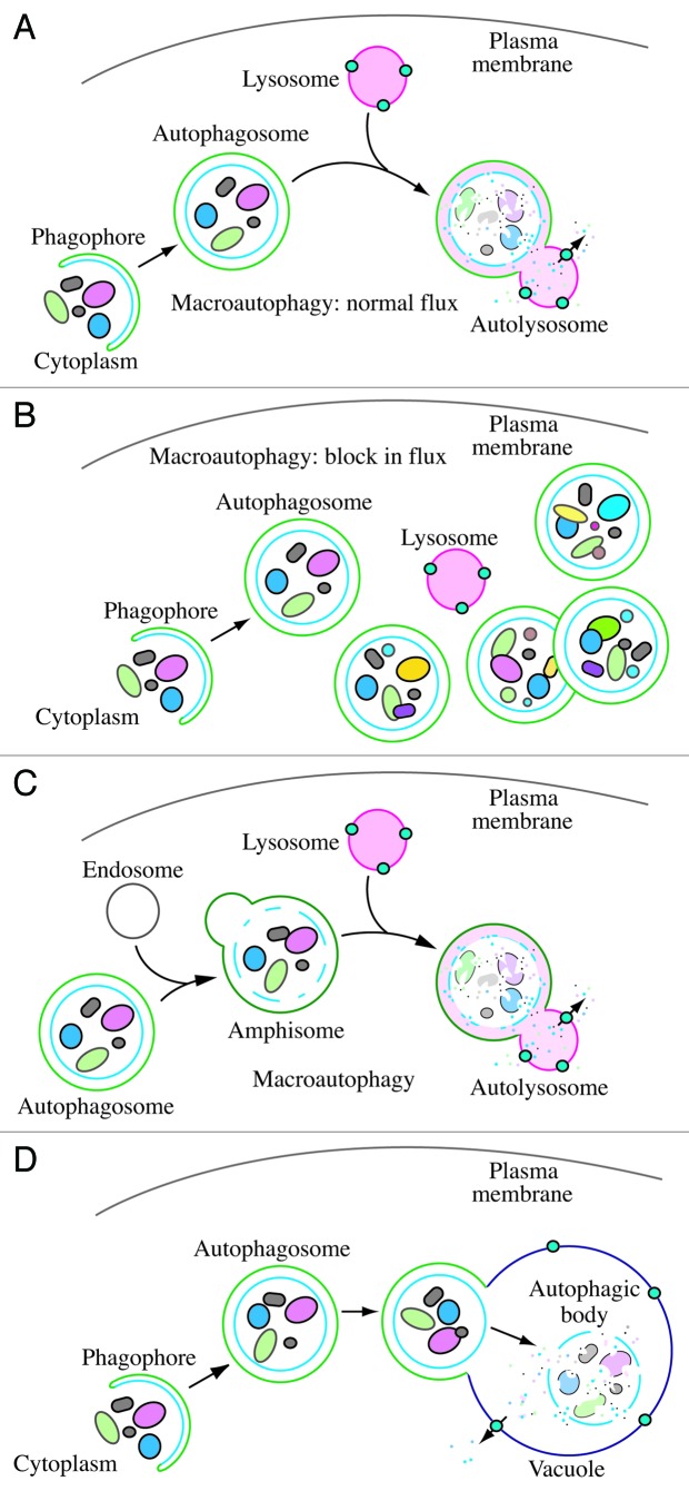
In 2008 we published the first set of guidelines for standardizing research in autophagy. Since then, research on this topic has continued to accelerate, and many new scientists have entered the field. Our knowledge base and relevant new technologies have also been expanding. Accordingly, it is important to update these guidelines for monitoring autophagy in different organisms. Various reviews have described the range of assays that have been used for this purpose. Nevertheless, there continues to be confusion regarding acceptable methods to measure autophagy, especially in multicellular eukaryotes. A key point that needs to be emphasized is that there is a difference between measurements that monitor the numbers or volume of autophagic elements (e.g., autophagosomes or autolysosomes) at any stage of the autophagic process vs. those that measure flux through the autophagy pathway (i.e., the complete process); thus, a block in macroautophagy that results in autophagosome accumulation needs to be differentiated from stimuli that result in increased autophagic activity, defined as increased autophagy induction coupled with increased delivery to, and degradation within, lysosomes (in most higher eukaryotes and some protists such as Dictyostelium) or the vacuole (in plants and fungi). In other words, it is especially important that investigators new to the field understand that the appearance of more autophagosomes does not necessarily equate with more autophagy. In fact, in many cases, autophagosomes accumulate because of a block in trafficking to lysosomes without a concomitant change in autophagosome biogenesis, whereas an increase in autolysosomes may reflect a reduction in degradative activity. Here, we present a set of guidelines for the selection and interpretation of methods for use by investigators who aim to examine macroautophagy and related processes, as well as for reviewers who need to provide realistic and reasonable critiques of papers that are focused on these processes. These guidelines are not meant to be a formulaic set of rules, because the appropriate assays depend in part on the question being asked and the system being used. In addition, we emphasize that no individual assay is guaranteed to be the most appropriate one in every situation, and we strongly recommend the use of multiple assays to monitor autophagy. In these guidelines, we consider these various methods of assessing autophagy and what information can, or cannot, be obtained from them. Finally, by discussing the merits and limits of particular autophagy assays, we hope to encourage technical innovation in the field.


Figures

None
Figure 1. Schematic model demonstrating the induction of autophagosome formation when turnover is blocked vs. normal autophagic flux, and illustrating the morphological intermediates of macroautophagy. (A) The initiation of autophagy includes the formation of the phagophore, the initial sequestering compartment, which expands into an autophagosome. Completion of the autophagosome is followed by fusion with lysosomes and degradation of the contents, allowing complete flux, or flow, through the entire pathway. This is a different outcome than the situation shown in (B) where induction results in the initiation of autophagy, but a defect in autophagosome turnover due, for example, to a block in fusion with lysosomes or disruption of lysosomal functions will result in an increased number of autophagosomes. In this scenario, autophagy has been induced, but there is no or limited autophagic flux. (C) An autophagosome can fuse with an endosome to generate an amphisome, prior to fusion with the lysosome. (D) Schematic drawing showing the formation of an autophagic body in plants and fungi. The large size of the plant and fungal vacuole relative to autophagosomes allows the release of the single-membrane autophagic body within the vacuole lumen. In cells that lack vacuolar hydrolase activity, or in the presence of inhibitors that block hydrolase activity, intact autophagic bodies accumulate within the vacuole lumen and can be detected by light microscopy. The lysosome of most higher eukaryotes is too small to allow the release of an autophagic body.
None
Figure 2. TEM images of autophagic vacuoles in isolated mouse hepatocytes. (A) One autophagosome or early autophagic vacuole (AVi) and one degradative autophagic vacuole (AVd) are shown. The AVi can be identified by its contents (morphologically intact cytoplasm, inlcuding ribosomes, and rough endoplasmic reticulum), and the limiting membrane that is partially visible as two bilayers separated by a narrow electron-lucent cleft, i. e., as a double membrane (arrow). The AVd can be identified by its contents, partially degraded, electron-dense rough endoplasmic reticulum. The vesicle next to the AVd is an endosomal/lysosomal structure containing 5-nm gold particles that were added to the culture medium to trace the endocytic pathway. (B) One AVi, containing rough endoplasmic reticulum and a mitochondrion, and one AVd, containing partially degraded rough endoplasmic reticulum, are shown. Note that the limiting membrane of the AVi is not clearly visible, possibly because it is tangentially sectioned. However, the electron-lucent cleft between the two limiting membranes is visible and helps in the identification of the AVi. The AVd contains a region filled by small internal vesicles (asterisk), indicating that the AVd has fused with a multivesicular endosome. mi, mitochondrion. Image provided by E.-L. Eskelinen.
None
Figure 3. Different autophagic vesicles observed after freeze fracturing in cultured osteosarcoma cells after treatment with the autophagy inducer voacamine. (A) Early autophagosome delimited by a double membrane. (B) Inner monolayer of an autophagosome membrane lacking protein particles. (C) Autolysosome delimited by a single membrane rich in protein particles. In the cross-fractured portion (on the right) the profile of the single membrane and the inner digested material are easily visible. Images provided by S. Meschini, M. Condello and A. Giuseppe.
None
Figure 4. Cryoelectron microscopy can be used as a three-dimensional approach to monitor the autophagic process. Two computed sections of an electron tomogram of the autophagic vesicle-rich cytoplasm in a hemophagocyte of a semi-thin section after high-pressure freezing preparation. The dashed area is membrane-free (A) but tomography reveals newly formed phagophore-like membranes (B). Image published previously and provided by M. Schneider and P. Walter.
None
Figure 5. LC3-I conversion and LC3-II turnover. (A) Expression levels of LC3-I and LC3-II during starvation. Atg5+/+ (wild-type) and Atg5−/− MEFs were cultured in DMEM without amino acids and serum for the indicated times, and then subjected to immunoblot analysis using anti-LC3 antibody and anti-tubulin antibody. E-64d (10 µg/ml) and pepstatin A (10 µg/ml) were added to the medium where indicated. Positions of LC3-I and LC3-II are indicated. The inclusion of lysosomal protease inhibitors reveals that the apparent decrease in LC3-II is due to lysosomal degradation as easily seen by comparing samples with and without inhibitors at the same time points (the overall decrease seen in the presence of inhibitors may reflect decreasing effectiveness of the inhibitors over time). Monitoring autophagy by following steady-state amounts of LC3-II without including inhibitors in the analysis can result in an incorrect interpretation that autophagy is not taking place (due to the apparent absence of LC3-II). Conversely, if there are high levels of LC3-II but there is no change in the presence of inhibitors this may indicate that induction has occurred but that the final steps of autophagy are blocked, resulting in stabilization of this protein. This figure was modified from data previously published in reference and is reproduced by permission of Landes Bioscience, copyright 2007. (B) Lysates of 4 human adipose tissue biopsies were resolved on two 12% polyacrylamide gels, as described previously. Proteins were transferred in parallel to either a PVDF or a nitrocellulose membrane, and blotted with anti-LC3 antibody, and then identified by reacting the membranes with an HRP-conjugated anti-rabbit IgG antibody, followed by ECL. The LC3-II/LC3-I ratio was calculated based on densitometry analysis of both bands. *p < 0.05. (C) HEK 293 and HeLa cells were cultured in nutrient-rich medium (DMEM containing10% FCS) or incubated for 4 h in starvation conditions (Krebs-Ringer medium) in the absence (-) or presence (+) of E-64d and pepstatin at 10 µg/ml each (Inhibitors). Cells were then lysed and the proteins resolved by SDS-PAGE. Endogenous LC3 was detected by immunoblotting. Positions of LC3-I and LC3-II are indicated. In the absence of lysosomal protease inhibitors, starvation results in a modest increase (HEK 293 cells) or even a decrease (HeLa cells) in the amount of LC3-II. The use of inhibitors reveals that this apparent decrease is due to lysosome-dependent degradation. This figure was modified from data previously published in reference and is reproduced by permission of Landes Bioscience, copyright 2005. (D) Sequence and schematic representation of the different forms of LC3B. The sequence for the nascent (proLC3) from mouse is shown. The glycine at position 120 indicates the cleavage site for ATG4. After this cleavage, the truncated LC3 is referred to as LC3-I, which is still a soluble form of the protein. Conjugation to PE generates the membrane-associated LC3-II (equivalent to Atg8–PE).
None
Figure 6. Effect of different inhibitors on LC3-II accumulation. SH-SY5Y human neuroblastoma cells were plated and allowed to adhere for a minimum of 24 h, then treated in fresh medium. Treatments were as follows: rapamycin (Rap), (A) 1 µM, 4 h or (B) 10 µM, 4 h; E-64d, final concentration 10 µg/ml from a 1 mg/ml stock in ethanol (ETOH); NH4Cl (NH4+), final concentration 10 mM from a 1 M stock in water; pepstatin A (Pst), final concentration 10 µg/ml from a 1 mg/ml stock in ethanol, or 68.6 µg/ml from a 6.86 mg/ml stock in DMSO; ethanol or DMSO, final concentration 1%. Pre-incubations in (B) were for 1 or 4 h as indicated. 10 mM NH4Cl (or 30 µM chloroquine, not shown) were the most effective compounds for demonstrating the accumulation of LC3-II. E-64d was also effective in preventing the degradation of LC3-II, with or without a preincubation, but ammomium chloride (or chloroquine) may be more effective. Pepstatin A at 10 µg/ml with a 1 h pre-incubation was not effective at blocking degradation, whereas a 100 µM concentration with 4 h pre-incubation had a partial effect. Thus, alkalinizing compounds are more effective in blocking LC3-II degradation, and pepstatin A must be used at saturating conditions to have any noticeable effect. Images provided by C. Isidoro. Note that the band running just below LC3-I at approximately 17.5 kDa may be a processing intermediate of LC3-I; it is detectable in freshly prepared homogenates, but is less visible after the sample is subjected to a freeze-thaw cycle.
None
Figure 7. GFP-LC3 processing can be used to monitor delivery of autophagosomal membranes. (A) Atg5−/− MEFs engineered to express Atg5 under the control of the Tet-off promoter were grown in the presence of doxycyline (10 ng/ml) for one week to suppress autophagy. Cells were then cultured in the absence of drug for the indicated times, with or without a final 2 h starvation. Protein lysates were analyzed by western blot using anti-LC3 and anti-GFP antibodies. The positions of untagged and GFP-tagged LC3-I and LC3-II, and free GFP are indicated. This figure was modified from data previously published in reference , FEBS Letters, 580, Hosokawa N, Hara Y, Mizushima N, Generation of cell lines with tetracycline-regulated autophagy and a role for autophagy in controlling cell size, pp. 2623–2629, copyright 2006, with permission from Elsevier. (B) Differential role of unsaturating and saturating concentrations of lysosomal inhibitors on GFP-LC3 cleavage. HeLa cells stably transfected with GFP-LC3 were treated with various concentrations of chloroquine (CQ) for 6 h. Total lysates were prepared and subjected to immunoblot analysis. (C) CQ-induced free GFP fragments require classical autophagy machinery. Wild-type and Atg5−/− MEFs were first infected with adenovirus GFP-LC3 (100 viral particles per cell) for 24 h. The cells were then either cultured in regular culture medium with or without CQ (10 µM), or subjected to starvation in EBSS buffer in the absence or presence of CQ for 6 h. Total lysates were prepared and subjected to immunoblot analysis. (B and C) are modified from the data previously published in reference .
None
Figure 8. Movement of activated pDendra2-hp62 (orange) from the nucleus (middle) to the aggregate in ARPE-19 cells, revealed by confocal microscopy. Cells were exposed to 5 µM MG132 for 24 h to induce the formation of perinuclear aggregates. The cells were then exposed to a UV pulse (the UV-induced area is shown by red lines that are inside of the nucleus) that converts Dendra2 from green to red, and the time shown after the pulse is indicated. SQSTM1/p62 is present in a small nuclear aggregrate, and is shuttled from the nucleus to a perinuclear large protein aggregate (detected as red). Scale bar, 5 µm. Image provided by K. Kaarniranta.
None
Figure 9. Changes in the detection and localization of GFP-LC3 upon the induction of autophagy. U87 cells stably expressing GFP-LC3 were treated with PBS, rapamycin (200 nM), or rapamycin in combination with 3-MA (2 mM) for 24 h. Representative fluorescence images of cells counterstained with DAPI (nuclei) are shown. Scale bar, 10 µm. This figure was modified from Figure 6 published in Badr et al. Lanatoside C sensitizes glioblastoma cells to tumor necrosis factor–related apoptosis-inducing ligand and induces an alternative cell death pathway. Neuro-Oncology 2011, 13:1213–24, by permission of Oxford University Press.
None
Figure 10. The GFP and mRFP signals of tandem fluorescent LC3 (tfLC3, mRFP-GFP-LC3) show different localization patterns. HeLa cells were cotransfected with plasmids expressing either tfLC3 or LAMP1-CFP. Twenty-four hours after the transfection, the cells were starved in Hanks’ solution for 2 h, fixed and analyzed by microscopy. The lower panels are a higher magnification of the upper panels. Bar, 10 µm in the upper panels and 2 µm in the lower panels. Arrows in the lower panels point to (or mark the location of) typical examples of colocalized signals of mRFP and LAMP1. Arrowheads point to (or mark the location of) typical examples of colocalized particles of GFP and mRFP signals. This figure was previously published in reference and is reproduced by permission of Landes Bioscience, copyright 2007.
None
Figure 11. GFP fluorescence in the autolysosome can be recovered upon neutralization of the pH. (A) GFP-LC3 emits green fluorescence in the autolysosomes of post-mortem processed heart sections. Cryosections of 3.8% paraformaldehyde fixed ventricular myocardium from 3-week old GFP-LC3 transgenic mice at the baseline (control) or starved for 24 h (starved) were processed for immunostaining using a standard protocol (buffered at pH 7.4). Most of the GFP-LC3 puncta are positive for LAMP1, suggesting that the autolysosomes had recovered GFP fluorescence. (B) Colocalization between GFP-LC3 direct fluorescence (green) and indirect immunostaining for GFP (red). Sections processed as in (A) were immunostained for GFP using a red fluorescence-tagged secondary antibody, and the colocalization with GFP fluorescence was examined by confocal microscopy. Almost all of the red puncta emit green fluorescence. Images provided by Xuejun Wang.
None
Figure 12. Saponin extraction allows quantification of LC3-II fluorescence by FACS. (A) Schematic diagram of the effects of the saponin wash. Due to the reorganization of the EGFP-LC3 reporter protein, induction of autophagosome formation does not change the total levels of fluorescence in EGFP-LC3-transfected cells. However, extraction of EGFP-LC3-I with saponin results in a higher level of fluorescence in cells with proportionally higher levels of EGFP-LC3-II-containing autophagosomes. This figure was previously published in reference (B) Saponin extraction can also be used to measure flux of endogenous LC3 protein. Human osteosarcoma cells were starved of amino acids and serum by incubation in EBSS, for the indicated times in the presence or absence of a 1 h chloroquine (50 µM) treatment. Cells were then washed with PBS containing 0.05% saponin and processed for FACS analysis for endogenous LC3. These data are provided by K.E. Eng and G.M. McInerney.
None
Figure 13. Assessing autophagy with multispectral imaging cytometry. (A) Bright Detail Intensity (BDI) measures the foreground intensity of bright puncta (that are three pixels or less) within the cell image. For each cell, the local background around the spots is removed before intensity calculation. Thus, autophagic cells with puncta have higher BDI values. (B) Media control (untreated wild type), rapamycin-treated wild-type and Atg5−/− MEFs were gated based on BDI. Representative images of cells with high or low BDI values. Scale bar, 10 µm. Images provided by M.L. Albert.
None
Figure 14. Regulation of the SQSTM1 protein during autophagy. (A) The level of SQSTM1 during starvation. Atg5+/+ and Atg5−/− MEFs were cultured in DMEM without amino acids and serum for the indicated times, and then subjected to immunoblot analysis using anti-SQSTM1/p62 antibody (Progen Biotechnik). This figure was previously published in reference and is reproduced by permission of Landes Bioscience, copyright 2007. (B) The level of SQSTM1 in the brain of neural cell-specific Atg5 knockout mice. This image was generously provided by Dr. Taichi Hara (Tokyo Medical and Dental University).
None
Figure 15.S. cerevisiae cells transformed with a plasmid encoding HA-Atg1 were cultured to mid-log phase and shifted to SD-N (minimal medium lacking nitrogen that induces a starvation response). Immunoblotting was done with anti-HA antibody. The upper band corresponds to autophosphorylation of Atg1. This figure was modified from data previously published in reference and is reproduced by permission of the American Society for Cell Biology, copyright 2011.
None
Figure 16. Confocal microscopy image of HCT116 cells immunostained with human-specific antibody to ATG12. Cells were starved for 8 h or treated with chloroquine (50 µM) for 3 h. Scale bar, 10 µm. Image provided by M. Llanos Valero, M.A. de la Cruz and R. Sanchez-Prieto.
None
Figure 17. Automated WIPI1 puncta image acquisition and analysis monitors the induction and inhibition of autophagy. Stable U2OS clones expressing GFP-WIPI1 were selected using 0.6 μg/ml G418 and then cultured in 96-well plates. Cells were treated for 3 h with nutrient-rich medium (control), nutrient-free medium (EBSS), or with 233 nM wortmannin. Cells were fixed in 3.7% paraformaldehyde and stained with DAPI (5 μg/ml in PBS). An automated imaging and analysis platform was used to determine the number of both GFP-WIPI1 puncta-positive cells and the number of GFP-WIPI1 puncta per individual cell. Cells without GFP-WIPI1 puncta are highlighted in red (cell detection) and purple (nuclei detection), whereas GFP-WIPI1 puncta-positive cells are highlighted in yellow (GFP-WIPI1 puncta detection), green (cell detection) and blue (nuclei detection). Bars, 20 µm. These images were provided by S. Pfisterer and T. Proikas-Cezanne.
None
Figure 18. pGFP-Atg8a can be used to monitor autophagy in Drosophila melanogaster. The autophagosome marker pGFP-Atg8a, results in expression of Atg8a fused to GFP from the endogenous Atg8a promoter. Live imaging of gastric caeca from Drosophila melanogaster midgut; pGFP-Atg8a puncta (green) and Hoechst 33342 (blue). Midgut from early third instar larvae prior to the onset of cell death (top) and from dying midgut at 2 h after puparium formation (bottom). Bar, 25 µm. Figure provided by D. Denton and S. Kumar.
None
Figure 19.S. cerevisiae cells were cultured to mid-log phase and shifted to SD-N for the indicated times. Samples were taken before (+) and at the indicated times after (–) nitrogen starvation. Immunoblotting was done with anti-phospho-Slt2 and anti-phospho-Hog1 antibody. This figure was modified from data previously published in reference and is reproduced by permission of the American Society for Cell Biology, copyright 2011.
None
Figure 20. Autophagosomes with recognizable cargo are rare in cells. (A) To assess relative rates of autophagosome formation, the fusion inhibitor bafilomycin A1 (10 nM) was applied for 2 h prior to fixation with 2% glutaraldehyde in order to trap newly formed autophagosomes. Two different PINK1 shRNA lines exhibit increased AV formation over 2 h compared with the control shRNA line. *p > 0.05 vs. Control. (B) Autophagosomes in bafilomycin A1-treated control cells contain a variety of cytoplasmic structures (left, arrow), while mitochondria comprise a prominent component of autophagosomes in A14 bafilomycin A1-treated (PINK1 shRNA) cells (right, arrow). Scale bar, 500 nm. These data indicate induction of selective mitophagy in PINK1-deficient cells. This figure was modified from Figure 2 published in Chu CT. A pivotal role for PINK1 and autophagy in mitochondrial quality control: implications for Parkinson disease. Hum Mol Genet 2010; 19:R28?37.
None
Figure 21. Human fibroblasts showing colocalization of mitochondria with lysosomes. The degree of colocalization of mitochondria with lysosomes in human fibroblasts was measured via live cell imaging microscopy at 37°C and 5% CO2 atmosphere using the ApoTome® technique. LysoTracker® Red DND-99 staining was applied to mark lysosomal structures (red), and MitoTracker® Green FM to visualize mitochondria (green). Hoechst 33342 dye was used to stain nuclei (blue). A positive colocalization is indicated by yellow signals (merge) due to the overlap of LysoTracker® Red and MitoTracker® Green staining (white arrows). Scale bar, 10 µm. Statistical evaluation is performed by calculating the Pearson’s coefficient for colocalizing pixels. Image provided by L. Burbulla and R. Krüger.
None
Figure 22. Detection of mitophagy in primary cortical neurons using mitochondria-targeted Keima. Neurons transfected with mito-Keima were visualized using 458 nm (green, mitochondria at neutral pH) and 561 nm (red, mitochondria in acidic pH) laser lines and 575 nm band pass filter. Compared with the control (A) wild-type PINK1 overexpression (B) increases the number of the mitochondria exposed to acidic conditions. Scale bar, 2 µm. (C) Quantification of red dots suggests increased mitophagy in wild-type PINK1 but not in the kinase dead PINK1K219M-overexpressing neurons. Figure provided by V. Choubey and A. Kaasik.
None
Figure 23. LysoTracker Red stains lysosomes and can be used to monitor autophagy in Drosophila. Live fat body tissues from Drosophila were stained with LysoTracker Red (red) and Hoechst 33342 (blue) to stain the nucleus. Tissues were isolated from fed (left) or 3 h starved (right) animals. Bar, 25 µm. This figure was modified from data presented in reference , Scott RC, Schuldiner O, Neufeld TP, Role and regulation of starvation-induced autophagy in the Drosophila fat body, Dev Cell 2004; 7:167–78, copyright 2004, with permission from Elsevier.
None
Figure 24. GFP::LGG-1 and GFP::LGG-2 are autophagy markers in C. elegans. (A–F) Animals were generated that carry an integrated transgene expressing a GFP-tagged version of lgg-1, the C. elegans ortholog of mammalian MAP1LC3. Representative green fluorescence images in the pharyngeal muscles of (A) control RNAi animals without starvation, (B) control RNAi animals after 9 d of starvation, (C) atg-7 RNAi animals after 9 d of starvation, (D) starvation-hypersensitive gpb-2 mutants without leucine after 3 d of starvation, and (E) gpb-2 mutants with leucine after 3 d of starvation. The arrows show representative GFP::LGG-1-positive punctate areas that label pre-autophagosomal and autophagosomal structures. (F) The relative levels of PE-conjugated and unconjugated GFP::LGG-1 were determined by western blotting. These figures were modified from data previously published in Kang C, You YJ, Avery L, Dual roles of autophagy in the survival of Caenorhabditis elegans during starvation. Genes Dev 2007, 21:2161–71, Copyright © 2007, Genes & Development by Cold Spring Harbor Laboratory Press and Kang C, Avery L, Systemic regulation of starvation response in Caenorhabditis elegans. Genes Dev 2009, 23:12–7, Copyright © 2011, Genes & Development by Cold Spring Harbor Laboratory Press, www.genesdev.org. (G and H) GFP:LGG-2 serves as a marker for autophagosomes in early C. elegans embryos. (G) GFP::LGG-2 expressed in the germline from an integrated transgene reveals the formation of autophagosomes (green) around sperm-inherited membranous organelles (red). DNA of the two pronuclei is stained (blue). (H) Later during development, GFP::LGG-2-positive structures are present in all cells of the embryo. Scale bar, 10 µm. Images provided by V. Galy.
None
Figure 25. A large dystrophic neurite from a brain biopsy of a patient with Gerstmann-Sträussler-Scheinker (GSS) disease not unlike those reported for Alzheimer disease. This structure is filled with innumerable autophagic vacuoles, some of which are covered by a double membrane. Electron dense lysosomal-like structures are also visible. The red arrow points to a double-membrane autophagic compartment. Scale bar, 200 nm. Image provided by P. Liberski.
None
Figure 26. A high-power electron micrograph from a brain biopsy showing autophagic vacuoles in a case of ganglioglioma. Scale bar, 200 nm. Image provided by P. Liberski.
None
Figure 27. Detection of macroautophagy in tobacco BY-2 cells. (A) Induction of autophagosomes in tobacco BY-2 cells expressing YFP-NtAtg8 (shown in green for ease of visualization) under conditions of nitrogen limitation (Induced). Arrowheads indicate autophagosomes that can be seen as a bright green dot. No such structure was found in cells grown in normal culture medium (Control). Bar, 10 µm. N, nucleus; V, vacuole. (B) Ultrastructure of an autophagosome in a tobacco BY-2 cell cultured for 24 h without a nitrogen source. Bar, 200 µm. AP, autophagosome; P, plastid; CW, cell wall. This image was provided by K. Toyooka (RIKEN Plant Science Center).

Comment in

Similar articles

  • Guidelines for the use and interpretation of assays for monitoring autophagy (4th edition)1.
    Klionsky DJ, Abdel-Aziz AK, Abdelfatah S, Abdellatif M, Abdoli A, Abel S, Abeliovich H, Abildgaard MH, Abudu YP, Acevedo-Arozena A, Adamopoulos IE, Adeli K, Adolph TE, Adornetto A, Aflaki E, Agam G, Agarwal A, Aggarwal BB, Agnello M, Agostinis P, Agrewala JN, Agrotis A, Aguilar PV, Ahmad ST, Ahmed ZM, Ahumada-Castro U, Aits S, Aizawa S, Akkoc Y, Akoumianaki T, Akpinar HA, Al-Abd AM, Al-Akra L, Al-Gharaibeh A, Alaoui-Jamali MA, Alberti S, Alcocer-Gómez E, Alessandri C, Ali M, Alim Al-Bari MA, Aliwaini S, Alizadeh J, Almacellas E, Almasan A, Alonso A, Alonso GD, Altan-Bonnet N, Altieri DC, Álvarez ÉMC, Alves S, Alves da Costa C, Alzaharna MM, Amadio M, Amantini C, Amaral C, Ambrosio S, Amer AO, Ammanathan V, An Z, Andersen SU, Andrabi SA, Andrade-Silva M, Andres AM, Angelini S, Ann D, Anozie UC, Ansari MY, Antas P, Antebi A, Antón Z, Anwar T, Apetoh L, Apostolova N, Araki T, Araki Y, Arasaki K, Araújo WL, Araya J, Arden C, Arévalo MA, Arguelles S, Arias E, Arikkath J, Arimoto H, Ariosa AR, Armstrong-James D, Arnauné-Pelloquin L, Aroca A, Arroyo DS, Arsov I, Artero R, Asaro DML, Aschner M, Ashrafizadeh M, Ashur-Fabian O, Atanasov AG, Au AK, Auberger P, Auner HW, Aurelian L, Autelli R… See abstract for full author list ➔ Klionsky DJ, et al. Autophagy. 2021 Jan;17(1):1-382. doi: 10.1080/15548627.2020.1797280. Epub 2021 Feb 8. Autophagy. 2021. PMID: 33634751 Free PMC article.
  • The Black Book of Psychotropic Dosing and Monitoring.
    DeBattista C, Schatzberg AF. DeBattista C, et al. Psychopharmacol Bull. 2024 Jul 8;54(3):8-59. Psychopharmacol Bull. 2024. PMID: 38993656 Free PMC article. Review.
  • Sexual Harassment and Prevention Training.
    Cedeno R, Bohlen J. Cedeno R, et al. 2024 Mar 29. In: StatPearls [Internet]. Treasure Island (FL): StatPearls Publishing; 2025 Jan–. 2024 Mar 29. In: StatPearls [Internet]. Treasure Island (FL): StatPearls Publishing; 2025 Jan–. PMID: 36508513 Free Books & Documents.
  • EORTC guidelines for the use of erythropoietic proteins in anaemic patients with cancer: 2006 update.
    Bokemeyer C, Aapro MS, Courdi A, Foubert J, Link H, Osterborg A, Repetto L, Soubeyran P; European Organisation for Research and Treatment of Cancer (EORTC) Taskforce for the Elderly. Bokemeyer C, et al. Eur J Cancer. 2007 Jan;43(2):258-70. doi: 10.1016/j.ejca.2006.10.014. Epub 2006 Dec 19. Eur J Cancer. 2007. PMID: 17182241
  • Short-Term Memory Impairment.
    Cascella M, Al Khalili Y. Cascella M, et al. 2024 Jun 8. In: StatPearls [Internet]. Treasure Island (FL): StatPearls Publishing; 2025 Jan–. 2024 Jun 8. In: StatPearls [Internet]. Treasure Island (FL): StatPearls Publishing; 2025 Jan–. PMID: 31424720 Free Books & Documents.

Cited by

References

    1. Klionsky DJ, Abeliovich H, Agostinis P, Agrawal DK, Aliev G, Askew DS, et al. Guidelines for the use and interpretation of assays for monitoring autophagy in higher eukaryotes. Autophagy. 2008;4:151–75. - PMC - PubMed
    1. Klionsky DJ. Autophagy: from phenomenology to molecular understanding in less than a decade. Nat Rev Mol Cell Biol. 2007;8:931–7. doi: 10.1038/nrm2245. - DOI - PubMed
    1. Klionsky DJ, Cuervo AM, Seglen PO. Methods for monitoring autophagy from yeast to human. Autophagy. 2007;3:181–206. - PubMed
    1. Mizushima N. Methods for monitoring autophagy. Int J Biochem Cell Biol. 2004;36:2491–502. doi: 10.1016/j.biocel.2004.02.005. - DOI - PubMed
    1. Mizushima N, Yoshimori T, Levine B. Methods in mammalian autophagy research. Cell. 2010;140:313–26. doi: 10.1016/j.cell.2010.01.028. - DOI - PMC - PubMed

Publication types

Grants and funding