Illegitimate recombination: an efficient method for random mutagenesis in Mycobacterium avium subsp. hominissuis
- PMID: 22966811
- PMCID: PMC3511198
- DOI: 10.1186/1471-2180-12-204
Illegitimate recombination: an efficient method for random mutagenesis in Mycobacterium avium subsp. hominissuis
Abstract
Background: The genus Mycobacterium (M.) comprises highly pathogenic bacteria such as M. tuberculosis as well as environmental opportunistic bacteria called non-tuberculous mycobacteria (NTM). While the incidence of tuberculosis is declining in the developed world, infection rates by NTM are increasing. NTM are ubiquitous and have been isolated from soil, natural water sources, tap water, biofilms, aerosols, dust and sawdust. Lung infections as well as lymphadenitis are most often caused by M. avium subsp. hominissuis (MAH), which is considered to be among the clinically most important NTM. Only few virulence genes from M. avium have been defined among other things due to difficulties in generating M. avium mutants. More efforts in developing new methods for mutagenesis of M. avium and identification of virulence-associated genes are therefore needed.
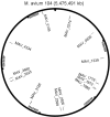
Results: We developed a random mutagenesis method based on illegitimate recombination and integration of a Hygromycin-resistance marker. Screening for mutations possibly affecting virulence was performed by monitoring of pH resistance, colony morphology, cytokine induction in infected macrophages and intracellular persistence. Out of 50 randomly chosen Hygromycin-resistant colonies, four revealed to be affected in virulence-related traits. The mutated genes were MAV_4334 (nitroreductase family protein), MAV_5106 (phosphoenolpyruvate carboxykinase), MAV_1778 (GTP-binding protein LepA) and MAV_3128 (lysyl-tRNA synthetase LysS).
Conclusions: We established a random mutagenesis method for MAH that can be easily carried out and combined it with a set of phenotypic screening methods for the identification of virulence-associated mutants. By this method, four new MAH genes were identified that may be involved in virulence.
Figures


Similar articles
-
Evidence for genes associated with the ability of Mycobacterium avium subsp. hominissuis to escape apoptotic macrophages.Front Cell Infect Microbiol. 2015 Aug 25;5:63. doi: 10.3389/fcimb.2015.00063. eCollection 2015. Front Cell Infect Microbiol. 2015. PMID: 26380226 Free PMC article.
-
Virulence of Mycobacterium avium Subsp. hominissuis Human Isolates in an in vitro Macrophage Infection Model.Int J Mycobacteriol. 2018 Jan-Mar;7(1):48-52. doi: 10.4103/ijmy.ijmy_11_18. Int J Mycobacteriol. 2018. PMID: 29516886
-
Abundance of Mycobacterium avium ssp. hominissuis in soil and dust in Germany - implications for the infection route.Lett Appl Microbiol. 2014 Jul;59(1):65-70. doi: 10.1111/lam.12243. Epub 2014 Mar 26. Lett Appl Microbiol. 2014. PMID: 24612016
-
Mycobacterium avium Subsp. hominissuis Interactions with Macrophage Killing Mechanisms.Pathogens. 2021 Oct 22;10(11):1365. doi: 10.3390/pathogens10111365. Pathogens. 2021. PMID: 34832521 Free PMC article. Review.
-
Genetic diversity and phylogeny of Mycobacterium avium.Infect Genet Evol. 2014 Jan;21:375-83. doi: 10.1016/j.meegid.2013.12.007. Epub 2013 Dec 15. Infect Genet Evol. 2014. PMID: 24345519 Review.
Cited by
-
Isolation, Identification, and Characterization of a New Highly Pathogenic Field Isolate of Mycobacterium avium spp. avium.Front Vet Sci. 2018 Jan 15;4:243. doi: 10.3389/fvets.2017.00243. eCollection 2017. Front Vet Sci. 2018. PMID: 29379790 Free PMC article.
-
Genetic Manipulation of Non-tuberculosis Mycobacteria.Front Microbiol. 2021 Feb 17;12:633510. doi: 10.3389/fmicb.2021.633510. eCollection 2021. Front Microbiol. 2021. PMID: 33679662 Free PMC article. Review.
-
Global Assessment of Mycobacterium avium subsp. hominissuis Genetic Requirement for Growth and Virulence.mSystems. 2019 Dec 10;4(6):e00402-19. doi: 10.1128/mSystems.00402-19. mSystems. 2019. PMID: 31822597 Free PMC article.
-
The many lives of nontuberculous mycobacteria.J Bacteriol. 2018 Feb 26;200(11):e00739-17. doi: 10.1128/JB.00739-17. Online ahead of print. J Bacteriol. 2018. PMID: 29483164 Free PMC article.
-
Mutation on lysX from Mycobacterium avium hominissuis impacts the host-pathogen interaction and virulence phenotype.Virulence. 2020 Dec;11(1):132-144. doi: 10.1080/21505594.2020.1713690. Virulence. 2020. PMID: 31996090 Free PMC article.
References
-
- Kirschner RA Jr, Parker BC, Falkinham III JO. Epidemiology of infection by nontuberculous mycobacteria: Mycobacterium avium, Mycobacterium intracellulare, and Mycobacterium scrofulaceum in acid, brown-water swamps of the Southeastern United States and their association with environmental variables. Am Rev Respir Dis. 1992;145:271–275. - PubMed
-
- Matlova L, Dvorska L, Palecek K, Maurenc L, Bartos M, Pavlik I. Impact of sawdust and wood shavings in bedding on pig tuberculous lesions in lymph nodes, and IS1245 RFLP analysis of Mycobacterium avium subsp. hominissuis of serotypes 6 and 8 isolated from pigs and environment. Vet Microbiol. 2004;102:227–236. doi: 10.1016/j.vetmic.2004.06.003. - DOI - PubMed
MeSH terms
Substances
LinkOut - more resources
Full Text Sources
Miscellaneous

