. 2012 Sep 11;43(1):65.
doi: 10.1186/1297-9716-43-65.

Natural Bagaza virus infection in game birds in southern Spain

Affiliations

Natural Bagaza virus infection in game birds in southern Spain

Virginia Gamino et al. Vet Res. .

Abstract

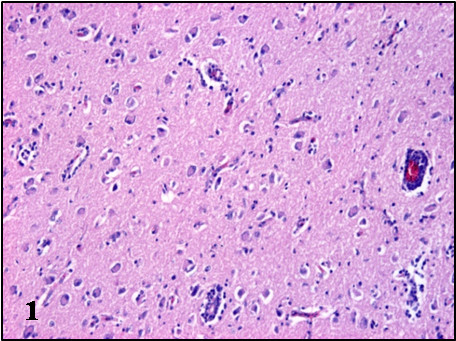
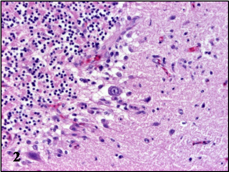
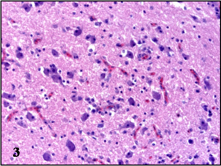
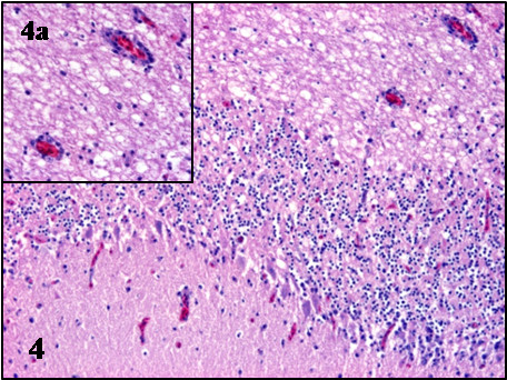
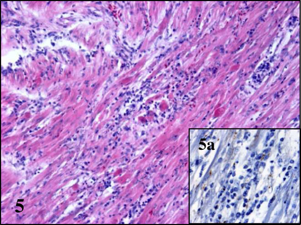
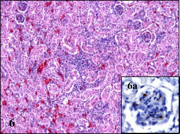
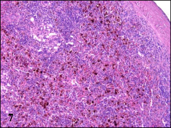
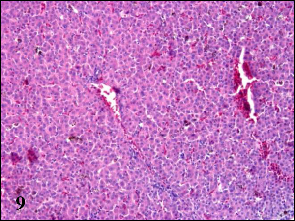
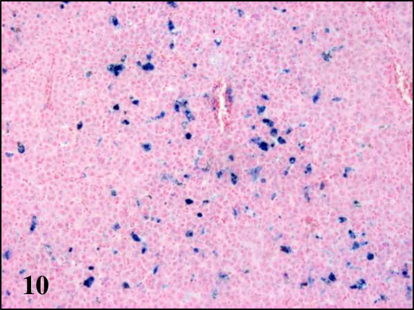
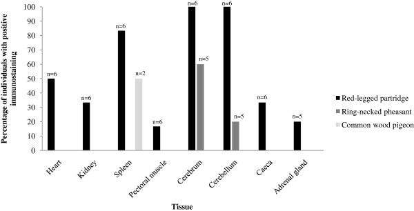
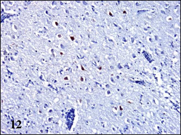
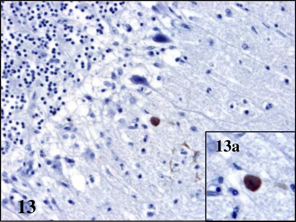
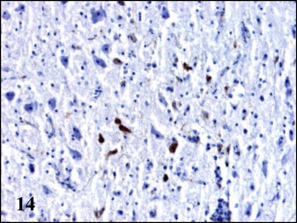
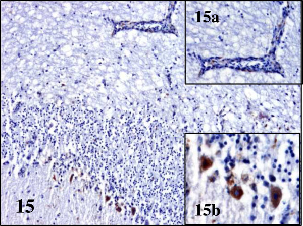
In late summer 2010 a mosquito born flavivirus not previously reported in Europe called Bagaza virus (BAGV) caused high mortality in red-legged partridges (Alectoris rufa) and ring-necked pheasants (Phasianus colchicus). We studied clinical findings, lesions and viral antigen distribution in naturally BAGV infected game birds in order to understand the apparently higher impact on red-legged partridges. The disease induced neurologic signs in the two galliform species and, to a lesser extent, in common wood pigeons (Columba palumbus). In red-legged partridges infection by BAGV caused severe haemosiderosis in the liver and spleen that was absent in pheasants and less evident in common wood pigeons. Also, BAGV antigen was present in vascular endothelium in multiple organs in red-legged partridges, and in the spleen in common wood pigeons, while in ring-necked pheasants it was only detected in neurons and glial cells in the brain. These findings indicate tropism of BAGV for endothelial cells and a severe haemolytic process in red-legged partridges in addition to the central nervous lesions that were found in all three species.


Figures

Figure 1
Figure 1
Microscopic lesions in the brain of BAGV infected game birds. Cerebrum, ring-necked pheasant. Neuronal necrosis and lymphoplasmacytic perivascular infiltrates. HE, ×100.
Figure 2
Figure 2
Microscopic lesions in the brain of BAGV infected game birds. Cerebellum, ring-necked pheasant. Purkinje cell necrosis and glial nodule. HE, ×200.
Figure 3
Figure 3
Microscopic lesions in the brain of BAGV infected game birds. Cerebrum, red-legged partridge. Neuronal necrosis and gliosis. HE, ×200.
Figure 4
Figure 4
Microscopic lesions in the brain of BAGV infected game birds. Cerebellum, red-legged partridge. Purkinje cell necrosis and phagocitosis and endothelial cell swelling (4a). HE, ×100.
Figure 5
Figure 5
Lesions and BAGV antigen distribution in the heart of BAGV infected red-legged partridges. Severe myocarditis with presence of BAGV antigen labeling in myofibres and capillary endothelial cells (5a). HE, ×100; immunohistochemistry for detection of flavivirus antigen, ×400.
Figure 6
Figure 6
Lesions and BAGV antigen distribution in the kidney of BAGV infected red- legged partridges. Focal necrosis and BAGV antigen labelling in capillary endothelial cells of the glomerular mesangium (6a). HE, ×100, immunohistochemistry for detection of flavivirus antigen, ×400.
Figure 7
Figure 7
Haemosiderosis in spleen section of red-legged partridges infected with BAGV. HE ×100.
Figure 8
Figure 8
Haemosiderosis in spleen section of red-legged partridges infected with BAGV. Perls’ stain, ×100.
Figure 9
Figure 9
Haemosiderosis in liver section of red-legged partridges infected with BAGV. HE, ×100.
Figure 10
Figure 10
Haemosiderosis in liver section of red-legged partridges infected with BAGV. Perls’ stain, ×100
Figure 11
Figure 11
Distribution of positive flavivirus (presumptive BAGV) immunostaining in naturally BAGV infected red-legged partridges, ring-necked pheasants and common wood pigeons. Each column represents the percentage of individuals that showed positive immunostaining in each organ and “n” is the number of individuals in which each tissue was tested.
Figure 12
Figure 12
Viral antigen distribution in the brain of BAGV infected game birds. Cerebrum, ring-necked pheasant. Labeling of BAGV antigen in the cytoplasm of neurons. Immunohistochemistry for detection of flavivirus antigen, ×100.
Figure 13
Figure 13
Viral antigen distribution in the brain of BAGV infected game birds. Cerebellum, ring-necked pheasant. Labeling of BAGV antigen in the cytoplasm of Purkinje cells (13a). Immunohistochemistry for detection of flavivirus antigen, ×200; ×400 (13a).
Figure 14
Figure 14
Viral antigen distribution in the brain of BAGV infected game birds. Cerebrum, red-legged partridge. Labeling of BAGV antigen in the cytoplasm of neurons and capillary endothelial cells. Immunohistochemistry for detection of flavivirus antigen, ×200.
Figure 15
Figure 15
Viral antigen distribution in the brain of BAGV infected game birds. Cerebellum, red-legged partridge. Labeling of BAGV antigen in the capillary endothelial cells (15a) and cytoplasm of Purkinje cells (15b). Immunohistochemistry for detection of flavivirus antigen, ×100; ×400 (15a and b).

References

    1. Aguero M, Fernandez-Pinero J, Buitrago D, Sanchez A, Elizalde M, San Miguel E, Villalba R, Llorente F, Jimenez-Clavero MA. Bagaza virus in partridges and pheasants, Spain, 2010. Emerg Infect Dis. 2011;17:1498–1501. - PMC - PubMed
    1. Digoutte JP. Bagaza (BAG) strain: Dak Ar B 209. Am J Trop Med Hyg. 1978;27:376–377.
    1. García-Bocanegra I, Zorrilla I, Rodríguez E, Rayas E, Camacho L, Redondo I, Gómez-Guillamón F. Monitoring of the Bagaza virus epidemic in wild bird species in Spain, 2010. Transbound Emerg Dis. in press. - DOI - PubMed
    1. Munster VJ, Baas C, Lexmond P, Waldenstrom J, Wallensten A, Fransson T, Rimmelzwaan GF, Beyer WEP, Schutten M, Olsen B, Osterhaus AD, Fouchier RA. Spatial, temporal, and species variation in prevalence of influenza A viruses in wild migratory birds. PLoS Pathog. 2007;3:e61. doi: 10.1371/journal.ppat.0030061. - DOI - PMC - PubMed
    1. Jimenez-Clavero MA, Aguero M, Rojo G, Gomez-Tejedor C. A new fluorogenic real-time RT-PCR assay for detection of lineage 1 and lineage 2 West Nile viruses. J Vet Diagn Invest. 2006;18:459–462. doi: 10.1177/104063870601800505. - DOI - PubMed

Publication types

MeSH terms

Substances

LinkOut - more resources