. 2012 Sep 11:11:66.
doi: 10.1186/1476-4598-11-66.

Integrin αvβ3 and CD44 pathways in metastatic prostate cancer cells support osteoclastogenesis via a Runx2/Smad 5/receptor activator of NF-κB ligand signaling axis

Affiliations

Integrin αvβ3 and CD44 pathways in metastatic prostate cancer cells support osteoclastogenesis via a Runx2/Smad 5/receptor activator of NF-κB ligand signaling axis

Aditi Gupta et al. Mol Cancer. .

Abstract

Background: Bone loss and pathological fractures are common skeletal complications associated with androgen deprivation therapy and bone metastases in prostate cancer patients. We have previously demonstrated that prostate cancer cells secrete receptor activator of NF-kB ligand (RANKL), a protein essential for osteoclast differentiation and activation. However, the mechanism(s) by which RANKL is produced remains to be determined. The objective of this study is to gain insight into the molecular mechanisms controlling RANKL expression in metastatic prostate cancer cells.

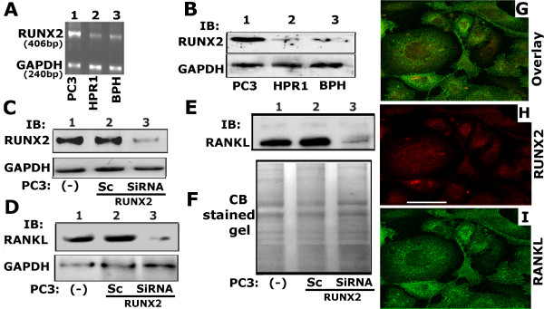
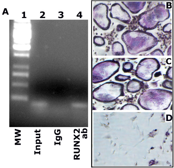
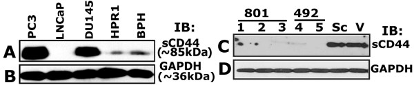
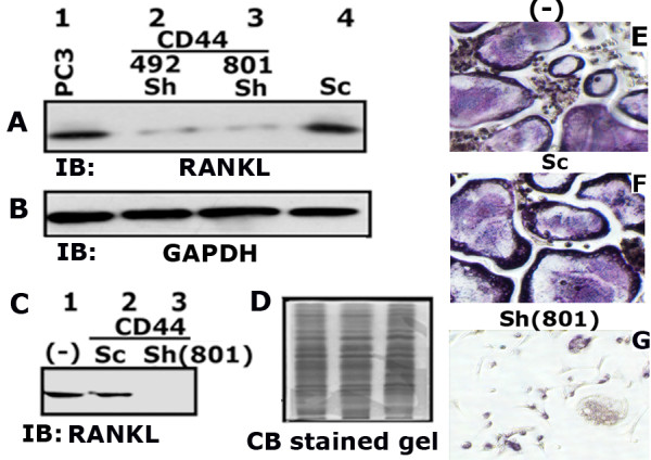
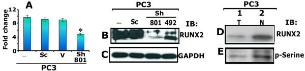
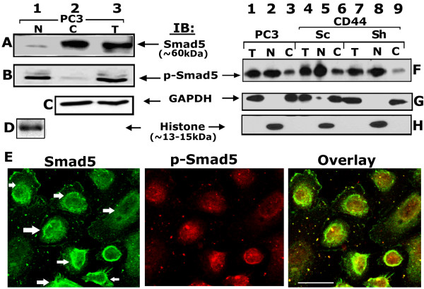
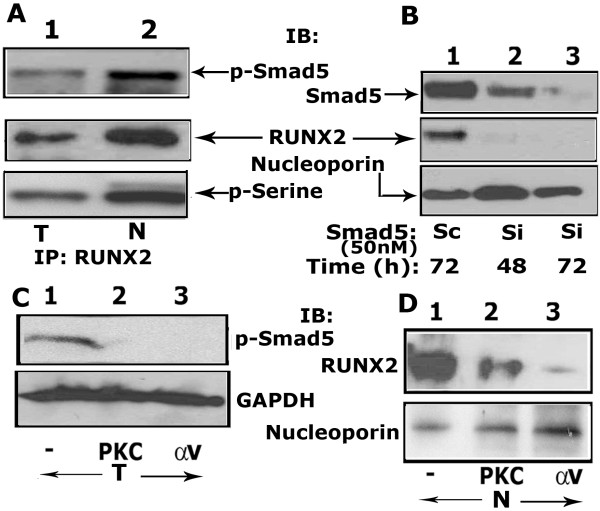
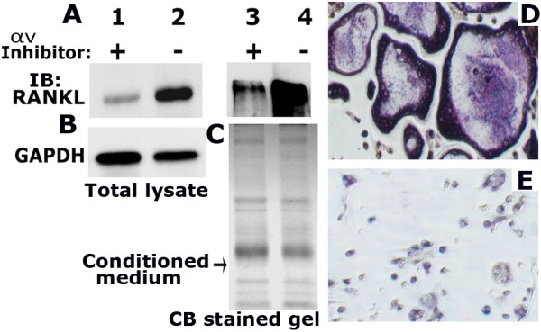
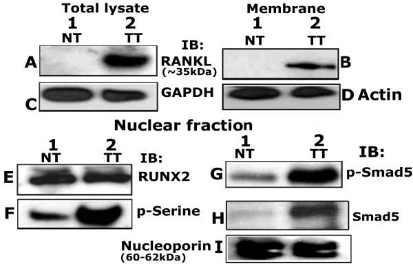
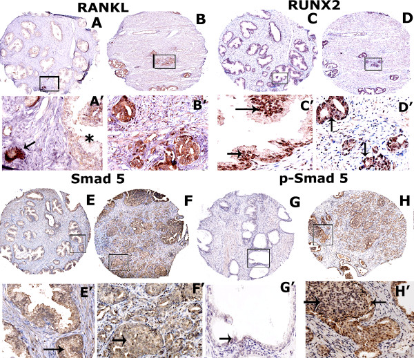
Results: We show here that phosphorylation of Smad 5 by integrin αvβ3 and RUNX2 by CD44 signaling, respectively, regulates RANKL expression in human-derived PC3 prostate cancer cells isolated from bone metastasis. We found that RUNX2 intranuclear targeting is mediated by phosphorylation of Smad 5. Indeed, Smad5 knock-down via RNA interference and inhibition of Smad 5 phosphorylation by an αv inhibitor reduced RUNX2 nuclear localization and RANKL expression. Similarly, knockdown of CD44 or RUNX2 attenuated the expression of RANKL. As a result, conditioned media from these cells failed to support osteoclast differentiation in vitro. Immunohistochemistry analysis of tissue microarray sections containing primary prostatic tumor (grade2-4) detected predominant localization of RUNX2 and phosphorylated Smad 5 in the nuclei. Immunoblotting analyses of nuclear lysates from prostate tumor tissue corroborate these observations.

Conclusions: Collectively, we show that CD44 signaling regulates phosphorylation of RUNX2. Localization of RUNX2 in the nucleus requires phosphorylation of Smad-5 by integrin αvβ3 signaling. Our results suggest possible integration of two different pathways in the expression of RANKL. These observations imply a novel mechanistic insight into the role of these proteins in bone loss associated with bone metastases in patients with prostate cancer.


Figures

Figure 1
Figure 1
Analysis of expression of RUNX2 and RANKL in PC3 cells. A and B. RT-PCR and immunoblotting analysis of expression of RUNX2 in PC3 (lanes 1), HPR1 (lane 2) and BPH (lane 3) cells is shown. C-E: The effects of SiRNA to RUNX2 on RUNX2 (C) and RANKL (D) protein levels in total cellular lysates (C and D) and conditioned medium (E). Immunoblotting analysis in conditioned medium represents the secreted levels of RANKL. Untransfected (−) or scrambled SiRNA (Sc) transfected PC3 cells were used as controls (B-E). GAPDH was used as a loading control for RT-PCR (A) and Western blot (B -D) analyses. The loading control for the conditioned medium is shown by the use of Coomassie blue staining of the blot (F). G - I: Immunostaining and confocal microscopy analysis of distribution of RUNX2 (red; H) and RANKL (green; I) in PC3 cells. Distribution of both RANKL (red) and RUNX2 (green) are shown in panel G. Results shown are representative of three independent experiments. Scale bar: 50 μm.
Figure 2
Figure 2
Analysis of binding of RUNX2 with RANKL promoter and the effect of RUNX2 knockdown on osteoclast differentiation. A. Chromatin immunoprecipitation (ChIP) assay. ChIP assay was used to determine the RUNX2 binding sites in RANKL promoter. Immunoprecipitates were made with an antibody (rabbit) to RUNX2 (lane 4) or rabbit IgG (lane 3) using lysates made from PC3 cells. DNA from the input (lane 2) and immunoprecipitates (lanes 3 and 4) was analyzed by RT-PCR using primers specific for RUNX2 binding sites on RANKL promoter. As expected, a product size 153 bp was observed in the RT-PCR analysis. The experiment was repeated twice and obtained similar results. B-D: The conditioned media (CM) from PC3 cells untreated (B) or treated with scrambled (C) and SiRNA (D) to RUNX2 were used for osteoclast differentiation in vitro. TRAP-positive osteoclasts are stained in dark purple. Cells were observed under an inverted phase contrast microscope and images were captured (X 200). The results shown are representative of three experiments.
Figure 3
Figure 3
Characterization of stable CD44 knockdown cell lines. A. Western blot analysis: Equal amount of protein lysates (50 μg) made from indicated cell lines were immunoblotted with a CD44 antibody to detect total cellular levels of CD44 protein. C. Immunoblotting analysis of the total cellular levels of CD44 in the stable clonal isolates derived from PC3 cells transfected with CD44 ShRNA constructs (801 and 492; lanes 1–5) is shown. PC3 cells transfected with vector DNA (V) and scrambled ShRNA construct (Sc) were used as controls. B and D: Equal loading of protein was verified with the GAPDH level in each lane. The experiment was carried out three times with similar results.
Figure 4
Figure 4
Analysis of RANKL expression level in PC3 cells knockdown of CD44. A and C. Equal amount of total cellular lysates (50 μg protein; A) and conditioned media (CM-20 μg protein; C) were immunoblotted with a RANKL antibody to detect RANKL protein. CM was used to detect the secreted RANKL protein. B and D. The blot in A was stripped and reprobed with a GAPDH antibody. Equal level of GAPDH protein was observed (B). The loading control for the CM was shown by the use of Coomassie blue staining of a gel ran in parallel (D). E-G. The effect of CM on osteoclast differentiation in vitro is shown. TRAP-positive osteoclasts are stained in dark purple. Images were captured (X 200) with an inverted phase contrast microscope. The results shown are representative of three independent experiments.
Figure 5
Figure 5
Effects of CD44 knockdown on RUNX2 expression (mRNA and protein) and distribution in PC3 cells. A. The expression levels of RUNX2 mRNA was determined by real-time PCR analysis and normalized relative to GAPDH expression. Bar represents the mean ± SEM of three different experiments. *p <0.01 vs. untransfected (−) and transfected PC3 cells with scrambled ShRNA construct (Sc) and vector DNA (V). B and C. Equal amount of lysates (20 μg protein) made from PC3 cells untransfected (−) and transfected with scramble (Sc) and ShRNA CD44 constructs (492 and 801) were used for immunoblotting analysis with an antibody to RUNX2. Immunoblotting with an antibody to GAPDH (C) was used as a loading control. D and E. PC3 cells were analyzed for the phosphorylation of RUNX2 in total cellular (T) and nuclear (N) lysates by immunoblotting of RUNX2 immunoprecipitates with antibodies to RUNX2 (D) and phospho-serine (E; p-Serine). The results shown are representative of three independent experiments.
Figure 6
Figure 6
Analysis of Smad 5 phosphorylation in PC3 cells. A and B; F-H. Protein and phosphorylation levels of Smad 5 were determined by Western blot analysis in nuclear (N), cytosolic (C) and total cellular (T) proteins isolated from PC3 cells (A and B) and PC3 cells knockdown of CD44 (F-H). 50 μg of indicated protein (A-D, F-H) was used for immunoblotting (IB) analyses. The blot in A was stripped and reprobed successively with p-Smad 5, GAPDH and histone antibodies (B-D). Similarly, the blot in F was stripped and reprobed twice simultaneously with GAPDH and histone antibodies (G and H). Immunoblotting with an antibody to GAPDH (C and G) and histone (D and H) was used as a control for normalization of cellular and nuclear protein, respectively. E. Confocal analysis of immunostained PC3 cells with Smad 5 (green) and p-Smad 5 (red) antibodies is shown. Distribution of both Smad 5 and p-Smad 5 is shown in the overlay panel. Scale bar-50 μm. The results shown are representative of three independent experiments.
Figure 7
Figure 7
The effect of PKC and integrin αv inhibitor on the phosphorylation of Smad 5 and RUNX2 localization in the nuclei. A. Analysis of interaction of p-Smad 5 with RUNX2. Equal amount of total cellular and nuclear proteins were immunoprecipitated with a RUNX2 antibody and immunoblotted with a p-Smad 5 antibody (A, top panel). Subsequently, the blot was reprobed sequentially with a RUNX2 (middle panel) and p-Serine (bottom panel) antibody after stripping. B. Effect of SiRNA to Smad 5 on the nuclear levels of RUNX2. Time-dependent effect of SiRNA (Si) nucleotides on Smad 5 levels at 48 and 72 h is shown. Equal amount of nuclear proteins were immunoblotted sequentially with antibodies to Smad 5, RUNX2 and nucleoporin after stripping. Scrambled RNAi nucleotide (Sc) transfected cells were used as controls (lane 1). C. Effects of PKC and integrin αv inhibitors (lanes 2 and 3) on the phosphorylation of Smad 5. Untreated (-) PC3 cells were used as control. Total cellular (T) lysate proteins were immunoblotted with a p-Smad 5 antibody. D. Effects of PKC and integrin αv inhibitors (lanes 2 and 3) on the nuclear localization of RUNX2. Untreated (-) PC3 cells were used as control. Nuclear lysate proteins (N) were immunoblotted with a RUNX2 antibody. B-D: Loading control antibodies to GAPDH (C) and nucleoporin (B and D) were used to estimate relative amounts of total and nuclear proteins loaded in each lane. The results shown are representative of three independent experiments.
Figure 8
Figure 8
The effect of integrin αv inhibitor on RANKL expression and osteoclast differentiation. A-C: Western blot analysis. Equal amount of total cellular lysates (50 μg protein; A, lanes 1 and 2) and conditioned media (CM-20 μg protein; lanes 3 and 4) were immunoblotted with a RANKL antibody. CM was used to detect the secreted levels of RANKL. The blot in A was reprobed with a GAPDH antibody after stripping (B). GAPDH level was used as a control for loading. The loading control for the CM is shown by the use of Coomassie blue staining of a gel ran in parallel (C). D and E. The effect of CM on osteoclast differentiation in vitro is shown. TRAP-positive osteoclasts are stained in dark purple. Images were captured (X 200) with an inverted phase contrast microscope. The results shown are representative of three independent experiments.
Figure 9
Figure 9
Western analyses in prostatic normal and tumor lysates. Total cellular (A and C), membrane (B and D) and nuclear (E to I) lysates from normal (NT) and prostatic tumor (TT) tissue (~20 μg protein) were immunoblotted (IB) with a RANKL (A and B), RUNX2 (E), phosphoserine (p-Serine; F), phospho-Smad 5 (p-Smad 5; G) and Smad 5 (H) antibody. Equal loading of the protein was shown in total cellular, membrane and nuclear lysates by relevant immunoblotting analysis with antibodies to GAPDH (C), actin (D) and nucleoporin (I). The results shown are representative of three independent experiments with three different lysates purchased from the vendor.
Figure 10
Figure 10
Immunohistochemistry on TMA derived from normal and cancerous prostate tissue. Immunohistochemical staining was performed in prostate cancer and normal tissue microarray with an antibody to RANKL (A and B), RUNX2 (C and D), Smad 5 (E and F) and p-Smad 5 (G-H). Normal tissue adjacent to prostate cancer are shown in A, C, E and G. Prostate carcinoma at grade 2–3 are shown in B, D, F and H. Arrows in C’, D’G’ and H’ point to nuclear localization of RUNX2 and p-Smad-5 proteins. An arrow in A’ points to a prostate carcinoma filled lumen (A’) adjacent to normal tissue (indicated by an asterisk; A’). Sections were immunostained (brown) with indicated primary antibody as described in the Methods section. Immunostained sections were counterstained with hematoxylin stain (blue). Magnification is 50X in A-H. Location of the high magnification (X200) regions shown in A’-H’ is indicated by a rectangle field in A-H. Staining was repeated two times.

Similar articles

Cited by

References

    1. van der Gulden JW, Kiemeney LA, Verbeek AL, Straatman H. Mortality trend from prostate cancer in The Netherlands (1950–1989) 7. Prostate. 1994;24:33–38. doi: 10.1002/pros.2990240108. - DOI - PubMed
    1. Brawley OW. Prostate cancer epidemiology in the United States. World J Urol. 2012;30:195–200. doi: 10.1007/s00345-012-0824-2. - DOI - PubMed
    1. Carlin BI, Andriole GL. The natural history, skeletal complications, and management of bone metastases in patients with prostate carcinoma 1. Cancer. 2000;88:2989–2994. doi: 10.1002/1097-0142(20000615)88:12+<2989::AID-CNCR14>3.0.CO;2-Q. - DOI - PubMed
    1. Sanchez-Sweatman OH, Orr FW, Singh G. Human metastatic prostate PC3 cell lines degrade bone using matrix metalloproteinases. Invasion Metastasis. 1998;18:297–305. doi: 10.1159/000024522. - DOI - PubMed
    1. Dougall WC. RANKL signaling in bone physiology and cancer. Curr Opin Support Palliat Care. 2007;1:317–322. doi: 10.1097/SPC.0b013e3282f335be. - DOI - PubMed

Publication types

MeSH terms