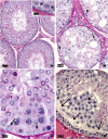
Late morfofunctional alterations of the Sertoli cell caused by doxorubicin administered to prepubertal rats
- PMID: 22967030
- PMCID: PMC3502149
- DOI: 10.1186/1477-7827-10-79
Late morfofunctional alterations of the Sertoli cell caused by doxorubicin administered to prepubertal rats
Abstract
Background: Doxorubicin is a potent chemotherapeutic drug used against a variety of cancers. It acts through interaction with polymerases and topoisomerase II and free radical production. Doxorubicin activity is not specific to cancer cells and can also damage healthy cells, especially those undergoing rapid proliferation, such as spermatogonia. In previous studies our group showed that etoposide, another topoisomarese II poison, causes irreversible damage to Sertoli cells. Thus, the aim of this study was to address the effects of doxorubicin on Sertoli cell morphology and function and on the seminiferous epithelium cycle when administered to prepubertal rats.
Methods: Prepubertal rats received the dose of 5 mg/Kg of doxorubicin, which was fractioned in two doses: 3 mg/Kg at 15dpp and 2 mg/Kg at 22 dpp. The testes were collected at 40, 64 and 127 dpp, fixed in Bouin's liquid and submitted to transferrin immunolabeling for Sertoli cell function analysis. Sertoli cell morphology and the frequency of the stages of the seminiferous epithelium cycle were analyzed in PAS + H-stained sections.
Results: The rats treated with doxorubicin showed reduction of transferrin labeling in the seminiferous epithelium at 40 and 64 dpp, suggesting that Sertoli cell function is altered in these rats. All doxorubicin-treated rats showed sloughing and morphological alterations of Sertoli cells. The frequency of the stages of the seminiferous epithelium cycle was also affected in all doxorubicin-treated rats.
Conclusions and discussion: These data show that doxorubicin administration during prepuberty causes functional and morphological late damage to Sertoli cells; such damage is secondary to the germ cell primary injury and contributed to enhance the spermatogenic harm caused by this drug. However, additional studies are required to clarify if there is also a direct effect of doxorubicin on Sertoli cells producing a primary damage on these cells.
Figures


Similar articles
-
Sertoli cell function in albino rats treated with etoposide during prepubertal phase.Histochem Cell Biol. 2006 Sep;126(3):353-61. doi: 10.1007/s00418-006-0168-3. Epub 2006 Mar 21. Histochem Cell Biol. 2006. PMID: 16550346
-
Sertoli cell morphological alterations in albino rats treated with etoposide during prepubertal phase.Microsc Microanal. 2008 Jun;14(3):225-35. doi: 10.1017/S1431927608080318. Microsc Microanal. 2008. PMID: 18482470
-
Apoptosis and testicular alterations in albino rats treated with etoposide during the prepubertal phase.Anat Rec A Discov Mol Cell Evol Biol. 2004 Jul;279(1):611-22. doi: 10.1002/ar.a.20045. Anat Rec A Discov Mol Cell Evol Biol. 2004. PMID: 15224403
-
Amifostine reduces the seminiferous epithelium damage in doxorubicin-treated prepubertal rats without improving the fertility status.Reprod Biol Endocrinol. 2010 Jan 10;8:3. doi: 10.1186/1477-7827-8-3. Reprod Biol Endocrinol. 2010. PMID: 20064221 Free PMC article.
-
Spermatogenesis by Sisyphus: proliferating stem germ cells fail to repopulate the testis after 'irreversible' injury.Adv Exp Med Biol. 2001;500:421-8. doi: 10.1007/978-1-4615-0667-6_64. Adv Exp Med Biol. 2001. PMID: 11764975 Review.
Cited by
-
Rat PRDM9 shapes recombination landscapes, duration of meiosis, gametogenesis, and age of fertility.BMC Biol. 2021 Apr 28;19(1):86. doi: 10.1186/s12915-021-01017-0. BMC Biol. 2021. PMID: 33910563 Free PMC article.
-
Cross-talk between ER stress and mitochondrial pathway mediated adriamycin-induced testicular toxicity and DA-9401 modulate adriamycin-induced apoptosis in Sprague-Dawley rats.Cancer Cell Int. 2019 Apr 4;19:85. doi: 10.1186/s12935-019-0805-2. eCollection 2019. Cancer Cell Int. 2019. PMID: 30992692 Free PMC article.
-
Irinotecan metabolite SN38 results in germ cell loss in the testis but not in the ovary of prepubertal mice.Mol Hum Reprod. 2016 Nov;22(11):745-755. doi: 10.1093/molehr/gaw051. Epub 2016 Jul 28. Mol Hum Reprod. 2016. PMID: 27470502 Free PMC article.
-
Autophagy, a critical element in the aging male reproductive disorders and prostate cancer: a therapeutic point of view.Reprod Biol Endocrinol. 2023 Sep 26;21(1):88. doi: 10.1186/s12958-023-01134-1. Reprod Biol Endocrinol. 2023. PMID: 37749573 Free PMC article. Review.
-
Chemotherapy induced damage to spermatogonial stem cells in prepubertal mouse in vitro impairs long-term spermatogenesis.Toxicol Rep. 2020 Dec 26;8:114-123. doi: 10.1016/j.toxrep.2020.12.023. eCollection 2021. Toxicol Rep. 2020. PMID: 33425685 Free PMC article.
References
-
- Hida H, Coudray C, Calop J, Favier A. Effect of antioxidants on adriamycin-induced microsomal lipid peroxidation. Biol Trace Elem Res. 1995;47:111–116. - PubMed
-
- Jahnukainen K, Hou M, Parvinen M, Eksborg S, Söder O. Stage-specific inhibition of deoxyribonucleic acid synthesis and induction of apoptosis by anthracyclines in cultured rat spermatogenic cells. Biol Reprod. 2000;63:482–487. - PubMed
-
- Suominen JS, Linderborg J, Nikula H, Hakovirta H, Parvinen M, Toppari J. The effects of mono-2-ethylhexyl phthalate, adriamycin and N-ethyl-Nitrosourea on stage-specific apoptosis and DNA synthesis in the mouse spermatogenesis. Toxicol Lett. 2003;143:163–173. - PubMed
-
- Calabresi P, Parks RE. , Jr. In: As bases farmacológicas da terapêutica. Gilman AG, Goodman LS, Rall TW, Murad F, editor. Rio de Janeiro, Guanabara-Koogan; 1985. Quimioterapia das doenças neoplásicas; pp. 813–856.
-
- Gewirtz DA. A critical evaluation of the mechanisms of action proposed for the antitumor effects of the anthracycline antibiotics adriamycin and daunorubicin. Biochem Pharmacol. 1999;57:727–741. - PubMed
Publication types
MeSH terms
Substances
LinkOut - more resources
Full Text Sources

