. 2012 Dec;32(6):631-9.
doi: 10.1042/BSR20120046.

Protein instability and functional defects caused by mutations of dihydro-orotate dehydrogenase in Miller syndrome patients

Affiliations

Protein instability and functional defects caused by mutations of dihydro-orotate dehydrogenase in Miller syndrome patients

JingXian Fang et al. Biosci Rep. 2012 Dec.

Abstract

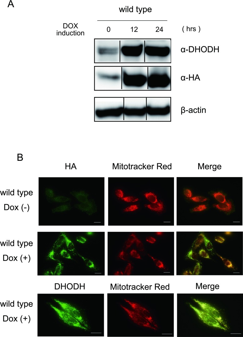
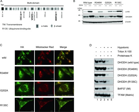
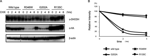
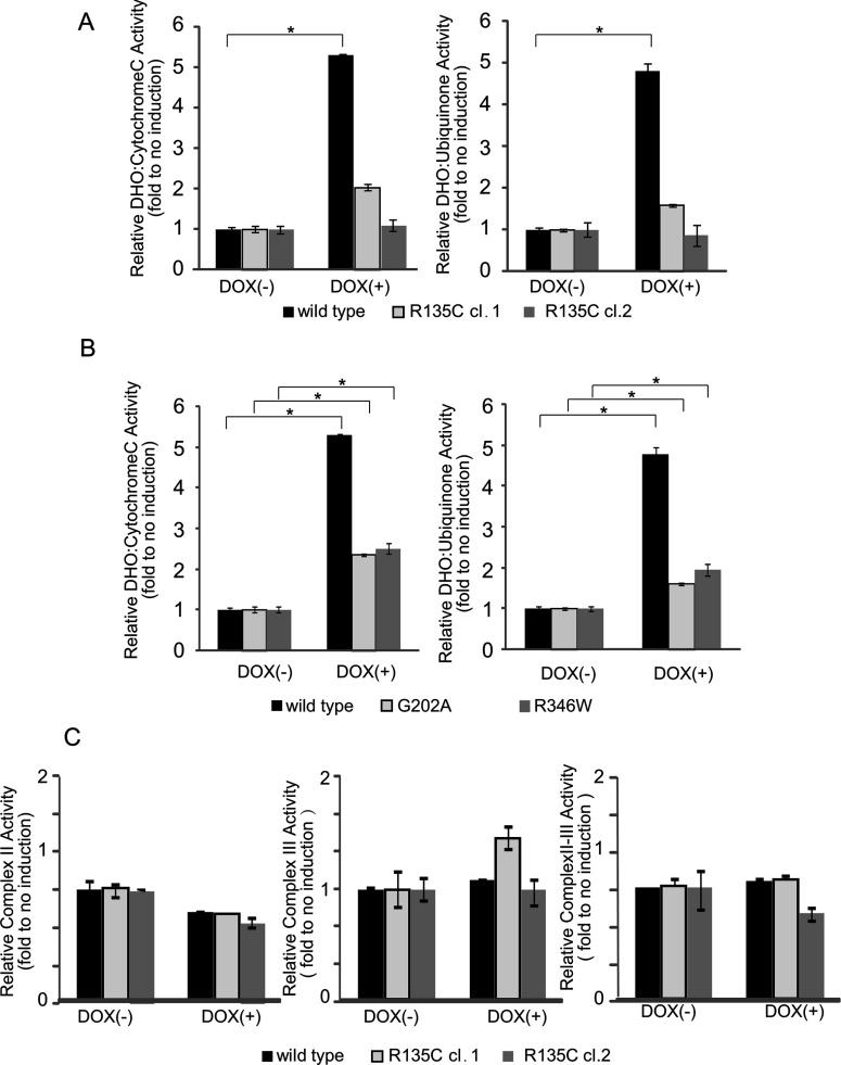
Miller syndrome is a recessive inherited disorder characterized by postaxial acrofacial dysostosis. It is caused by dysfunction of the DHODH (dihydroorotate dehydrogenase) gene, which encodes a key enzyme in the pyrimidine de novo biosynthesis pathway and is localized at mitochondria intermembrane space. We investigated the consequence of three missense mutations, G202A, R346W and R135C of DHODH, which were previously identified in patients with Miller syndrome. First, we established HeLa cell lines stably expressing DHODH with Miller syndrome-causative mutations: G202A, R346W and R135C. These three mutant proteins retained the proper mitochondrial localization based on immunohistochemistry and mitochondrial subfractionation studies. The G202A, R346W DHODH proteins showed reduced protein stability. On the other hand, the third one R135C, in which the mutation lies at the ubiquinone-binding site, was stable but possessed no enzymatic activity. In conclusion, the G202A and R346W mutation causes deficient protein stability, and the R135C mutation does not affect stability but impairs the substrate-induced enzymatic activity, suggesting that impairment of DHODH activity is linked to the Miller syndrome phenotype.


Figures

Figure 1
Figure 1. Expression and localization of DHODH-HA protein
(A) Inducible expression of recombinant DHODH-HA. DHODH-HA was inducibly expressed in HeLa cells using a Tet-on system. DHODH was detected by immunoblotting with anti-DHODH and anti-HA antibodies. β-Actin was the loading control. Lane 1, no induction; lane 2, 12 h; lane 3, 24 h after DOX treatment. (B) Immunocytochemistry of recombinant DHODH. DHODH-HA-transfected cells were cultured in the absence or presence of DOX for 24 h. Mitochondria and recombinant DHODH were visualized with the mitochondria-staining dye MitoTracker Red (centre panels) and anti-HA or anti-DHODH antibodies (left panels) respectively. The right panels show merged images. The scale bars represent 10 μm.
Figure 2
Figure 2. Expression and localization of Miller syndrome mutations
(A) Schematic representation of the primary structure of DHODH and the location of Miller syndrome-associated mutations. Arg135 is the ubiquinone-binding site. TM indicates a transmembrane domain. The 12 α-helices and 13 β-sheet regions are shown as boxes and arrows respectively. We analysed three Miller syndrome mutations R135C, G202A and R346W. (B) Inducible expression of wild-type mutant HA-tagged DHODH. Wild-type and mutant HA-tagged DHODH were inducibly expressed in HeLa cells using a Tet-on system. Endogenous and exogenous DHODH were detected by immunoblotting with anti-DHODH and anti-HA antibodies. β-actin was used as the loading control. (C) Immunocytochemistry of recombinant mutant DHODH. Mutant DHODH-HA-transfected cells were cultured in the presence of DOX for 24 h. Mitochondria and mutant DHODH were visualized with the mitochondria-staining dye, MitoTracker Red (centre panels) and anti-HA antibodies (left panels) respectively. The right panels show merged images. Scale bar represent 10 μm. (D) Submitochondrial localization of exogenous mutant DHODH-HA. Mitochondria were incubated in hypotonic buffer to disrupt the outer membranes without (lanes 3 and 4) or with (lane 5) the non-ionic detergent Triton X-100. Then, the mitochondria were digested with proteinase K (lanes 2, 4 and 5). The indicated proteins were detected by immunoblotting. BAP37 and TFAM are markers of the mitochondrial inner membrane (IM) and matrix respectively.
Figure 3
Figure 3. Protein unstability of two mutation of Miller syndrome mutations
(A) HeLa cells stably expressing wild-type or mutant DHODH-HA were treated with vehicle (0 h) or 100 μM CHX for 2, 4 or 8 h. Immunoblotting was performed using anti-DHODH and anti-HA antibodies. β-Actin was used as the loading control. (B) Relative intensity of α-HA which is shown in (A) were plotted after CHX treatment. Treatment of G202A and R346W DHODH-transfected cells with the translation inhibitor CHX significantly decreased the expression levels of mature DHODH protein.
Figure 4
Figure 4. Functional analysis of Miller syndrome mutations
(A) Reduced DHO-dependent reductase activities in R135C DHODH-transfected cells. DHO: ubiquinone oxidoreductase activity and DHO: cytochrome c oxidoreductase activity were measured using cell lysates as described in the Experimental section. Increased DHO-dependent reductase activity was observed upon the induction of wild-type DHODH; however, induction of R135C DHODH did not result in increased enzymatic activities. The results represent the means±S.D. of three independent experiments. *P<0.05 compared with controls. (B) DHO-dependent reductase activity in G202C and R246W DHODH-expressing cells. DHO: ubiquinone oxidoreductase activity and DHO: cytochrome c oxidoreductase activity were measured using cell lysates. A 2-fold increase in DHO-dependent oxidoreductase activity was observed upon the induction of G202A or R346W DHODH. The results represent the means±S.D. of three independent experiments. *P<0.05 compared with controls. (C) The complex II and III activities in R135C and wild-type DHODH were measured using cell lysates as described in the Experimental section. We observed that no difference in the activities of complex II, complex III and the combined complex II–III between R135C and wild-type DHODH.

Similar articles

Cited by

References

    1. McBride H. M., Neuspiel M., Wasiak S. Mitochondria: more than just a powerhouse. Curr. Biol. 2006;16:R551–R560. - PubMed
    1. DiMauro S., Schon E. A. Mitochondrial disorders in the nervous system. Annu. Rev. Neurosci. 2008;31:91–123. - PubMed
    1. Frenzel M., Rommelspacher H., Sugawa M. D., Dencher N. A. Ageing alters the supramolecular architecture of OxPhos complexes in rat brain cortex. Exp. Gerontol. 2010;45:563–572. - PubMed
    1. Wallace D. C. Mitochondrial DNA mutations in disease and aging. Environ. Mol. Mutagen. 2010;51:440–450. - PubMed
    1. Tuppen H. A., Blakely E. L., Turnbull D. M., Taylor R. W. Mitochondrial DNA mutations and human disease. Biochim. Biophys. Acta. 2010;1797:113–128. - PubMed

Publication types

MeSH terms

Substances

Supplementary concepts