. 2012 Nov;53(11):1898-906.
doi: 10.1111/j.1528-1167.2012.03665.x. Epub 2012 Sep 11.

Albumin is taken up by hippocampal NG2 cells and astrocytes and decreases gap junction coupling

Affiliations

Albumin is taken up by hippocampal NG2 cells and astrocytes and decreases gap junction coupling

Oliver Braganza et al. Epilepsia. 2012 Nov.

Abstract

Purpose: Dysfunction of the blood-brain barrier (BBB) and albumin extravasation have been suggested to play a role in the etiology of human epilepsy. In this context, dysfunction of glial cells attracts increasing attention. Our study was aimed to analyze in the hippocampus (1) which cell types internalize albumin injected into the lateral ventricle in vivo, (2) whether internalization into astrocytes impacts their coupling and expression of connexin 43 (Cx43), and (3) whether expression of Kir4.1, the predominating astrocytic K(+) channel subunit, is altered by albumin.

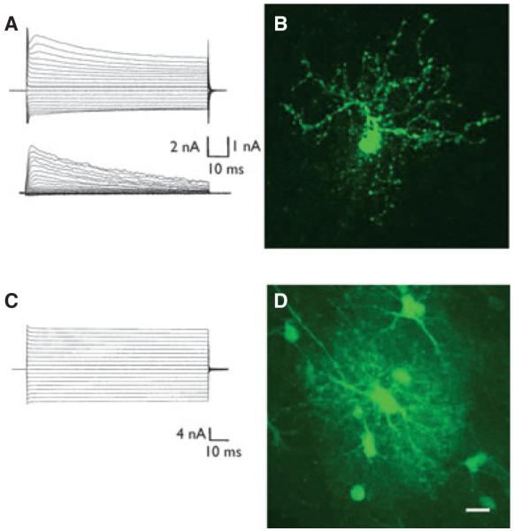
Methods: The patch-clamp method was combined with single cell tracer filling to investigate electrophysiologic properties and gap junction coupling (GJC). For cell identification, mice with cell type-specific expression of a fluorescent protein (NG2kiEYFP mice) and immunohistochemistry were employed. Semiquantitative real time polymerase chain reaction (RT-PCR) allowed analysis of Kir4.1 and Cx43 transcript levels.

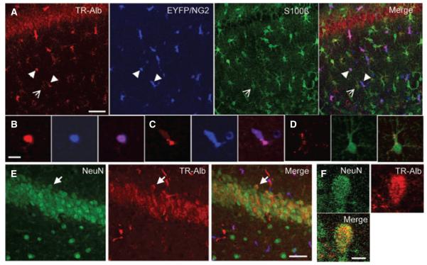
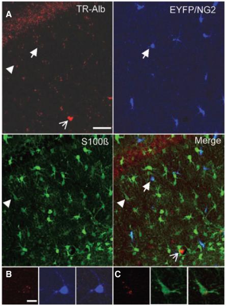
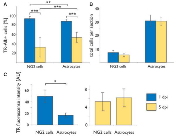
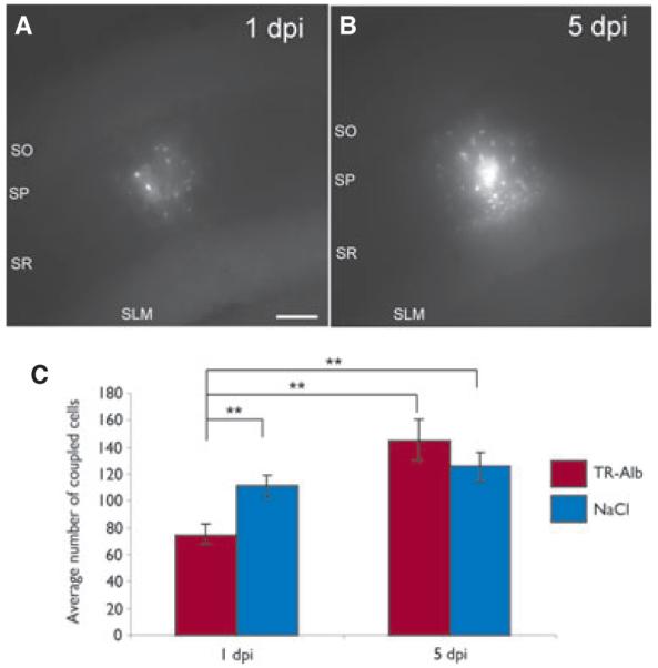
Key findings: We show that fluorescently labeled albumin is taken up by astrocytes, NG2 cells, and neurons, with NG2 cells standing out in terms of the quantity of uptake. Within 5 days postinjection (dpi), intracellular albumin accumulation was largely reduced suggesting rapid degradation. Electrophysiologic analysis of astrocytes and NG2 cells revealed no changes in their membrane properties at either time point. However, astrocytic GJC was significantly decreased at 1 dpi but returned to control levels within 5 dpi. We found no changes in hippocampal Cx43 transcript expression, suggesting that other mechanisms account for the observed changes in coupling. Kir4.1 mRNA was regulated oppositely in the CA1 stratum radiatum, with a strong increase at 1 dpi followed by a decrease at 5 dpi.

Significance: The present study demonstrates that extravasal albumin in the hippocampus induces rapid changes of astrocyte function, which can be expected to impair ion and transmitter homeostasis and contribute to hyperactivity and epileptogenesis. Therefore, astrocytes may represent alternative targets for antiepileptic therapeutic approaches.


Figures

Figure 1
Figure 1
TR-Alb is taken up by astrocytes, NG2-cells, and neurons in the CA1 region of the hippocampus. NG2kiEYFP mice were injected with TR-Alb, i.c.v., sacrificed at 1 dpi, stained against EYFP and S100β or NeuN, and confocally imaged. (A) Maximum projection of 26 optical sections through the SR of the CA1 in the rostral hippocampus. Fluorescence panels from left to right display TR-Alb (red), EYFP (blue), S100β (green), and merge. (B, C) Blow-ups of two single optical sections of two NG2 cells from A (arrowheads). (D) Single optical section of an astrocyte shown in A (open arrow). (E) Maximum projection of a NeuN staining in the CA1 pyramidal cell layer. (F) Blow-up of a single optical section of a neuron in E (arrow). Scale bars A, E: 40 μm; B–D, F: 10 μm. Epilepsia © ILAE
Figure 2
Figure 2
Cellular TR-Alb fluorescence has largely decreased at 5 dpi. NG2kiEYFP mice were injected with TR-Alb, i.c.v., sacrificed at 5 dpi, and stained against EYFP and S100β. (A) Maximum projection of 26 optical sections through the SR of the CA1 region in the rostral hippocampus. Fluorescence panels from left to right display TR-Alb (red), EYFP (blue), S100β (green) and merge. Open arrow indicates a blood vessel. (B, C) Single optical sections of an NG2-cell (arrow) and an astrocyte (arrow-head) shown in A. Scale bars A: 40 μm; B, C: 10 μm. Epilepsia © ILAE
Figure 3
Figure 3
Quantitative analysis of TR-Alb uptake and fluorescence loss in astrocytes and NG2 cells. Light and dark grey bars represent cells of mice sacrificed at 1 and 5 dpi, respectively. (A) Percentage of TR-Alb positive cells among NG2 cells and astrocytes. (B) Average number of cells within the CA1 SR per 40 μm section. Cell numbers were determined stereologically by confocal laser scanning microscopy. One day post injection: n = 32 sections, four mice; 5 dpi: n = 25 sections, three mice (C) Average fluorescence intensity per cell in AU. n = 15 cells in three sections of three mice for each condition; *p < 0.05, **p < 0.01, ***p < 0.001. Epilepsia © ILAE
Figure 4
Figure 4
Functional and morphological characteristics of TR-Alb positive glial cells at 1 dpi. TR-Alb positive cells in the CA1 SR were identified by fluorescence and IR DIC microscopy. During patch-clamp recording cells were filled with 0.5% biocytin. (A) Complex whole cell current pattern of a TR-Alb positive cell (de- and hyperpolarization between −160 and +20 mV; holding potential −80 mV, 10 mV voltage step increments) (top panel). Subtraction of corresponding currents after applying prepulse protocols (see Methods) isolated transient outwardly rectifying K+ currents (bottom). (B) After 20 min of recording the slice was fixated and stained against biocytin. The cell in (A) shows the typical morphology of an NG2 cell. (C) The stimulus protocol described in (A) was applied to another TR-Alb positive cell that showed a passive current pattern. The cell was filled with biocytin during recording. (D) Postrecording biocytin immunocytochemistry of the cell in (C) revealed a typical astroglial morphology and GJC. Scale bars: 10 μm. Epilepsia © ILAE
Figure 5
Figure 5
TR-Alb induced a transient decrease of astrocytic GJC. (A, B) Biocytin spread after 20 min of biocytin filling of TR-Alb positive astrocytes at 1 and 5 dpi. Maximum projections of representative coupling clouds. (C) Quantification of tracer coupling per loaded astrocyte. As a control, NaCl instead of TR-Alb was injected i.c.v. **p < 0.01; scale bar 100 μm. Epilepsia © ILAE

References

    1. Alonso G. NG2 proteoglycan-expressing cells of the adult rat brain: possible involvement in the formation of glial scar astrocytes following stab wound. Glia. 2005;49:318–338. - PubMed
    1. Bergles DE, Jabs R, Steinhäuser C. Neuron-glia synapses in the brain. Brain Res Rev. 2010;63:130–137. - PMC - PubMed
    1. Bordey A, Sontheimer H. Postnatal development of ionic currents in rat hippocampal astrocytes in situ. J Neurophysiol. 1997;78:461–477. - PubMed
    1. Boucsein C, Kettenmann H, Nolte C. Electrophysiological properties of microglial cells in normal and pathologic rat brain slices. Eur J Neurosci. 2000;12:2049–2058. - PubMed
    1. Cacheaux LP, Ivens S, David Y, Lakhter AJ, Bar-Klain G, Shapira M, Heinemann U, Friedman A, Kaufer D. Transcriptome profiling reveals TGF-ß signaling involvement in epileptogenesis. J Neurosci. 2009;29:8927–8935. - PMC - PubMed

Publication types