. 2012 Nov;221(5):465-76.
doi: 10.1111/j.1469-7580.2012.01565.x. Epub 2012 Sep 12.

A three-dimensional axis for the study of femoral neck orientation

Affiliations

A three-dimensional axis for the study of femoral neck orientation

Noémie Bonneau et al. J Anat. 2012 Nov.

Abstract

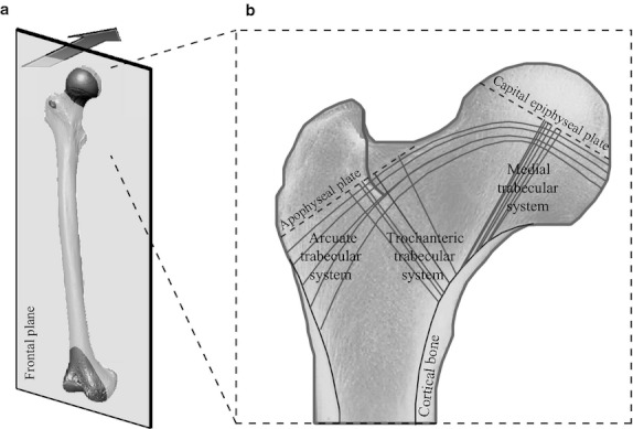
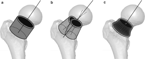
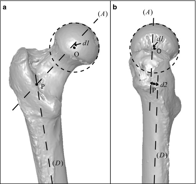
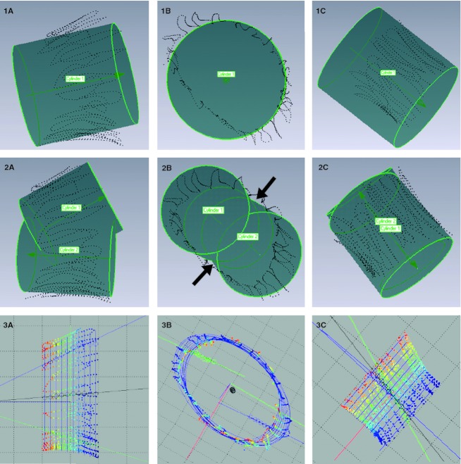
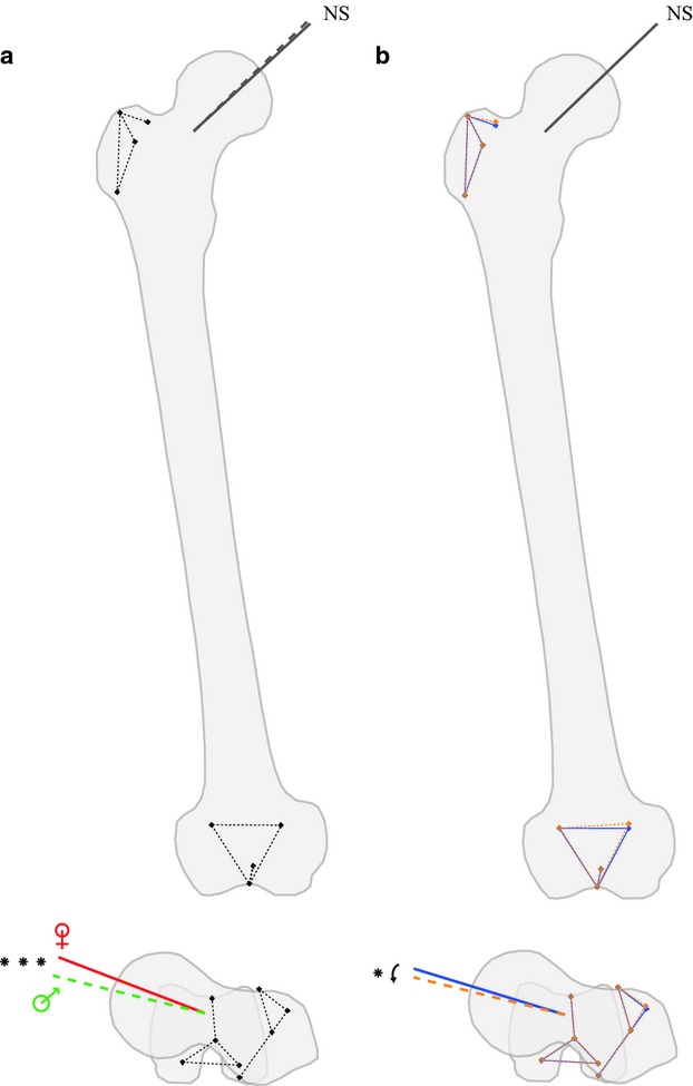
A common problem in the quantification of the orientation of the femoral neck is the difficulty to determine its true axis; however, this axis is typically estimated visually only. Moreover, the orientation of the femoral neck is commonly analysed using angles that are dependent on anatomical planes of reference and only quantify the orientation in two dimensions. The purpose of this study is to establish a method to determine the three-dimensional orientation of the femoral neck using a three-dimensional model. An accurate determination of the femoral neck axis requires a reconsideration of the complex architecture of the proximal femur. The morphology of the femoral neck results from both the medial and arcuate trabecular systems, and the asymmetry of the cortical bone. Given these considerations, two alternative models, in addition to the cylindrical one frequently assumed, were tested. The surface geometry of the femoral neck was subsequently used to fit one cylinder, two cylinders and successive cross-sectional ellipses. The model based on successive ellipses provided a significantly smaller average deviation than the two other models (P < 0.001) and reduced the observer-induced measurement error. Comparisons with traditional measurements and analyses on a sample of 91 femora were also performed to assess the validity of the model based on successive ellipses. This study provides a semi-automatic and accurate method for the determination of the functional three-dimensional femoral neck orientation avoiding the use of a reference plane. This innovative method has important implications for future studies that aim to document and understand the change in the orientation of the femoral neck associated with the acquisition of a bipedal gait in humans. Moreover, the precise determination of the three-dimensional orientation has implications in current research involved in developing clinical applications in diagnosis, hip surgery and rehabilitation.


Figures

Fig. 1
Fig. 1
(A) The femoral neck is well developed in Homo sapiens, with a global orientation slanting upwards and forwards. (B) The proximal femur presents a complex three-dimensional architecture. The cortical bone presents an asymmetric cross-sectional distribution along the femoral neck: a thick cortex is observed in the inferior part, while the cortex in the superior part is thin. The trabecular bone is organised along three major trabecular systems – medial, trochanteric and arcuate – surrounding an area of less resistance named Ward's triangle.
Fig. 2
Fig. 2
Three three-dimensional models were tested to determine the three-dimensional orientation of the femoral neck. The most intuitive model was the one based on a single cylinder (A). The superior part of the femoral neck, composed of both a thin cortex and the arcuate trabecular system, presents a different orientation compared with the inferior part, which is composed of both a thick cortex and the medial trabecular system. Thus, based on two cylinders, the two parts were modelled independently (B). Finally, the femoral neck was modelled based on successive cross-sectional ellipses (C). An ellipse is defined by two foci, a feature that was exploited here to obtain, in a single object, the two orientations of the superior and inferior parts of the femoral neck.
Fig. 3
Fig. 3
The position of the three-dimensional axis of the femoral neck was analysed in the general geometry of the femur as illustrated here in a frontal view (A) and a sagittal view (B). Both the distance of the centre of the femoral head and the femoral neck axis (d1), and the distance of the shaft axis and the femoral neck axis (d2) were calculated. (A), femoral neck axis; (D), femoral shaft axis; O, centre of the femoral head; P, pseudo-intersection between the femoral neck axis and the shaft axis.
Fig. 4
Fig. 4
Homologous landmarks used in the study and further described in Table 1. The scan of the right femur was performed using a Breuckmann® surface scanner. (A) Frontal view of the proximal part; (B) posterior view of the distal part; (C) proximal view; (D) distal view.
Fig. 5
Fig. 5
Results of the three different regression approaches are presented in the frontal (A), transverse (B) and proximal (C) views of the femoral neck. The femoral neck is poorly modelled by a circular cylinder (1): this model overestimates the antero-posterior diameter of the femoral neck. The regression based on two cylinders (2) has the advantage to consider the antero-posterior flattening of the neck and provides a more realistic model. However, an artefact between the two cylinders is created at the intersection of the two surfaces (arrows). Moreover, during the processing, the selection of the anterior and posterior parts of the femoral neck has to be done manually and, thus, involves the intervention of an operator. With the model based on successive cross-sectional ellipses (3), the artefact formed by the two surfaces is absent and the antero-posterior flattening is accurately modelled due to the long axis of the ellipses oriented in the supero-inferior direction of the neck. Moreover, this model considers the antero-posterior narrowing at the middle of the femoral neck, which is ignored by the two other models. Thus, the structure of the femoral neck was better described by the model based on successive cross-sectional ellipses.
Fig. 6
Fig. 6
Significant variations in both the three-dimensional orientation of the femoral neck and the femoral shape are illustrated in colour. (A) A significant difference in the orientation of the femoral neck between men (in dotted lines) and women (in solid lines) was detected. In the transverse plane, the anteversion of the femoral neck tends to be greater in women than in men, while in the frontal plane no difference was observed on the inclination of the femoral neck. No significant effect of sex was detected on the femoral shape and, thus, the mean shape conformation is illustrated. (B) The angle of anteversion tends to decrease with age. Orange dotted lines illustrate changes with age in both the shape conformation and the three-dimensional orientation of the femoral neck compared with the mean shape conformation as illustrated in blue solid lines.

References

    1. Anderson JY, Trinkaus E. Patterns of sexual, bilateral and interpopulational variation in human femoral neck-shaft angles. J Anat. 1998;192:279–285. - PMC - PubMed
    1. Baylac M. Rmorph a morphometric library for R. 2010. Available from the author: baylac(at)mnhn.fr [accessed 1 January 2010]
    1. Beck TJ, Ruff CB, Scott WW, et al. Sex differences in geometry of the femoral neck with aging: a structural analysis of bone mineral data. Calcif Tiss Intl. 1992;50:24–29. - PubMed
    1. Bonneau N, Simonis C, Seringe R, et al. Study of femoral torsion during prenatal growth: interpretation associated with the effects of intrauterine pressure. Am J Phys Anthropol. 2011;145:438–445. - PubMed
    1. Bookstein FL. Morphometric Tools for Landmark Data (Geometry and Biology) Cambridge: Cambridge University Press; 1991.

Publication types

LinkOut - more resources