. 2012 Sep 25;109(39):15894-9.
doi: 10.1073/pnas.1208324109. Epub 2012 Sep 11.

Collaborative interplay between FGF-2 and VEGF-C promotes lymphangiogenesis and metastasis

Affiliations

Collaborative interplay between FGF-2 and VEGF-C promotes lymphangiogenesis and metastasis

Renhai Cao et al. Proc Natl Acad Sci U S A. .

Abstract

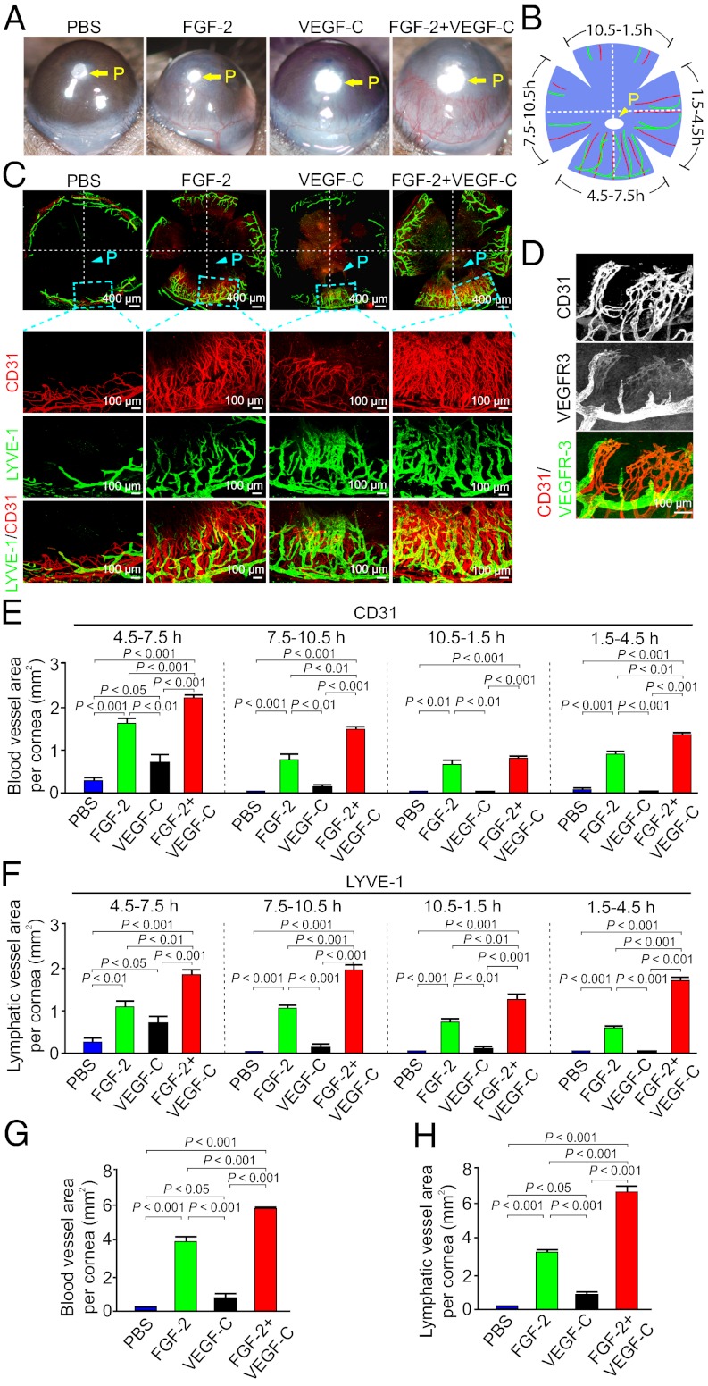
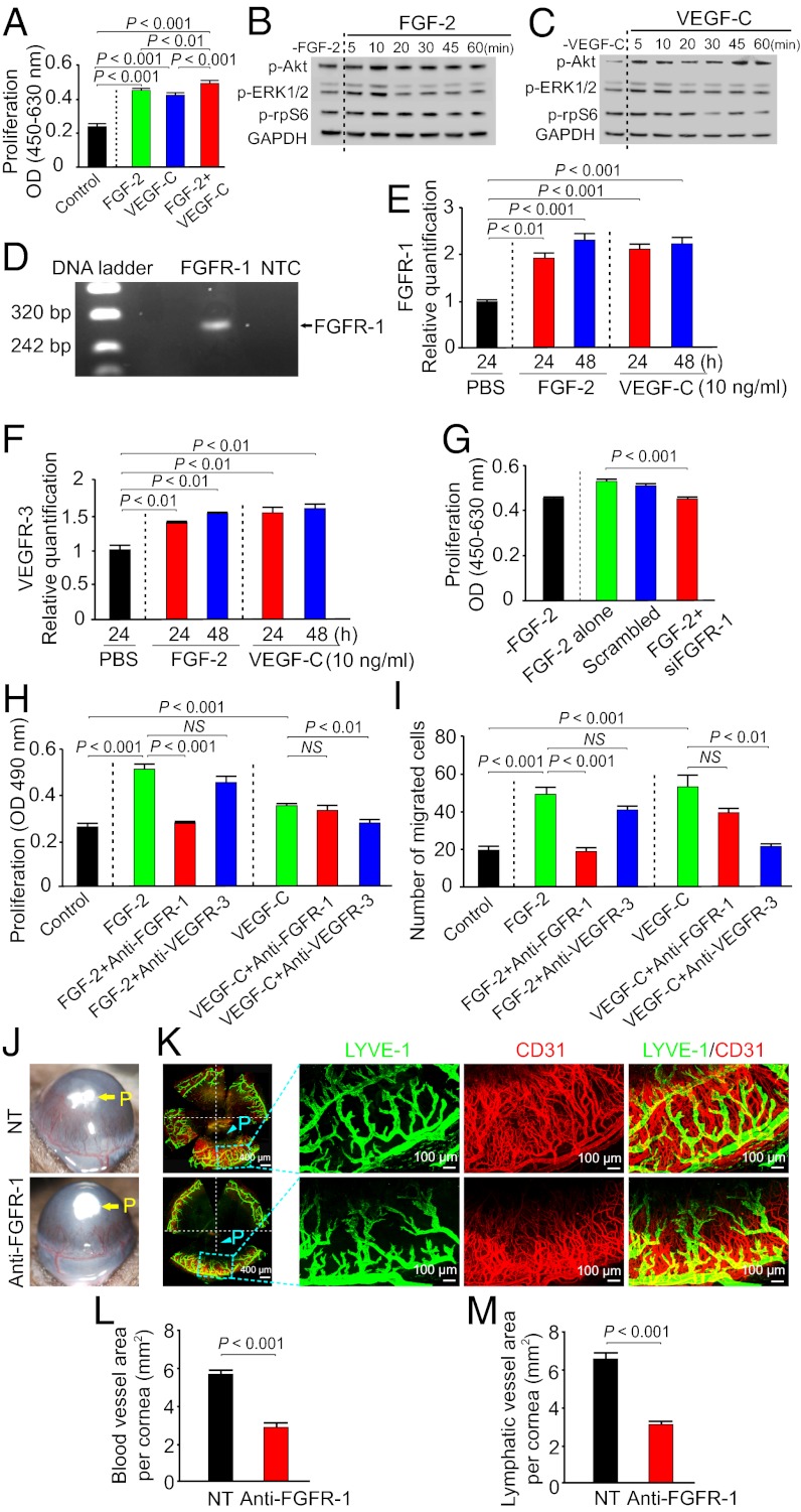
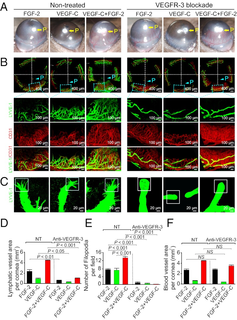
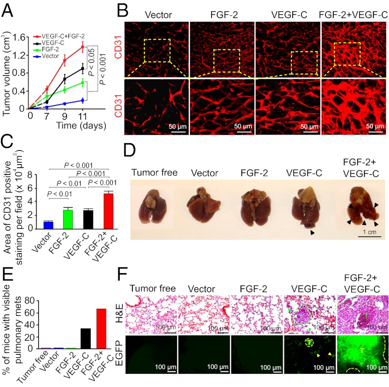
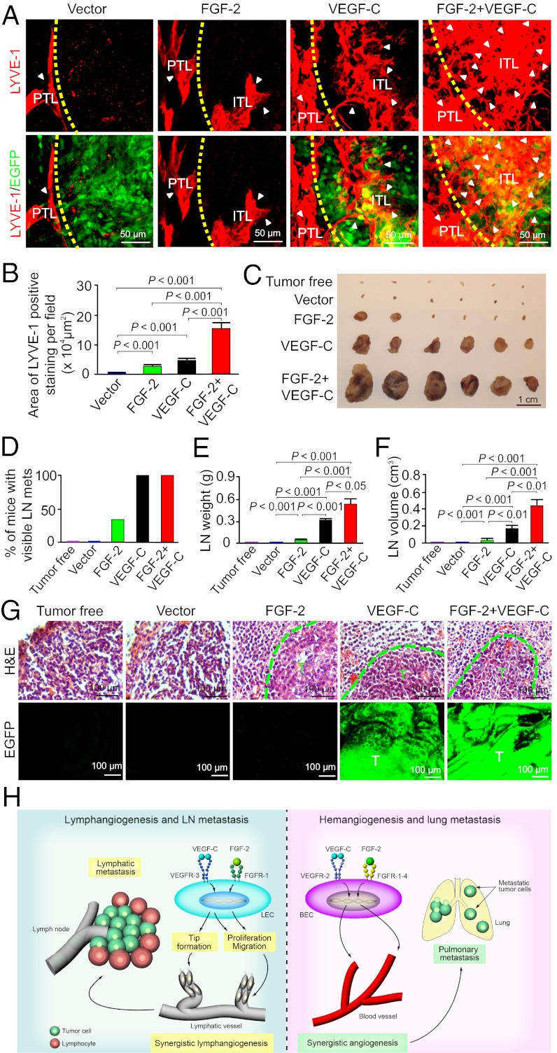
Interplay between various lymphangiogenic factors in promoting lymphangiogenesis and lymphatic metastasis remains poorly understood. Here we show that FGF-2 and VEGF-C, two lymphangiogenic factors, collaboratively promote angiogenesis and lymphangiogenesis in the tumor microenvironment, leading to widespread pulmonary and lymph-node metastases. Coimplantation of dual factors in the mouse cornea resulted in additive angiogenesis and lymphangiogenesis. At the molecular level, we showed that FGFR-1 expressed in lymphatic endothelial cells is a crucial receptor that mediates the FGF-2-induced lymphangiogenesis. Intriguingly, the VEGFR-3-mediated signaling was required for the lymphatic tip cell formation in both FGF-2- and VEGF-C-induced lymphangiogenesis. Consequently, a VEGFR-3-specific neutralizing antibody markedly inhibited FGF-2-induced lymphangiogenesis. Thus, the VEGFR-3-induced lymphatic endothelial cell tip cell formation is a prerequisite for FGF-2-stimulated lymphangiogenesis. In the tumor microenvironment, the reciprocal interplay between FGF-2 and VEGF-C collaboratively stimulated tumor growth, angiogenesis, intratumoral lymphangiogenesis, and metastasis. Thus, intervention and targeting of the FGF-2- and VEGF-C-induced angiogenic and lymphangiogenic synergism could be potentially important approaches for cancer therapy and prevention of metastasis.


Conflict of interest statement

The authors declare no conflict of interest.

Figures

Fig. 1.
Fig. 1.
FGF-2 and VEGF-C collaboratively stimulate hem- and lymphangiogensis. (A) Micropellets containing FGF-2, VEGF-C, or FGF-2 plus VEGF-C together with the slow-release polymer were implanted into micropockets of mouse corneas (n = 5–6 per group). At day 6 after implantation, corneal neovascularization was examined and photographed. Slow-release polymer containing PBS was used as a negative control. P indicates the position of the implanted pellet. (B) Schematic diagram demonstrates the flat-mounted cornea and clock-hours of the circumferential cornea. (C) A representative cornea from each group that were double immunostained with CD31 (red) and LYVE-1 (green), which showed no overlapping signals. (D) Double immunostaining of corneal blood and lymphatic vessels using VEGFR-3 (green) and CD31 (red) specific antibodies. (E) Quantification of corneal CD31+ blood vessels at different clock hours (n = 5–6 /group). (F) Quantification of corneal LYVE-1 positive lympghatic vessels at different clockhours (n = 5–6 per group). (G) Quantification of the total CD31+ vessels in the entire of circumferential area of each cornea (n = 5–6 per group). (H) Quantification of the total LYVE-1+ lymphatic vessels in the entire of circumferential area of each cornea (n = 5–6 per group).
Fig. 2.
Fig. 2.
In vitro LEC activity and signaling and suppression of hem- and lymphangiogenesis by FGFR-1 blockade. (A) Stimulation of hLEC proliferation by FGF-2, VEGF-C, FGF-2 plus VEGF-C or buffer alone (16 samples per group). (B) Immunoblot analysis of activation of signaling components in hLECs by FGF-2. GDPDH showed the standard level of sample loading. (C) Immunoblot analysis of activation of signaling components in hLECs by VEGF-C. GDPDH showed the standard level of sample loading. (D) Amplification of Fgfr-1 by reverse-transcription PCR using cDNA extracted from LECs. (E) qPCR analysis of Fgfr-1 mRNA expression levels in hLECs stimulated by FGF-2 or VEGF-C. PBS-stimulated cells were used as a control. (F) Quantitative PCR analysis of Vegfr-3 mRNA expression levels in hLECs stimulated by FGF-2 or VEGF-C. PBS-stimulated cells were used as a control. (G) Fgfr-1 specific siRNA substantially inhibited FGF-2–induced LEC proliferation. A scrambled nucleotide sequence was used as a control. (H) Inhibition of FGF-2– or VEGF-C–induced mLEC proliferation by FGFR-1 or VEGFR-3 blockade (six samples per group). (I) Inhibition of FGF-2– or VEGF-C–induced mLEC migration by FGFR-1 or VEGFR-3 blockade (six samples per group). (J) Suppression of FGF-2 plus VEGF-C–induced corneal neovascularization by FGFR-1 blockade. (K) Suppression of FGF-2 plus VEGF-C–induced corneal hemangiogenesis (CD31+ blood vessels) and lymphangiogenesis (LYVE-1+ lymphatic vessels) by FGFR-1 blockade. P, pellet. (L) Quantification of antiangiogenic activity by FGFR-1 blockade (n = 5–6 per group). (M) Quantification of anti-lymphangiogenic activity by FGFR-1 blockade (n = 5–6 per group).
Fig. 3.
Fig. 3.
Anti-VEGFR-3 blocks FGF-2–induced lymphangiogenesis and lymphatic tip formation. (A) FGF-2–, VEGF-C–, or FGF-2 plus VEGF-C–induced corneal neovascularization was treated with or without VEGFR-3 blockade. (B) FGF-2–, VEGF-C–, or FGF-2 plus VEGF-C–induced corneas in response to anti–VEGFR-3 treatment were double immunostained with CD31 (red) and LYVE-1 (green). VEGFR-3 blockade almost completely suppressed FGF-2–, VEGF-C–, or FGF-2 plus VEGF-C–induced corneal lymphangiogenesis but not hemangiogenesis. (C) Suppression of FGF-2–, VEGF-C–, or FGF-2 plus VEGF-C–induced tip formation of lymphatic vessels by VEGFR-3 blockade. (D) Quantification of LYVE-1+ lymphatic vessels in various groups treated with or without VEGFR-3 blockade (n = 4–6 per group). (E) Quantification of lymphatic tips in various groups treated with or without VEGFR-3 blockade (n = 4–6 per group). (F) Quantification of CD31+ blood vessels in various groups treated with or without VEGFR-3 blockade (n = 4–6 per group).
Fig. 4.
Fig. 4.
FGF-2 and VEGF-C collaboratively promote tumor angiogenesis and hematogenous metastasis. (A) Growth rates of vector, FGF-2–, VEGF-C–, and FGF-2 plus VEGF-C–expressing mouse fibrosarcomas. (B) Detection of tumor microvessels in various tumor groups by CD31 immunostaining. (C) Quantification of CD31+ vessel density in various tumor groups (n = 6 per group). (D) Lung morphology of a representative mouse from each tumor-bearing group (n = 6 per group). Arrowheads point to lung surface metastatic nodules. (E) Quantification of percentage of tumor-bearing mice that had lung metastases (n = 6 per group). (F) Histological examination of pulmonary metastases in mice bearing various tumors. Dashed lines encircle metastatic tumors that express EGFP (green). T, tumor.
Fig. 5.
Fig. 5.
FGF-2 and VEGF-C collaboratively promotes tumor lymphangiogenesis and lymphatic metastasis. (A) Detection of peritumoral and intratumoral lymphatic vessels (PTL and ITL) in various tumor groups by LYVE-1 immunostaining. Dashed line defines the rim between the tumor and peritumoral regions. Arrowheads indicate the tumoral lymphatics. (B) Quantification of intratumoral LYVE-1+ lymphatic vessel density in various tumor groups (n = 6 per group). (C) Sentinel lymph node morphology of various tumor-bearing groups (n = 6 per group). (D) Quantification of percentage of tumor-bearing mice that had sentinel lymph-node metastases (n = 6 per group). (E) Average weight of sentinel lymph nodes in various tumor-bearing mice (n = 6 /group). (F) Average volume of sentinel lymph nodes in various tumor-bearing mice (n = 6 per group). (G) Sentinel lymph node histology showed the presence of lymphatic metastases in FGF-2, VEGF-C, and FGF-2 plus VEGF-C tumor-mice. Dashed lines encircle metastatic tumors that express EGFP (green). T, tumor. (H) Schematic diagram of molecular mechanisms by which FGF-2 and VEGF-C collaboratively induce hem-/lymphangiogenesis and metastasis. VEGF-C activates VEGFR-3 receptor on LECs, leading to LEC tip formation, proliferation, and migration. FGF-2 via activation of FGFR-1 stimulates LEC proliferation and migration. However, VEGFR-3–triggered tip cell formation is a prerequisite for FGF-2–induced lymphangiogenesis. Blockade of the VEGFR-3 signaling system completely inhibits FGF-2–induced lymphangiogenesis. Lymhangiogenic interplay between FGF-2 and VEGF-C in the tumor environment promoted lymphatic metastasis. On blood vessel endothelial cells (BVECs), VEGF-C binds to VEGFR-2 and induced hem-angiogenic signals. FGF-2 directly induces hemangiogenesis by activation of FGFR-1 to -4 expressed on BVECs. The FGF-2 plus VEGF-C–induced high numbers of disorganized tumor blood vessels facilitate hematogenous metastasis.

Similar articles

Cited by

References

    1. Fidler IJ, Ellis LM. The implications of angiogenesis for the biology and therapy of cancer metastasis. Cell. 1994;79:185–188. - PubMed
    1. Hanahan D, Weinberg RA. Hallmarks of cancer: The next generation. Cell. 2011;144:646–674. - PubMed
    1. Rouhi P, et al. Hypoxia-induced metastasis model in embryonic zebrafish. Nat Protoc. 2010;5:1911–1918. - PubMed
    1. Lee SL, et al. Hypoxia-induced pathological angiogenesis mediates tumor cell dissemination, invasion, and metastasis in a zebrafish tumor model. Proc Natl Acad Sci USA. 2009;106:19485–19490. - PMC - PubMed
    1. Cao Y. Opinion: Emerging mechanisms of tumour lymphangiogenesis and lymphatic metastasis. Nat Rev Cancer. 2005;5:735–743. - PubMed

Publication types

MeSH terms

Substances