. 2012 Oct 16;109(42):16847-51.
doi: 10.1073/pnas.1211508109. Epub 2012 Sep 11.

Rhythmic ring-ring stacking drives the circadian oscillator clockwise

Affiliations

Rhythmic ring-ring stacking drives the circadian oscillator clockwise

Yong-Gang Chang et al. Proc Natl Acad Sci U S A. .

Abstract

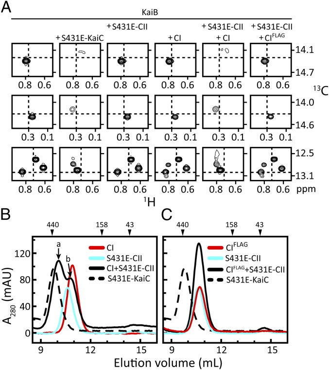
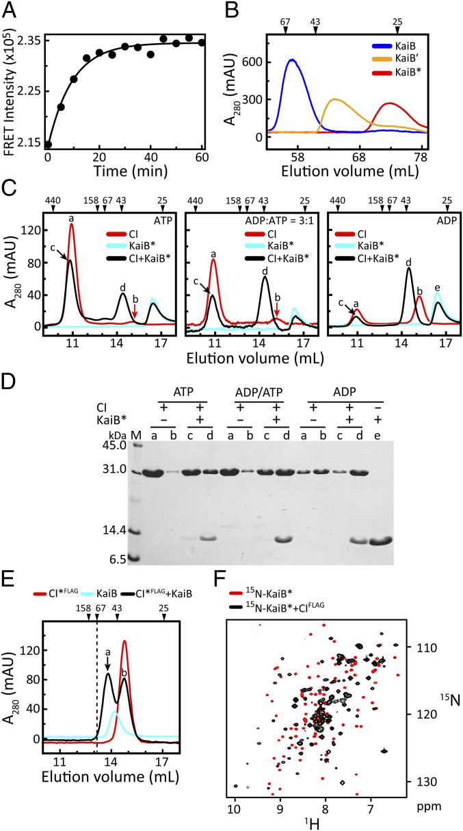
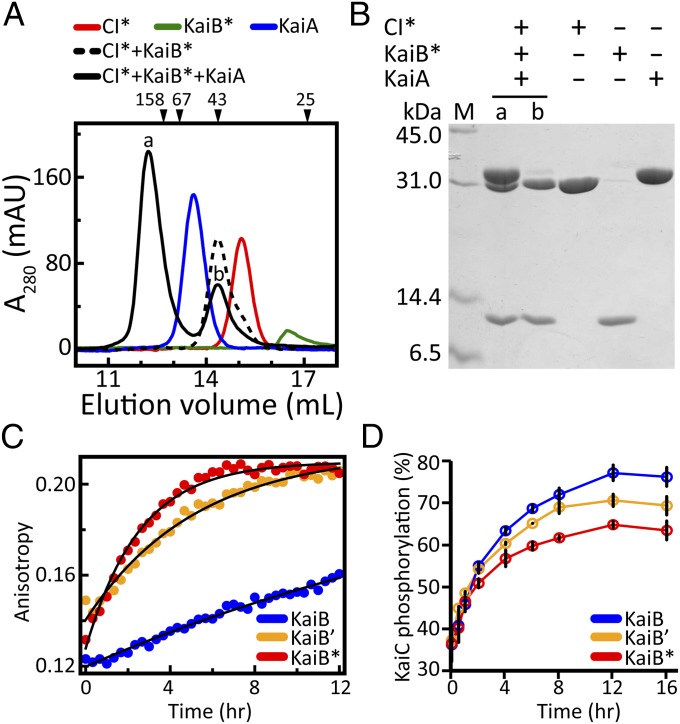
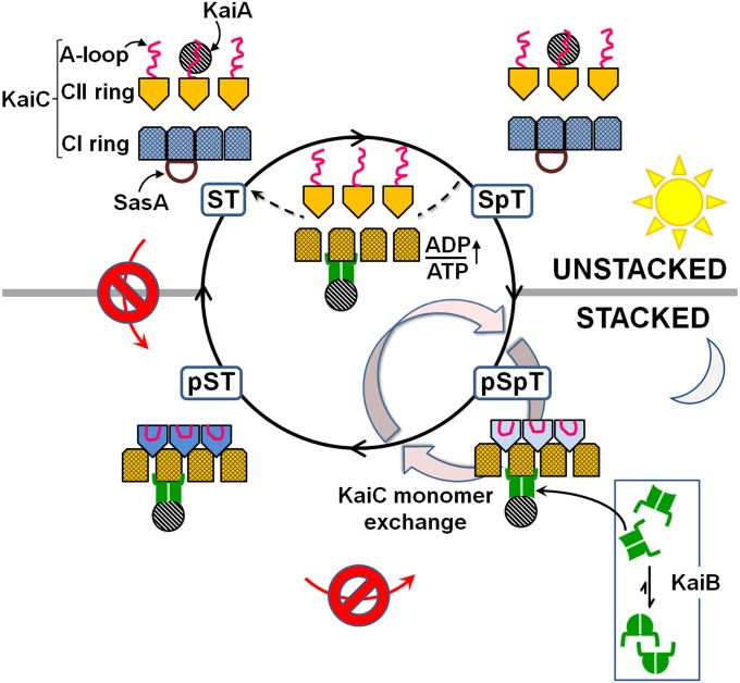
The oscillator of the circadian clock of cyanobacteria is composed of three proteins, KaiA, KaiB, and KaiC, which together generate a self-sustained ∼24-h rhythm of phosphorylation of KaiC. The mechanism propelling this oscillator has remained elusive, however. We show that stacking interactions between the CI and CII rings of KaiC drive the transition from the phosphorylation-specific KaiC-KaiA interaction to the dephosphorylation-specific KaiC-KaiB interaction. We have identified the KaiB-binding site, which is on the CI domain. This site is hidden when CI domains are associated as a hexameric ring. However, stacking of the CI and CII rings exposes the KaiB-binding site. Because the clock output protein SasA also binds to CI and competes with KaiB for binding, ring stacking likely regulates clock output. We demonstrate that ADP can expose the KaiB-binding site in the absence of ring stacking, providing an explanation for how it can reset the clock.


Conflict of interest statement

The authors declare no conflict of interest.

Figures

Fig. 1.
Fig. 1.
Stacking of CI and CII rings of KaiC is required for KaiC–KaiB binding. (A) Selected regions from methyl-TROSY spectra (26, 27) of U-[15N, 2H]-Ile-δ1-[13C, 1H]–labeled KaiB alone (column 1) or in the presence of S431E-KaiC (column 2), S431E-CII (column 3), CI (column 4), S431E-CII and CI (column 5), or S431E-CII and CIFLAG (column 6). Complete spectra are shown in SI Appendix, Fig. S1. (B and C) Gel-filtration profiles of mixtures of CI (B) and CIFLAG (C) with S431E-CII. Full-length S431E-KaiC was run as a control. Peaks denoted by a and b were checked by SDS/PAGE (SI Appendix, Fig. S2). The additional FLAG tag at the C terminus of CIFLAG was included to maximize potential repulsive interactions with the N-terminal FLAG tag on isolated S431E-CII domains. The same runs of S431E-CII and S431E-KaiC are shown in B and C. Details on the protein constructs are provided in SI Appendix, Table S1.
Fig. 2.
Fig. 2.
KaiB binds to monomeric CI. (A) FRET measurements of KaiB subunit exchange after mixing two samples of KaiB separately labeled with the donor and acceptor fluorophores 1,5-IAEDANS and 6-IAF. Excitation and emission wavelengths were set to 347 nm and 530 nm, respectively. (B) Gel-filtration profiles of KaiB and KaiB variants KaiB′ and KaiB* (SI Appendix, Table S1). KaiB′ differs from KaiB by two amino acyl substitutions, Y8A and Y94A. KaiB* differs from KaiB′ by being truncated after residue Y94A. (C) Gel-filtration profiles of mixtures of CI and KaiB* in the presence of [ATP] = 0.25 mM (Left), [ADP]/[ATP] = 0.75 mM/0.25 mM (Middle), and [ADP] = 0.25 mM (Right). Peaks denoted by a, b, c, d, and e were checked by SDS/PAGE. For clarity, a red arrow indicates peak b. (D) SDS/PAGE gel of peaks a, b, c, d, and e in C. (E) Gel-filtration profile of a mixture of a monomeric variant CI*FLAG (SI Appendix, Table S1) with KaiB. Peaks denoted by a and b were checked by SDS/PAGE (SI Appendix, Fig. S6). The dashed line indicates the position of a 67-kDa marker. The size of a complex of CI* and a KaiB tetramer would exceed 67 kDa. (F) 15N, 1H-HSQC spectra of 15N-labeled KaiB* free (red contours) and bound (black contours) to CIFLAG. The NMR spectra were recorded at 37 °C (SI Appendix, Table S2). Note that CIFLAG exists as a monomer in an ATP-free buffer (SI Appendix, Fig. S5). Details on the purification of these proteins are provided in SI Appendix, Table S3.
Fig. 3.
Fig. 3.
Formation of the CI*–KaiB*–KaiA complex. (A) Gel-filtration profiles of CI* alone (red line), KaiB* alone (green line), KaiA alone (blue line), mixtures of CI* + KaiB* (dashed black line), and CI* + KaiB* + KaiA (solid black line). Elution profiles of mixtures of CI* + KaiA and KaiB* + KaiA are shown in SI Appendix, Fig. S7. (B) SDS/PAGE of peaks denoted by a and b in A. (C) Fluorescence anisotropy of the binding kinetics of 6-IAF–labeled KaiB and KaiB variants (KaiB′ or KaiB*) to S431E-KaiC (blue, KaiB; orange, KaiB′; red, KaiB*). (D) Phosphorylation kinetics profiles of KaiC in the presence of KaiA and KaiB or KaiB variants (KaiB′ or KaiB*). Color-coding is the same as in C. Each data point represents the average of two experiments. Error bars represent SEM. Values were determined from densitometric analysis of SDS/PAGE gels stained with Coomassie blue (SI Appendix, Fig. S8).
Fig. 4.
Fig. 4.
Allosteric model of how ring stacking in KaiC orchestrates the moving parts of the circadian oscillator of cyanobacteria. The model predicts that rhythmic ring stacking drives phosphorylation clockwise, reorganizes protein–protein interactions, provides a temporal window for ADP-induced resetting, and regulates clock output. Only three subunits of the CII ring and four subunits of the CI ring are shown for clarity. Likewise, the linkers connecting the CI and CII domains are not shown.

Comment in

Similar articles

Cited by

References

    1. Hogenesch JB, Ueda HR. Understanding systems-level properties: Timely stories from the study of clocks. Nat Rev Genet. 2011;12:407–416. - PubMed
    1. Ishiura M, et al. Expression of a gene cluster kaiABC as a circadian feedback process in cyanobacteria. Science. 1998;281:1519–1523. - PubMed
    1. Tomita J, Nakajima M, Kondo T, Iwasaki H. No transcription-translation feedback in circadian rhythm of KaiC phosphorylation. Science. 2005;307:251–254. - PubMed
    1. Nakajima M, et al. Reconstitution of circadian oscillation of cyanobacterial KaiC phosphorylation in vitro. Science. 2005;308:414–415. - PubMed
    1. Rust MJ, Markson JS, Lane WS, Fisher DS, O’Shea EK. Ordered phosphorylation governs oscillation of a three-protein circadian clock. Science. 2007;318:809–812. - PMC - PubMed

Publication types

MeSH terms

Substances