. 2012 Nov 14;31(22):4258-75.
doi: 10.1038/emboj.2012.261. Epub 2012 Sep 11.

Arginine methylation next to the PY-NLS modulates Transportin binding and nuclear import of FUS

Affiliations

Arginine methylation next to the PY-NLS modulates Transportin binding and nuclear import of FUS

Dorothee Dormann et al. EMBO J. .

Abstract

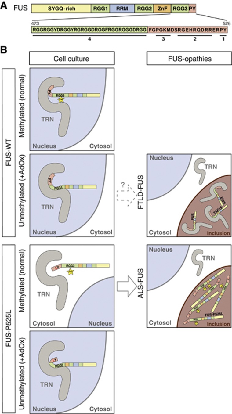
Fused in sarcoma (FUS) is a nuclear protein that carries a proline-tyrosine nuclear localization signal (PY-NLS) and is imported into the nucleus via Transportin (TRN). Defects in nuclear import of FUS have been implicated in neurodegeneration, since mutations in the PY-NLS of FUS cause amyotrophic lateral sclerosis (ALS). Moreover, FUS is deposited in the cytosol in a subset of frontotemporal lobar degeneration (FTLD) patients. Here, we show that arginine methylation modulates nuclear import of FUS via a novel TRN-binding epitope. Chemical or genetic inhibition of arginine methylation restores TRN-mediated nuclear import of ALS-associated FUS mutants. The unmethylated arginine-glycine-glycine domain preceding the PY-NLS interacts with TRN and arginine methylation in this domain reduces TRN binding. Inclusions in ALS-FUS patients contain methylated FUS, while inclusions in FTLD-FUS patients are not methylated. Together with recent findings that FUS co-aggregates with two related proteins of the FET family and TRN in FTLD-FUS but not in ALS-FUS, our study provides evidence that these two diseases may be initiated by distinct pathomechanisms and implicates alterations in arginine methylation in pathogenesis.


Conflict of interest statement

The authors declare that they have no conflict of interest.

Figures

Figure 1
Figure 1
Cytoplasmic mislocalization of ALS-associated FUS mutants is abrogated upon inhibition of methylation. (A) Schematic diagram showing the domain structure of FUS. Sequence of the C-terminal PY-NLS and ALS-causing point mutations within the NLS are given below. ALS-associated mutations outside the PY-NLS are described elsewhere (Mackenzie et al, 2010b). SYGQ-rich=serine, tyrosine, glycine, glutamine-rich domain; RRM=RNA recognition motif; ZnF=zinc finger. (B) Localization of HA-tagged FUS WT or the indicated ALS-associated FUS mutants in untreated (upper panels) or AdOx-treated (lower panels) HeLa cells. ALS-associated FUS mutants become predominantly nuclear upon AdOx treatment, suggesting that methylation modulates nuclear import of FUS. Scale bars: 20 μm. (C) Quantification of nuclear and cytosolic fluorescence intensities. Values are means across n cells, error bars indicate standard deviations (s.d.). Statistical significance between untreated and AdOx-treated is displayed as *** (P<0.001; one-way ANOVA). (D) HA–FUS protein levels in untreated and AdOx-treated HeLa cells were analysed by immunoblotting with an HA-specific antibody (upper panel). Actin served as a loading control (lower panel). AdOx treatment does not affect expression of HA–FUS constructs. (E) Localization of HA-tagged FUS-WT or P525L (red) in untreated or AdOx-treated primary rat hippocampal neurons. YFP (green) served as a cytosolic filler protein to visualize neuronal morphology, nuclei were visualized with DAPI. In contrast to untreated neurons, AdOx-treated neurons rarely show cytoplasmic mislocalization of FUS-P525L. Scale bars: 20 μm. Figure source data can be found with the Supplementary data.
Figure 2
Figure 2
Methylation affects nuclear localization of EWS and TAF15 mutants. (A) Schematic diagram showing the domain structures of FUS, EWS and TAF15 (FET proteins). (B) Localization of HA-tagged FUS, EWS or TAF15 (WT or with mutant PY-NLS) in untreated or AdOx-treated HeLa cells. Cytoplasmic FET protein mutants become predominantly nuclear upon AdOx treatment, suggesting that methylation affects nuclear import of all FET family members in a similar fashion. Scale bars: 20 μm. (C) Quantification of nuclear and cytosolic fluorescence intensities. Values are means across n cells, error bars indicate s.d. Statistical significance is displayed as *** (P<0.001) (one-way ANOVA).
Figure 3
Figure 3
PRMT1 silencing causes increased nuclear localization of FUS-P525L. (A) Protein N-arginine methyltransferase 1 (PRMT1) expression was silenced in HeLa cells by transfection of two different siRNAs (PRMT1#1 and PRMT1#2), a control (ctrl.) siRNA was used as a negative control. In all, 48 h after siRNA delivery, cells were transfected with HA-tagged FUS-WT or P525L and localization of these proteins was examined by HA immunostaining (green) and confocal microscopy. PRMT1 knockdown causes a predominantly nuclear localization of the FUS-P525L mutant, suggesting that arginine methylation by PRMT1 modulates nuclear import of FUS. Scale bars: 20 μm. Immunoblots on the right show PRMT1 knockdown efficiency. Total cell lysates were examined with a PRMT1-specific antibody (upper panel) and a tubulin-specific antibody (lower panel). (B) Quantification of nuclear and cytosolic fluorescence intensities. Values are means across n cells, error bars indicate s.d. Statistical significance is displayed as *** (P<0.001) (one-way ANOVA). Figure source data can be found with the Supplementary data.
Figure 4
Figure 4
Nuclear import of FUS-P525L upon AdOx treatment is dependent on TRN. (A) Localization of HA–FUS-P525L in untreated or AdOx-treated HeLa cells after co-expression of GFP as a control, a competitor of the TRN pathway (GFP–M9M) or a competitor of the Importin α pathway (GFP–Bimax) (green). After HA immunostaining (red), localization of mutant FUS was examined by confocal microscopy. The bottom row shows a higher magnification of the boxed regions. GFP–M9M expressing cells with stress granules are labelled with an arrowhead, cells without stress granules are labelled with an asterisk. Both in cells with and without stress granules, GFP–M9M prevents nuclear import of FUS-P525L upon AdOx treatment, demonstrating that import of FUS-P525L is TRN dependent. Scale bars: 20 μm. (B) Localization of a FUS deletion mutant lacking the core amino acids of the C-terminal PY-NLS (Δ514–526, see schematic diagram) in untreated or AdOx-treated HeLa cells. Nuclear relocalization upon AdOx treatment requires the FUS PY-NLS, suggesting that it requires direct TRN binding. Scale bars: 20 μm. Quantification shows nuclear and cytosolic fluorescence intensities. Values are means across n cells, error bars indicate s.d. Statistical significance is displayed as *** (P<0.001) (one-way ANOVA); NS=not significant.
Figure 5
Figure 5
Arginine residues in the RGG3 domain of FUS are required for nuclear import of mutant FUS. (A) GST–GFP reporter proteins with the indicated FUS sequences at the C-terminus were transiently expressed in untreated or AdOx-treated HeLa cells. Localization of reporter constructs was examined after GFP (green) immunostaining by confocal microscopy. Arginine residues in the PY-NLS (amino acids 514–526) are not sufficient for restoring nuclear import of the mutant reporter protein upon AdOx treatment, while arginines in the RGG3 domain (amino acids 455–505) are necessary and sufficient for this effect. Scale bars: 20 μm. (B) Quantification shows nuclear and cytosolic fluorescence intensities. Values are means across n cells, error bars indicate s.d. Statistical significance is displayed as *** (P<0.001) (one-way ANOVA); NS=not significant.
Figure 6
Figure 6
Both the RGG3 domain and the PY-NLS of FUS interact with TRN. (A) Schematic diagram of recombinant FUS proteins analysed by NMR spectroscopy. (B) Overlay of 2D 1H,15N HSQC NMR spectra recorded for FUS454–526WT (black) and FUS454–526P525L (magenta). Regions characteristic for glycine residues, position R524 and the C-terminal Y526 are encircled (dotted line). The distribution of NMR signals in regions characteristic for random coil proteins indicates that both proteins are intrinsically disordered in solution. (C) Overlay of selected regions (glycine and Y526) of 2D 1H,15N HSQC NMR spectra recorded for FUS454–526WT (black) and FUS454–526P525L (magenta) in isolation and in the presence of an equimolar stoichiometric equivalent of TRN (blue). NMR signals characteristic for glycine residues disappear upon addition of TRN, indicating binding of RGG repeats to TRN. The NMR signal of Y526 disappears upon addition of TRN in the WT protein, but is only slightly affected in the P525L mutant, indicating tight binding of TRN to the WT PY but not the mutant LY. (D) Schematic diagram of recombinant FUS proteins and synthetic FUS peptides analysed for TRN binding by ITC. Binding constants (KdITC) are shown on the right. Both the PY-NLS and the N-terminal RGG repeats contribute to TRN binding. The RGG3 repeat domain can bind TRN in the absence of a C-terminal PY-NLS and can compensate for the lack of binding of the mutant C-terminus in the P525L mutant.
Figure 7
Figure 7
Arginine methylation in the RGG3 domain of FUS weakens TRN binding. In vitro pull-down assays with unmethylated and methylated (me) synthetic FUS peptides (see schematic diagrams for sequences, asterisks indicate asymmetric dimethyl groups) and recombinant TRN. Biotinylated peptides were immobilized on streptavidin beads and were incubated with the indicated amount of TRN–His6 or His6–GST as a control. Bound TRN was visualized by SDS–PAGE after Coomassie staining (upper panel). The middle panel shows the amount of streptavidin and peptide that was boiled off the streptavidin beads. Lower panel shows input of TRN. Plots on the right show a quantification of the TRN pulldown efficiency. The band with the highest intensity was set to 1.0 AU (arbitrary units) and relative intensities of other bands were calculated. Plots show means from two independent experiments, error bars indicate s.d. (A) The unmethylated RGG3 domain (FUS473–503) binds TRN in the absence of a PY-NLS and methylation abrogates this interaction. (B) Methylation strongly impairs TRN binding of the FUS489–526P525L peptide. (C) Methylation slightly reduces TRN binding of the FUS489–526WT peptide (∼3-fold difference in Kd). Note that both FUS489–526WT and meFUS489–526WT bind TRN more tightly than FUS489–526P525L (reference lane labelled ‘P525L’ on the right). Figure source data can be found with the Supplementary data.
Figure 8
Figure 8
meFUS-specific monoclonal antibodies label ALS-associated FUS mutants in stress granules. (A) Schematic diagram of the peptide epitope (meFUS473–503) used for generation of meFUS-specific monoclonal antibodies. Asterisks denote asymmetric dimethyl groups. (B) Immunoblots show that monoclonal antibodies 14H5 and 9G6 are specific for methylated FUS, since staining is abrogated upon AdOx treatment and FUS knockdown. Open arrowhead indicates a non-specific methylated protein recognized by 14H5. Lower panels show FUS knockdown efficiency and Tubulin as a loading control. (C) Double-label immunocytochemistry of untransfected HeLa cells with meFUS-specific monoclonal antibodies 14H5 or 9G6 (green) and a polyclonal pan-FUS antibody (red). Both 14H5 and 9G6 specifically stain endogenous methylated FUS in the nuclei (blue). This staining is abrogated upon AdOx treatment and FUS knockdown. Scale bars: 20 μm. (D) HeLa cells stably expressing HA-tagged FUS-WT or HA–FUS-P525L were exposed to heat shock (44 °C) for 1 h prior to fixation or were kept at 37°C (untreated). Localization of methylated FUS was examined by confocal microscopy by co-labelling with a meFUS-specific antibody 9G6 (green), an HA-specific antibody (red), a TIA-1-specific antibody (white) and a nuclear counterstain (blue). Methylated FUS-P525L is recruited to TIA-1-positive stress granules after heat shock. Scale bars: 20 μm. Figure source data can be found with the Supplementary data.
Figure 9
Figure 9
meFUS-specific monoclonal antibodies label FUS inclusions in ALS-FUS, but not in FTLD-FUS. (A) Double-label immunofluorescence of spinal cord sections from four ALS patients carrying different FUS mutations with a monoclonal antibody against meFUS (9G6, green), a polyclonal pan-FUS antibody (red) and nuclear counterstaining (blue). The characteristic FUS-positive neuronal cytoplasmic inclusions (rows 1–4) and glial inclusions (insert in row 3) in all ALS-FUS cases were intensely co-labelled by the meFUS-specific antibody. Scale bar: 20 μm. (B) Double-label immunofluorescence with a monoclonal antibody against meFUS (9G6, green), a polyclonal pan-FUS antibody (red) and nuclear counterstaining (blue) in the spectrum of FTLD-FUS, including aFTLD-U, BIBD and NIFID. In striking contrast to ALS-FUS, the meFUS-specific antibody did not label FUS-positive neuronal cytoplasmic inclusions (arrows in row 1 and 2) and neuronal intranuclear inclusions (arrowheads in row 1 and 2), as shown in dentate granule cells in aFTLD-U and the brainstem in BIBD and NIFID. Scale bar: 50 μm (row 1) or 20 μm (rows 2–4).
Figure 10
Figure 10
Model of the FUS–TRN interaction in cellular models and human FUSopathies. (A) Schematic diagram of FUS with sequences of the C-terminal PY-NLS (light red) and RGG3 domain (light green). Numbers indicate epitopes that contribute to TRN binding: 1=C-terminal PY motif; 2=central basic motif forming a polarized helix; 3=N-terminal hydrophobic motif; 4=RGG repeat region as a novel TRN-binding epitope. (B) Panels on the left show the interaction of methylated and unmethylated FUS-WT and FUS-P525L with TRN and the consequences for nuclear import in cultured cells. The PY-NLS of FUS is shown in light red and the RGG3 repeat region in light green. The yellow star denotes asymmetric dimethylation of the RGG3 domain. Panels on the right show the pathological situation in post mortem brains of FTLD-FUS and ALS-FUS patients. In FTLD-FUS, neuronal cytoplasmic inclusions contain all three FET proteins and TRN, but are not immunoreactive with meFUS-specific antibodies, suggesting that hypomethylation of the FET proteins and thus increased TRN binding may possibly be involved in the co-deposition of these proteins in FTLD-FUS. In contrast, ALS-FUS caused by FUS mutations is characterized by neuronal cytoplasmic inclusions that contain methylated FUS, but are negative for EWS, TAF15 and TRN. This suggests that the selective nuclear import defect of the FUS protein is caused by combination of a genetic defect (point mutation in TRN-binding epitopes 1–3) and post-translational modification (arginine methylation in TRN-binding epitope 4).

Comment in

Similar articles

Cited by

References

    1. Aoki K, Ishii Y, Matsumoto K, Tsujimoto M (2002) Methylation of Xenopus CIRP2 regulates its arginine- and glycine-rich region-mediated nucleocytoplasmic distribution. Nucleic Acids Res 30: 5182–5192 - PMC - PubMed
    1. Aoki N, Higashi S, Kawakami I, Kobayashi Z, Hosokawa M, Katsuse O, Togo T, Hirayasu Y, Akiyama H (2012) Localization of fused in sarcoma (FUS) protein to the post-synaptic density in the brain. Acta Neuropathol 124: 383–394 - PubMed
    1. Araya N, Hiraga H, Kako K, Arao Y, Kato S, Fukamizu A (2005) Transcriptional down-regulation through nuclear exclusion of EWS methylated by PRMT1. Biochem Biophys Res Commun 329: 653–660 - PubMed
    1. Bartel RL, Borchardt RT (1984) Effects of adenosine dialdehyde on S-adenosylhomocysteine hydrolase and S-adenosylmethionine-dependent transmethylations in mouse L929 cells. Mol Pharmacol 25: 418–424 - PubMed
    1. Baumer D, Hilton D, Paine SM, Turner MR, Lowe J, Talbot K, Ansorge O (2010) Juvenile ALS with basophilic inclusions is a FUS proteinopathy with FUS mutations. Neurology 75: 611–618 - PMC - PubMed

Publication types

MeSH terms