. 2012 Dec;31(12):2335-42.
doi: 10.1109/TMI.2012.2217979. Epub 2012 Sep 7.

Relaxation in x-space magnetic particle imaging

Affiliations

Relaxation in x-space magnetic particle imaging

Laura R Croft et al. IEEE Trans Med Imaging. 2012 Dec.

Abstract

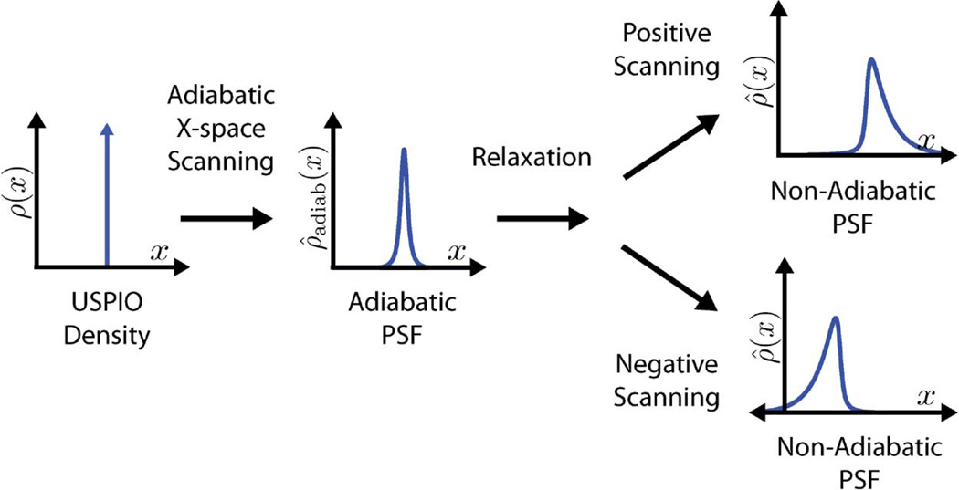
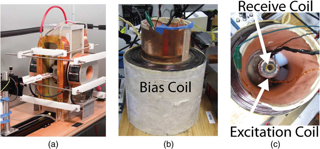
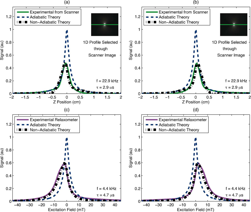
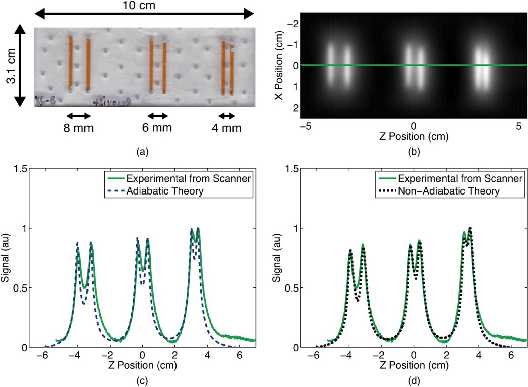
Magnetic particle imaging (MPI) is a new imaging modality that noninvasively images the spatial distribution of superparamagnetic iron oxide nanoparticles (SPIOs). MPI has demonstrated high contrast and zero attenuation with depth, and MPI promises superior safety compared to current angiography methods, X-ray, computed tomography, and magnetic resonance imaging angiography. Nanoparticle relaxation can delay the SPIO magnetization, and in this work we investigate the open problem of the role relaxation plays in MPI scanning and its effect on the image. We begin by amending the x-space theory of MPI to include nanoparticle relaxation effects. We then validate the amended theory with experiments from a Berkeley x-space relaxometer and a Berkeley x-space projection MPI scanner. Our theory and experimental data indicate that relaxation reduces SNR and asymmetrically blurs the image in the scanning direction. While relaxation effects can have deleterious effects on the MPI scan, we show theoretically and experimentally that x-space reconstruction remains robust in the presence of relaxation. Furthermore, the role of relaxation in x-space theory provides guidance as we develop methods to minimize relaxation-induced blurring. This will be an important future area of research for the MPI community.


Figures

Fig. 1
Fig. 1
Adiabatic x-space scanning blurs the SPIO density input according to the Langevin magnetization of the SPIOs. Relaxation effects further blur the image and create an asymmetrical shape to the PSF. This blurring effect occurs in the scanning direction, which results in nonidentical PSFs for the two scanning directions.
Fig. 2
Fig. 2
Berkeley x-space projection MPI scanner (a) acquires 2-D images. A 2.3 T/m magnetic gradient creates a FFL, and the excitation coil scans this FFL at 22.9 kHz with field strengths up to 35 mT-pp. The Berkeley x-space relaxometer, shown with side (b) and top (c) views, measures the point spread function of a particle sample. The excitation coil generates a sinusoidal magnetic field of 10–200 mT-pp strength at frequencies of 1.5–11.5 kHz. The signal received from the gradiometric receive coil is digitized at 10 MSPS without filtering. The bias coil can add ± 180 mT field for partial FOV scanning.
Fig. 3
Fig. 3
Experimentally measured PSFs displayed alongside theoretical PSFs calculated from the adiabatic and the non-adiabatic x-space theories for each scanning direction. The inclusion of relaxation into the theory predicted a significant loss in predicted resolution and peak signal and produced a shape which more closely resembled that of the experimental signal. (a), (b): We compared the x-space theoretical PSFs to a 1-D profile through (inset) a positive-velocity scan image of 2 µL of undiluted Resovist acquired in the Berkeley x-space projection MPI scanner with an excitation field of 20 mT-pp at 22.9 kHz. Scan time was 37 s and FOV was 6.6 cm × 5 cm. Non-adiabatic calculations used a measured time constant of 2.9 µs. (c), (d): We measured a PSF of Resovist in the Berkeley x-space relaxometer with an excitation field of 60 mT-pp at 4.4 kHz. Non-adiabatic calculations used a measured time constant of 4.7. (a) Resovist in scanner: negative scanning. (b) Resovist in scanner: positive scanning. (c) Resovist in relaxometer: negative scanning. (d) Resovist in relaxometer: positive scanning.
Fig. 4
Fig. 4
A line resolution phantom (a) was constructed with 1.75-mm-wide wells which were filled with 20 × diluted Resovist. We acquired a positive-velocity scan image (b) of this phantom in the Berkeley x-space projection MPI scanner at 20 mT-pp for a scan time of 59 s and a FOV of 13 cm 5 cm. No deconvolution was performed. We visualized a 1-D profile through the center of this image and compared this profile to the image predicted by the adiabatic x-space theory (c) and by the non-adiabatic x-space theory (d). The experimentally measured image showed better agreement with the non-adiabatic x-space theory than with the adiabatic x-space theory. Non-adiabatic calculations used a measured relaxation time constant of 2.9 µs.

References

    1. Ix JH, Mercado N, Shlipak MG, Lemos P, Boersma E, Linde-boom W, O’Neill WW, Wijns W, Serruys PW. “Association of chronic kidney disease with clinical outcomes after coronary revascularization: The Arterial Revascularization Therapies Study (ARTS),”. Am. Heart J. 2005;vol. 149(no. 3):512–519. - PubMed
    1. Reddan DN. “Chronic kidney disease, mortality, and treatment strategies among patients with clinically significant coronary artery disease,”. J. Am. Soc. Nephrol. 2003;vol. 14(no. 9):2373–2380. - PubMed
    1. Coresh J, Selvin E, Stevens LA, Manzi J, Kusek JW, Eggers P, Van Lente F, Levey AS. “Prevalence of chronic kidney disease the United States,”. J. Am. Med. Assoc. 2007;vol. 298(no. 17):2038–2047. - PubMed
    1. Ferrucci JT, Stark DD. “Iron oxide-enhanced MR imaging the liver and spleen: Review of the first 5 years,”. Am. J. Roentgenol. 1990;vol. 155(no. 5):943–950. - PubMed
    1. Weissleder R, Stark DD, Engelstad BL, Bacon BR, Compton CC, White DL, Jacobs P, Lewis J. “Superparamagnetic iron oxide: Pharmacokinetics and toxicity,”. Am. J. Roentgenol. 1989;vol. 152(no. 1):167–173. - PubMed

Publication types

Substances