. 2012 Nov;20(11):2134-42.
doi: 10.1038/mt.2012.178. Epub 2012 Sep 11.

Exon 45 skipping through U1-snRNA antisense molecules recovers the Dys-nNOS pathway and muscle differentiation in human DMD myoblasts

Affiliations

Exon 45 skipping through U1-snRNA antisense molecules recovers the Dys-nNOS pathway and muscle differentiation in human DMD myoblasts

Valentina Cazzella et al. Mol Ther. 2012 Nov.

Abstract

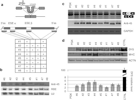
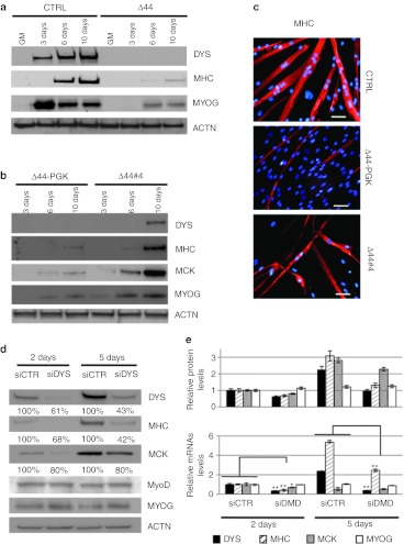
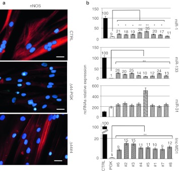
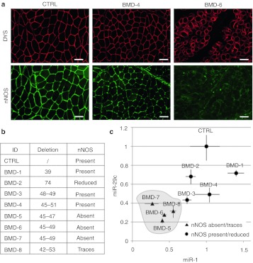
Exon skipping has been demonstrated to be a successful strategy for the gene therapy of Duchenne muscular dystrophy (DMD): the rational being to convert severe Duchenne forms into milder Becker ones. Here, we show the selection of U1 snRNA-antisense constructs able to confer effective rescue of dystrophin synthesis in a Δ44 Duchenne genetic background, through skipping of exon 45; moreover, we demonstrate that the resulting dystrophin is able to recover timing of myogenic marker expression, to relocalize neuronal nitric oxide synthase (nNOS) and to rescue expression of miRNAs previously shown to be sensitive to the Dystrophin-nNOS-HDAC2 pathway. Becker mutations display different phenotypes, likely depending on whether the shorter protein is able to reconstitute the wide range of wild-type functions. Among them, efficient assembly of the dystrophin-associated protein complex (DAPC) and nNOS localization are important. Comparing different Becker deletions we demonstrate the correlation between the ability of the mutant dystrophin to relocalize nNOS and the expression levels of two miRNAs, miR-1 and miR29c, known to be involved in muscle homeostasis and to be controlled by the Dys-nNOS-HDAC2 pathway.


Figures

Figure 1
Figure 1
Exon 45 skipping. (a) Schematic representation of the exon-skipping strategy for the human Δ44 Duchenne muscular dystrophy (DMD) mutation. The table summarizes the eight different constructs produced together with the corresponding target regions on exon 45 and flanking intron (ss, splice site; ESE, exonic splicing enhancer); sequences are indicated in Supplementary Table S1. (b) C27 myoblasts were transfected with the different antisense constructs together with the U16-RBE plasmid [expressing a 143-nucleotide (nt) long modified U16 snoRNA] Northern blot analysis was performed with probes against the 3′ splice site (anti-45), U16-RBE (RBE), and U2 small nuclear RNA (snRNA) (U2). The two latter hybridizations are used to normalize for transfection efficiency and as loading control, respectively. (c) Exon skipping activity. The amount of exon skipping was calibrated by Nested reverse transcription-PCR (RT-PCR) on RNA extracted from Δ44 cells infected with the antisense-expressing lentiviruses. Unskipped and skipped products are indicated on the right. Anti-45 refers to the expression of the antisense molecules; GAPDH is used as an internal control. (d) Rescue of dystrophin synthesis. Upper panel: western blot on proteins from CTRL (10 µg + 40 µg PGK), mock infected Δ44 (PGK- 50 µg) and exon-skipping Δ44 treated cells (#1 to #8–50 µg) probed with anti-dystrophin (DYS), anti-myosin heavy chain (MHC) and anti-actinin (ACTN) antibodies. Lower panel: the histogram indicates dystrophin levels normalized on actinin signals and expressed as percentage of CTRL. Error bars: means ± SD.
Figure 2
Figure 2
Analysis of muscle differentiation markers. (a) Western blot on proteins (10 µg) extracted from CTRL and Δ44 cells probed with anti-dystrophin (DYS), anti-myosin heavy chain (MHC), and anti-myogenin (MYOG) antibodies in growth medium (GM) and at different days upon shift to differentiation conditions (3, 6, and 10 days). Actinin (ACTN) was used as a loading control. (b) Western blot on proteins (50 µg) extracted from Δ44 cells, infected with lentivirus expressing GFP (Δ44-PGK) or with the #4 construct (Δ44#4), at different days upon shift to differentiation conditions (3, 6, and 10 days). Western blot was probed with anti-dystrophin (DYS), anti-MHC, anti-muscle creatine kinase (MCK), and anti-myogenin (MYOG) antibodies. Actinin (ACTN) was used as a loading control. (c) Immunofluorescence for MHC localization (red) on control myoblasts (CTRL) at 8 days of differentiation, in parallel with mock infected (Δ44-PGK) and exon-skipping treated (Δ44#4) Δ44 Duchenne muscular dystrophy (DMD) cells at 12 days of differentiation. DAPI staining for nuclei detection is also shown. Original magnification, ×20. Bar = 50 µm. (d) RNA interference against dystrophin. Control human myoblasts were transfected with scramble (siCTR) or with anti-dystrophin (siDYS) siRNAs and differentiated for 2 and 5 days. Western blot was performed with anti-DYS, anti-MHC, anti-MCK, anti-MyoD, and anti-MYOG antibodies. Actinin (ACTN) was used as loading control. (e) Upper panel: The histogram indicates DYS, MHC, MCK, and MyoG protein levels normalized on actinin signals. Error bars: means ± SD. Lower panel: DYS, MHC, MCK, and MyoG mRNAs relative expression in mock (siCTR) MCK, and DMD-siRNA treated (siDYS) human myoblasts after 2 and 5 days of differentiation assessed by quantitative reverse transcriptase-PCR (qRT-PCR). GAPDH was used as internal control. Relative expressions are shown with respect to mock (siCTR) cells, set to a value of 1. *P < 0.05, **P < 0.01.
Figure 3
Figure 3
Analysis of neuronal nitric oxide synthase (nNOS) localization and ncRNAs expression upon exon-skipping treatment. (a) nNOS localization analyzed by immunofluorescence with nNOS antibodies on CTRL, mock-infected Δ44 (Δ44-PGK) and exon-skipping Δ44-treated cells (Δ44#4) after 10 days of differentiation. Original magnification, ×40. Bar = 25 µm. (b) miRNAs and linc-MD1 expression analysis performed by quantitative reverse transcriptase-PCR (qRT-PCR) on RNA extracted from CTRL, mock-infected Δ44 (PGK), and Δ44 cells treated with the different antisense constructs (#1–#8) after 10 days of differentiation. U6 small nuclear RNA (snRNA) is used as endogenous control. Relative expressions are shown with respect to CTRL cells, set to a value of 100 and ncRNA relative quantifications are shown on the top of each lane. *P < 0.05, **P < 0.01.
Figure 4
Figure 4
Neuronal nitric oxide synthase (nNOS) localization and miRNAs expression in Becker biopsies. (a) Dystrophin (DYS) and neuronal NOS localization analyzed by immunofluorescence on control (CTRL), BMD-4, and BMD-6 human biopsies. Original magnification, ×10. Bar = 100 µm. (b) Table summarizes exon deletions of the different Becker muscular dystrophy (BMD) patients and corresponding nNOS levels. (c) Scatterplot showing the expression levels of miR-29c (Y axis) and miR-1 (X axis) analyzed by quantitative reverse transcriptase-PCR (qRT-PCR) on RNAs extracted from the different BMD biopsies. U6 small nuclear RNA (snRNA) is used as endogenous control. Relative expressions are shown with respect to healthy individual (CTRL), set to a value of 1. Error bars: means ± SD.

References

    1. Blake DJ, Weir A, Newey SE., and, Davies KE. Function and genetics of dystrophin and dystrophin-related proteins in muscle. Physiol Rev. 2002;82:291–329. - PubMed
    1. Ervasti JM. Dystrophin, its interactions with other proteins, and implications for muscular dystrophy. Biochim Biophys Acta. 2007;1772:108–117. - PubMed
    1. Chao DS, Gorospe JR, Brenman JE, Rafael JA, Peters MF, Froehner SC.et al. (1996Selective loss of sarcolemmal nitric oxide synthase in Becker muscular dystrophy J Exp Med 184609–618. - PMC - PubMed
    1. Wells KE, Torelli S, Lu Q, Brown SC, Partridge T, Muntoni F.et al. (2003Relocalization of neuronal nitric oxide synthase (nNOS) as a marker for complete restoration of the dystrophin associated protein complex in skeletal muscle Neuromuscul Disord 1321–31. - PubMed
    1. Lai Y, Thomas GD, Yue Y, Yang HT, Li D, Long C.et al. (2009Dystrophins carrying spectrin-like repeats 16 and 17 anchor nNOS to the sarcolemma and enhance exercise performance in a mouse model of muscular dystrophy J Clin Invest 119624–635. - PMC - PubMed

Publication types

MeSH terms