

Revisiting the clinical anatomy of the alar ligaments
- PMID: 22968541
- PMCID: PMC3540300
- DOI: 10.1007/s00586-012-2496-4
Revisiting the clinical anatomy of the alar ligaments
Abstract
Purpose: The morphology of the alar ligaments has been inconsistently described, particularly with regard to the existence of an atlantal portion. Despite these inconsistencies, these descriptions have been used to develop physical tests for the integrity of these ligaments in patients with cervical spine problems. The purpose of this study was to describe the detailed macrostructure of the alar ligaments.
Methods: The alar ligaments of 11 cervical spine specimens from embalmed adult cadavers were examined by fine dissection. A detailed description of the macrostructure of these ligaments and their attachment sites was recorded. Measurements were performed with respect to ligament dimensions and relations with selected bony landmarks.
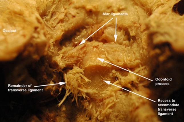
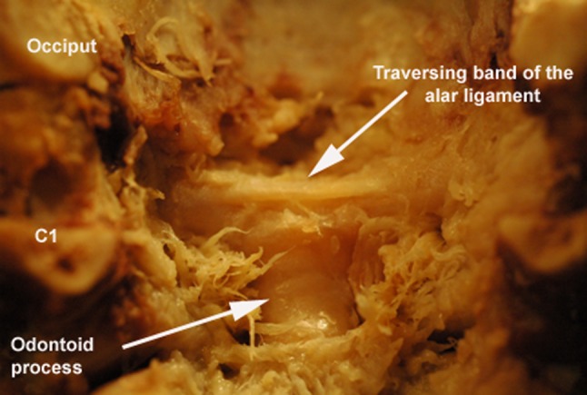
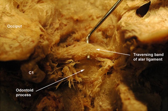
Results: No atlantal portion of the alar ligament was viewed in any specimen. The attachment of the ligaments on the odontoid process occurred on its lateral and posterolateral aspects, frequently below the level of the apex. The occipital attachment was on the medial surface of the occipital condyles in close proximity to the atlanto-occipital joints. The orientation of the ligaments was primarily horizontal. The presence of transverse bands extending occiput to occiput with minimal or no attachment to the odontoid process was a common variant.
Conclusions: The absence of findings with respect to the atlantal portion of the alar ligament suggests that it may be considered an anatomical variant, not an essential component for stability of the craniocervical complex. These findings may inform the use and interpretation of clinical tests for alar ligament integrity.
Figures
References
-
- Dvorak J, Dvorak V, Gilliar W, Schneider W, Spring H, Tritschler T. Musculoskeletal manual medicine. diagnosis and treatment. Stuttgart: Theime; 2008.
-
- White AAI, Panjabi MM. Clinical biomechanics of the spine. 2. Philadelphia: J.B. Lippincott Company; 1990.
-
- Aspinall W. Clinical testing for the craniovertebral hypermobility syndrome. J Orthop Sports Phys Ther. 1990;12(2):47–54. - PubMed
MeSH terms
LinkOut - more resources
Full Text Sources
Medical
Miscellaneous
