. 2012 Nov 1;44(21):1042-51.
doi: 10.1152/physiolgenomics.00052.2012. Epub 2012 Sep 11.

MiR-351 transiently increases during muscle regeneration and promotes progenitor cell proliferation and survival upon differentiation

Affiliations

MiR-351 transiently increases during muscle regeneration and promotes progenitor cell proliferation and survival upon differentiation

Yongxin Chen et al. Physiol Genomics. .

Abstract

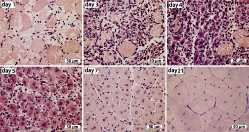
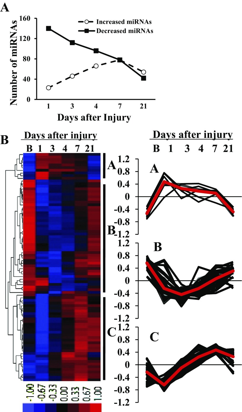
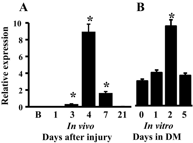
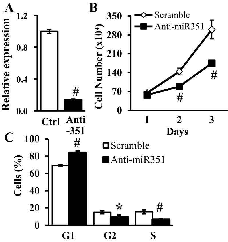
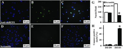
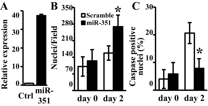
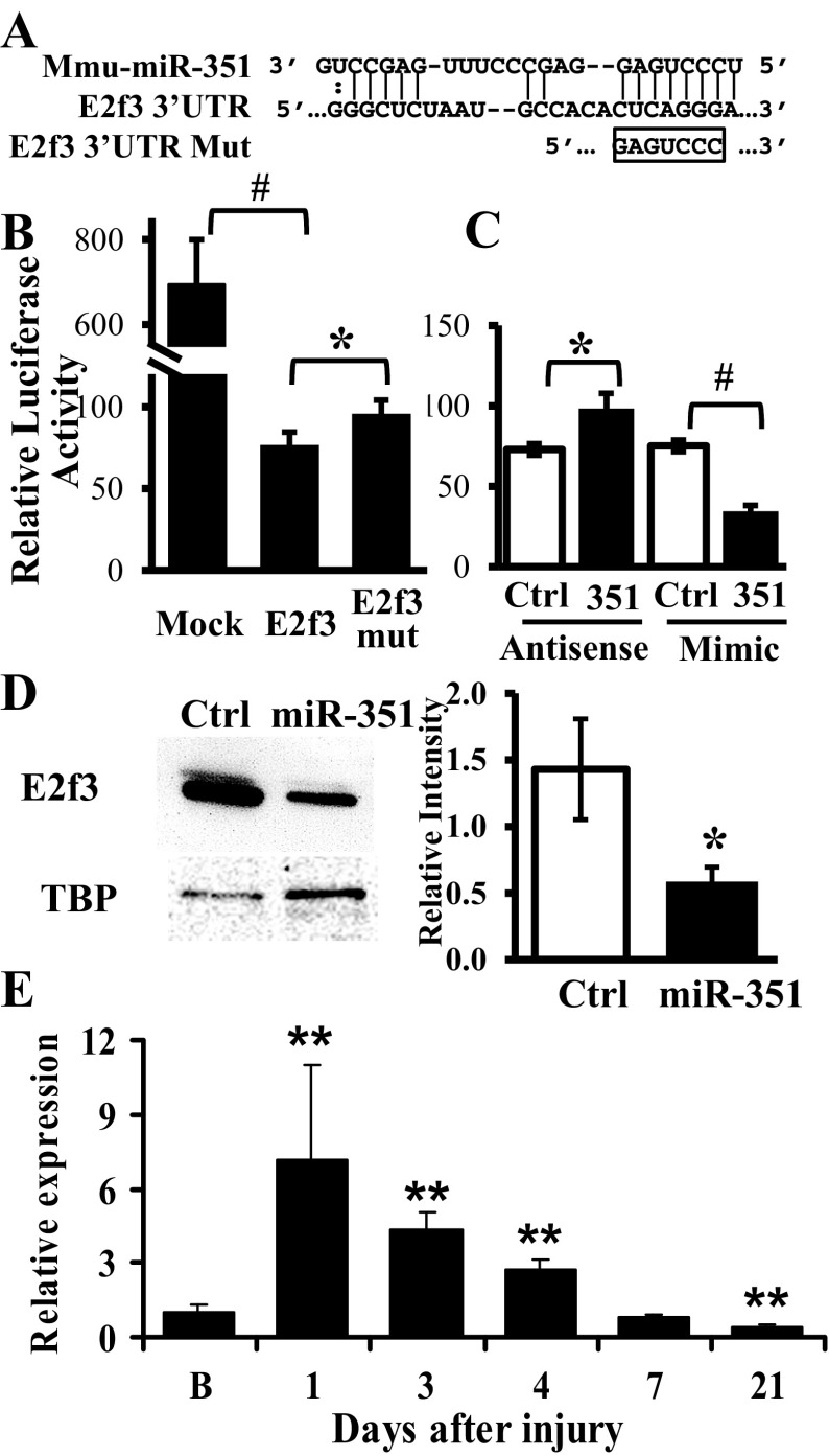
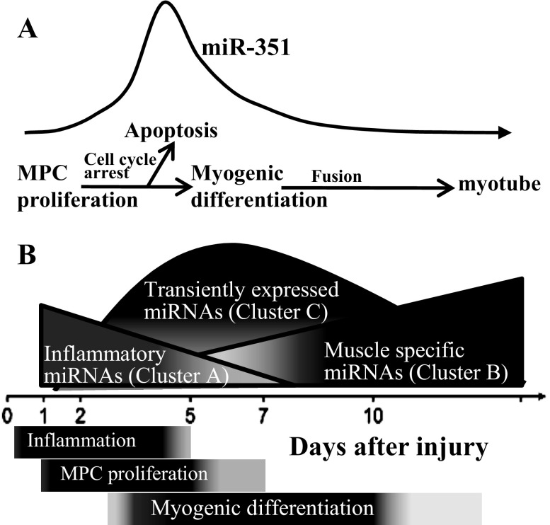
MicroRNAs (miRNAs) regulate many biological processes including muscle development. However, little is known regarding miRNA regulation of muscle regeneration. Murine tibialis anterior muscle was evaluated after cardiotoxin-induced injury and used for global miRNA expression analysis. From day 1 through day 21 following injury, 298 miRNAs were significantly changed at least at one time point, including 86 miRNAs that were altered >10-fold compared with uninjured skeletal muscle. Temporal miRNA expression patterns included inflammation-related miRNAs (miR-223 and -147) that increased immediately after injury; this pattern contrasted to that of mature muscle-specific miRNAs (miR-1, -133a, and -499) that abruptly decreased following injury followed by upregulation in later regenerative events. Another cluster of miRNAs were transiently increased in the early days of muscle regeneration including miR-351, a miRNA that was also transiently expressed during myogenic progenitor cell (MPC) differentiation in vitro. Based on computational predictions, further studies demonstrated that E2f3 was a target of miR-351 in myoblasts. Moreover, knockdown of miR-351 expression inhibited MPC proliferation and promoted apoptosis during MPC differentiation, whereas miR-351 overexpression protected MPC from apoptosis during differentiation. Collectively, these observations suggest that miR-351 is involved in both the maintenance of MPC proliferation and the transition into differentiated myotubes. Thus, a novel, time-dependent sequence of molecular events during muscle regeneration has been identified; miR-351 inhibits E2f3 expression, a key regulator of cell cycle progression and proliferation, and promotes MPC proliferation and protects early differentiating MPC from apoptosis, important events in the hostile tissue environment after acute muscle injury.


Figures

Fig. 1.
Fig. 1.
Time course of cardiotoxin (CTX)-induced tibialis anterior muscle injury and regeneration. Representative images; paraffin sections (4 μm) with hematoxylin and eosin stain. Day after CTX injection indicated on each image.
Fig. 2.
Fig. 2.
Distinct miRNA expression patterns during skeletal muscle injury and regeneration. A: number of miRNAs differentially expressed with >2-fold change during muscle regeneration compared with baseline, noninjured muscle. Data derived from 4 mice at each time point. B: hierarchical cluster analysis of differentially expressed miRNAs with >10-fold change during the time course of muscle injury and regeneration. Cluster analysis was performed for 86 differentially expressed miRNAs with >10-fold change after data adjustment (log transformation, median center and normalization). The color codes of red, black, and blue represent expression levels of high, average, and low, respectively. A–C, temporal expression of individual miRNA in cluster A (n = 10), cluster B (n = 43), and cluster C (n = 33) during muscle regeneration. The average value of miRNA expression in each cluster is presented as a red line; B, baseline, noninjured muscle. Data derived from 4 mice at each time point.
Fig. 3.
Fig. 3.
miR-351 expression patterns during in vivo muscle regeneration (A) and in vitro myogenic progenitor cell (MPC) differentiation (B). MiRNA expression was determined by qRT-PCR-array. Data represent the mean ± SD of 4 mice/time point in vivo and 4 different MPC cultures, *P ≤ 0.01 compared with day 0 in vitro or baseline muscle in vivo; B, baseline noninjured muscle; DM, differentiation medium.
Fig. 4.
Fig. 4.
Effects of inhibition of endogenous miR-351 in proliferating MPC. MPC were transfected with either miR-351 antisense or scramble control (Ctrl) oligonucleotides on day 0 and cultured in growth medium. A: miR-351 expression was measured by quantitative real time-PCR 2 days after transfection. B: total cell number determined at the indicated time. C: percent of cells at stages of the cell cycle determined at 3 days after transfection. Data represent means ± SD, n = 3 different cultures/assay. *P ≤ 0.01 compared with scramble oligonucleotide.
Fig. 5.
Fig. 5.
Effects of inhibition of miR-351 on apoptosis during MPC differentiation. MPC were subcultured and transfected with miRNA antisense (top) or scramble (bottom) oligonucleotide. Differentiation medium was added 1 day after transfection (differentiation day 0, Dif-D0). Caspase activity was measured at differentiation day 1 (Dif-D1) and labeled with a green fluorescence probe (FLICA; B and E); and nuclear integrity was monitored by Hoechst 33342 counterstaining (blue, A and D). C and F show merged images from green and blue fluorescence. Cell number was determined using images obtained with a fluorescent microscope to determine the nuclei/field (G) and the percent of caspase-positive cells (H). Data represent means ± SD of 3 different MPC cultures. *P < 0.001 compared with the scramble oligonucleotide.
Fig. 6.
Fig. 6.
Effects of miR-351 overexpression on MPC apoptosis in serum-free conditions. MPC were subcultured and transfected with miR-351 mimic or scramble control (Ctrl) oligonucleotide. Serum-free medium was added 1 day after transfection (day 0). A: miR-351 expression was measured by qRT-PCR in serum-free medium (day 2). Caspase activity in individual nuclei was assessed and the cells identified in images obtained from a fluorescent microscope to determine the nuclei/field (B) and the percent of caspase-positive cells (C). Data represent means ± SD of 3 different MPC cultures; *P ≤ 0.01 compared with scramble oligonucleotide.
Fig. 7.
Fig. 7.
E2f3 is a putative target of miR-351 in proliferating C2C12 myoblasts. A: predicted target site of miR-351 in the 3′-untranslated region (UTR) of mouse E2f3. An E2f3 3′-UTR mutant (E2f3 mut) was produced by introducing mutation in the seed sequence as indicated. B and C: luciferase assays were performed at day 2 after transfection of a mock control or luciferase reporters bearing intact or mutant E2f3 3′-UTR into myoblasts. To measure the effect of miR-351 on luciferase activity, the luciferase reporter fused to an intact E2f3 3′-UTR was cotransfected with either miRNA antisense or mimic and compared with that of scramble control (Ctrl). The results were expressed as firefly luciferase activity relative to the renilla luciferase expressed from a cotransfected plasmid and serving as a transfection control. D: endogenous E2f3 protein levels were determined by Western blot 2 days after transfection of miR-351 mimic or a scramble control (Ctrl) in myoblasts. Quantification of Western blot results was performed using scanning and ImageJ software. Results are expressed as integrated optical density normalized to TATA box binding protein (TBP) content; *P ≤ 0.04, #P ≤ 0.004. E: E2f3 mRNA expression was determined during in vivo muscle regeneration at 1, 3, 4, 7, and 21 days postinjury by qRT-PCR. Data are reported as fold change compared with uninjured baseline (B) muscle, 95% confidence intervals, 4–6 per mice/time point, and **P < 0.001.
Fig. 8.
Fig. 8.
Model of miRNA temporal expression during muscle injury and regeneration. A: the transient upregulation of miR-351 protects MPC from apoptosis during muscle regeneration. B: relative changes in the amount of different clusters of miRNAs during muscle regeneration. The inflammatory miRNAs (cluster A) were upregulated immediately after muscle injury; muscle-specific and other miRNAs (cluster B) were upregulated in the late phase of muscle regeneration. A group of miRNAs transiently expressed during regeneration (cluster C) regulates cell cycle and the switch from MPC proliferation to differentiation and protects early differentiating cells from apoptosis.

Similar articles

Cited by

References

    1. Anderson JE, Murray L. Barr Award Lecture. Studies of the dynamics of skeletal muscle regeneration: the mouse came back! Biochem Cell Biol 76: 13–26, 1998. - PubMed
    1. Antonini D, Russo MT, De Rosa L, Gorrese M, Del Vecchio L, Missero C. Transcriptional repression of miR-34 family contributes to p63-mediated cell cycle progression in epidermal cells. J Invest Dermatol 130: 1249–1257, 2010. - PubMed
    1. Asp P, Acosta-Alvear D, Tsikitis M, van Oevelen C, Dynlacht BD. E2f3b plays an essential role in myogenic differentiation through isoform-specific gene regulation. Genes Dev 23: 37–53, 2009. - PMC - PubMed
    1. Cam H, Dynlacht BD. Emerging roles for E2F: beyond the G1/S transition and DNA replication. Cancer Cell 3: 311–316, 2003. - PubMed
    1. Chen JF, Callis TE, Wang DZ. microRNAs and muscle disorders. J Cell Sci 122: 13–20, 2009. - PMC - PubMed

Publication types