

MHC mismatch results in neural progenitor cell rejection following spinal cord transplantation in a model of viral-induced demyelination
- PMID: 22969049
- PMCID: PMC3479361
- DOI: 10.1002/stem.1234
MHC mismatch results in neural progenitor cell rejection following spinal cord transplantation in a model of viral-induced demyelination
Abstract
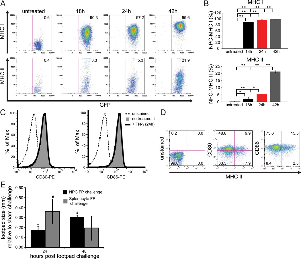
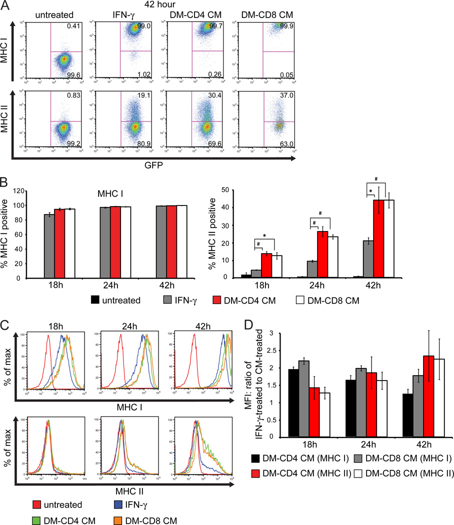
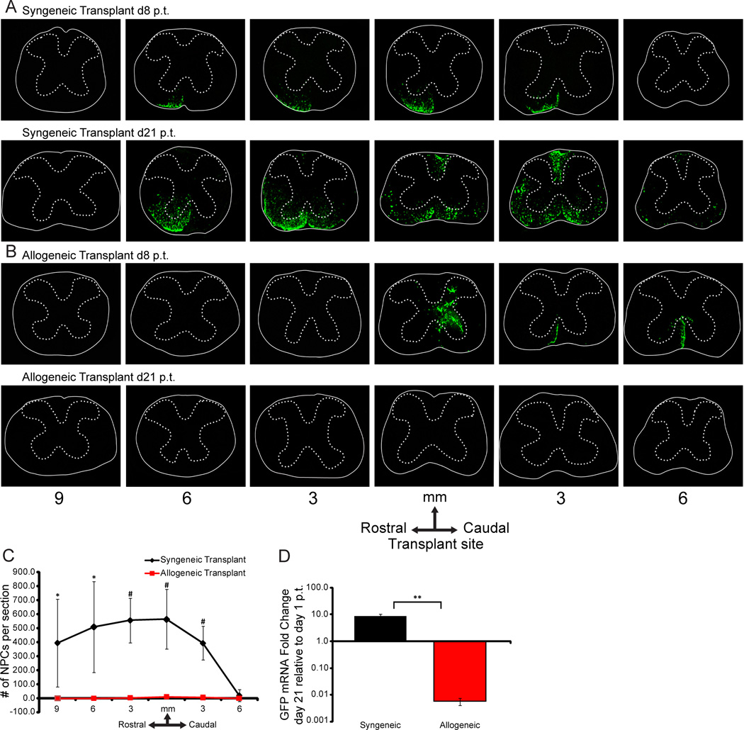
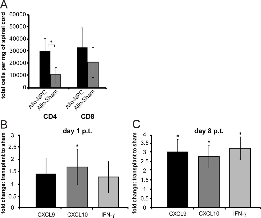
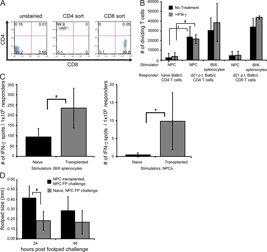
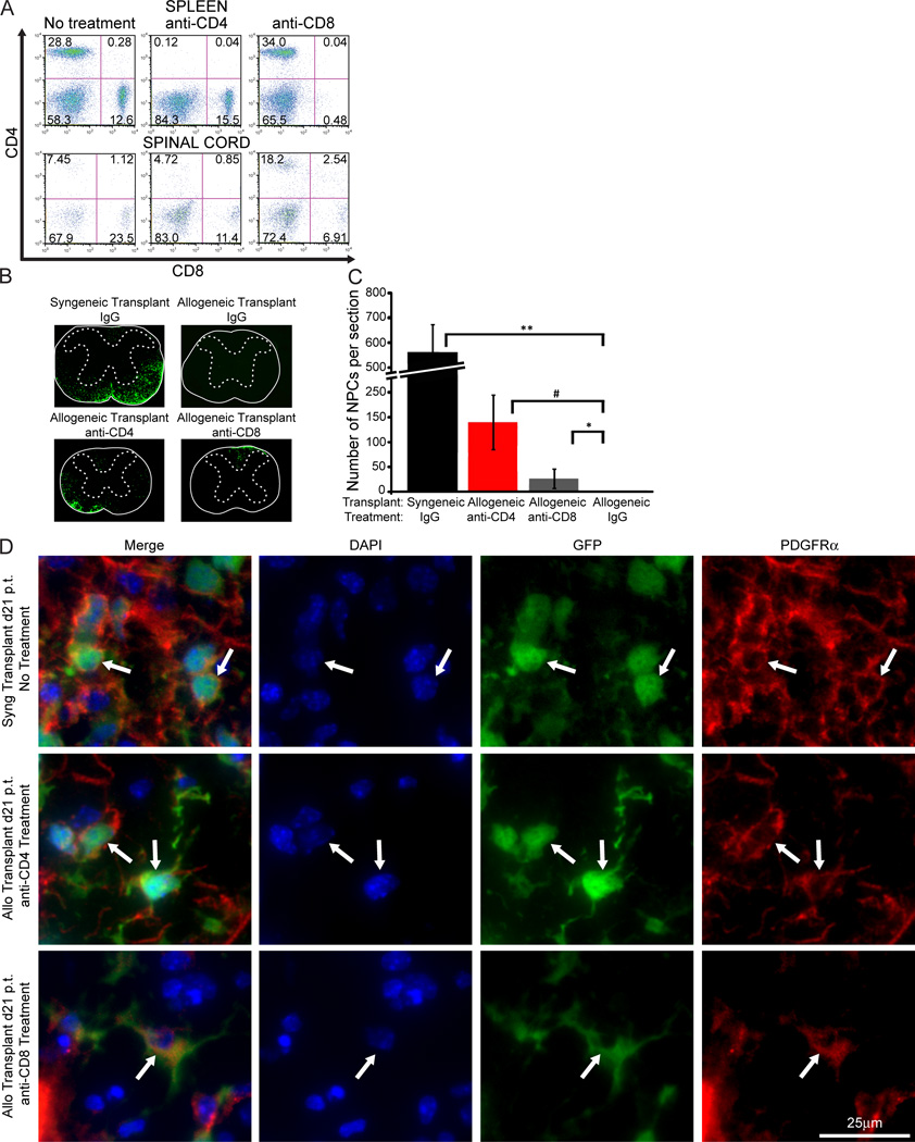
Transplantation of syngeneic neural progenitor cells (NPCs) into mice persistently infected with the JHM strain of mouse hepatitis virus (JHMV) results in enhanced differentiation into oligodendrocyte progenitor cells that is associated with remyelination, axonal sparing, and clinical improvement. Whether allogeneic NPCs are tolerated or induce immune-mediated rejection is controversial and poorly defined under neuroinflammatory demyelinating conditions. We have used the JHMV-induced demyelination model to evaluate the antigenicity of transplanted allogeneic NPCs within the central nervous system (CNS) of mice with established immune-mediated demyelination. Cultured NPCs constitutively expressed the costimulatory molecules CD80/CD86, and IFN-γ treatment induced expression of MHC class I and II antigens. Injection of allogeneic C57BL/6 NPCs (H-2b background) led to a delayed type hypersensitivity response in BALB/c (H-2d background) mice associated with T-cell proliferation and IFN-γ secretion following coculture with allogeneic NPCs. Transplantation of MHC-mismatched NPCs into JHMV-infected mice resulted in increased transcripts encoding the T-cell chemoattractant chemokines CXCL9 and CXCL10 that correlated with increased T-cell infiltration that was associated with NPC rejection. Treatment of MHC-mismatched mice with T-cell subset-specific depleting antibodies increased survival of allogeneic NPCs without affecting commitment to an oligodendrocyte lineage. Collectively, these results show that allogeneic NPCs are antigenic, and T-cells contribute to rejection following transplantation into an inflamed CNS suggesting that immunomodulatory treatments may be necessary to prolong survival of allogeneic cells.
Copyright © 2012 AlphaMed Press.
Figures
Similar articles
-
Activating receptor NKG2D targets RAE-1-expressing allogeneic neural precursor cells in a viral model of multiple sclerosis.Stem Cells. 2014 Oct;32(10):2690-701. doi: 10.1002/stem.1760. Stem Cells. 2014. PMID: 24898518 Free PMC article.
-
Olig1 function is required for remyelination potential of transplanted neural progenitor cells in a model of viral-induced demyelination.Exp Neurol. 2012 May;235(1):380-7. doi: 10.1016/j.expneurol.2012.03.003. Epub 2012 Mar 17. Exp Neurol. 2012. PMID: 22449475 Free PMC article.
-
Cystatin F attenuates neuroinflammation and demyelination following murine coronavirus infection of the central nervous system.J Neuroinflammation. 2024 Jun 15;21(1):157. doi: 10.1186/s12974-024-03153-0. J Neuroinflammation. 2024. PMID: 38879499 Free PMC article.
-
Comparative study of the role of professional versus semiprofessional or nonprofessional antigen presenting cells in the rejection of vascularized organ allografts.Transpl Immunol. 1995 Dec;3(4):273-89. doi: 10.1016/0966-3274(95)80013-1. Transpl Immunol. 1995. PMID: 8665146 Review.
-
Cell replacement therapies to promote remyelination in a viral model of demyelination.J Neuroimmunol. 2010 Jul 27;224(1-2):101-7. doi: 10.1016/j.jneuroim.2010.05.013. Epub 2010 Jun 2. J Neuroimmunol. 2010. PMID: 20627412 Free PMC article. Review.
Cited by
-
Role of Intrinsic (Graft) Versus Extrinsic (Host) Factors in the Growth of Transplanted Organs Following Allogeneic and Xenogeneic Transplantation.Am J Transplant. 2017 Jul;17(7):1778-1790. doi: 10.1111/ajt.14210. Epub 2017 Mar 3. Am J Transplant. 2017. PMID: 28117931 Free PMC article.
-
Neural stem cell treatment for perinatal brain injury: A systematic review and meta-analysis of preclinical studies.Stem Cells Transl Med. 2021 Dec;10(12):1621-1636. doi: 10.1002/sctm.21-0243. Epub 2021 Sep 20. Stem Cells Transl Med. 2021. PMID: 34542242 Free PMC article.
-
Promoting remyelination through cell transplantation therapies in a model of viral-induced neurodegenerative disease.Dev Dyn. 2019 Jan;248(1):43-52. doi: 10.1002/dvdy.24658. Epub 2018 Sep 6. Dev Dyn. 2019. PMID: 30067309 Free PMC article. Review.
-
Scutellarin Alleviates Behavioral Deficits in a Mouse Model of Multiple Sclerosis, Possibly Through Protecting Neural Stem Cells.J Mol Neurosci. 2016 Feb;58(2):210-20. doi: 10.1007/s12031-015-0660-0. Epub 2015 Oct 29. J Mol Neurosci. 2016. PMID: 26514969
-
Promoting remyelination: utilizing a viral model of demyelination to assess cell-based therapies.Expert Rev Neurother. 2014 Oct;14(10):1169-79. doi: 10.1586/14737175.2014.955854. Expert Rev Neurother. 2014. PMID: 25245576 Free PMC article. Review.
References
-
- Weinshenker BG. The natural history of multiple sclerosis: update 1998. Semin Neurol. 1998;18:301–307. - PubMed
-
- Rothman KJ. Epidemilogy, an introduction. New York: Oxford Univeristy Press; 2002. What is causation.
-
- Ebers GC, Sadovnick AD, Risch NJ. A genetic basis for familial aggregation in multiple sclerosis. Canadian Collaborative Study Group. Nature. 1995;377:150–151. - PubMed
-
- Sospedra M, Martin R. Immunology of multiple sclerosis. Annu Rev Immunol. 2005;23:683–747. - PubMed
-
- Ascherio A, Munger KL. Environmental risk factors for multiple sclerosis. Part I: the role of infection. Ann Neurol. 2007;61:288–299. - PubMed
Publication types
MeSH terms
Substances
Grants and funding
LinkOut - more resources
Full Text Sources
Other Literature Sources
Research Materials
