Review
. 2012 Nov;21(11):1597-619.
doi: 10.1002/pro.2156.

The enzymes of biotin dependent CO₂ metabolism: what structures reveal about their reaction mechanisms

Affiliations
Review

The enzymes of biotin dependent CO₂ metabolism: what structures reveal about their reaction mechanisms

Grover L Waldrop et al. Protein Sci. 2012 Nov.

Abstract

Biotin is the major cofactor involved in carbon dioxide metabolism. Indeed, biotin-dependent enzymes are ubiquitous in nature and are involved in a myriad of metabolic processes including fatty acid synthesis and gluconeogenesis. The cofactor, itself, is composed of a ureido ring, a tetrahydrothiophene ring, and a valeric acid side chain. It is the ureido ring that functions as the CO₂ carrier. A complete understanding of biotin-dependent enzymes is critically important for translational research in light of the fact that some of these enzymes serve as targets for anti-obesity agents, antibiotics, and herbicides. Prior to 1990, however, there was a dearth of information regarding the molecular architectures of biotin-dependent enzymes. In recent years there has been an explosion in the number of three-dimensional structures reported for these proteins. Here we review our current understanding of the structures and functions of biotin-dependent enzymes. In addition, we provide a critical analysis of what these structures have and have not revealed about biotin-dependent catalysis.


Figures

Figure 1
Figure 1
Structure of biotin. The enolate form of the cofactor is shown in (b).
Scheme 1
Scheme 1
Scheme 2
Scheme 2
Scheme 3
Scheme 3
Figure 2
Figure 2
Structure of the E. coli biotin carboxylase. A stereo-view of one subunit of the enzyme is shown in (a). The A-, B-, and C-domains are displayed in blue, wheat, and violet, respectively, and the bound phosphate ion is shown in a sphere representation. The quaternary structure of the enzyme is depicted in (b). The two-fold rotational axis relating one subunit to another is nearly perpendicular to the plane of the page. X-ray coordinates were obtained from the Protein Data Bank (1BNC). All structure figures were prepared with the software package PyMOL.
Figure 3
Figure 3
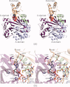
Structure of the E. coli biotin carboxylase in complex with ATP. A stereo-view of one subunit of the enzyme is presented in (a). The color-coding is the same as in Figure 2. The bound ATP is depicted in sphere representation. Note how the B-domain closes down upon nucleotide binding. Those amino acid residues important for ATP binding are shown in (b). The structure shown is that of the E288K mutant protein. Potential hydrogen bonds are indicated by the dashed lines. X-ray coordinates were obtained from the Protein Data Bank (1DV2).
Figure 4
Figure 4
General overall reaction of enzymes belonging to the ATP-grasp superfamily. Most members employ an acylphosphate intermediate in their reaction mechanisms.
Figure 5
Figure 5
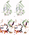
Elements of the ATP-grasp fold. The three-dimensional structure of PurT-encoded glycinamide ribonucleotide transformylase serves as a prototype for ATP-grasp superfamily members., This enzyme, from E. coli, is involved in the de novo pathway for purine biosynthesis. All ATP-grasp superfamily members have B-domains that change their positions depending upon the presence or absence of bound nucleotides. In addition, as indicated in (a), they all have a characteristic helix-turn-helix motif that connects the A- and B-domains (light green) and a T-loop that surrounds the phosphoryl oxygens of the nucleotide triphosphate (wheat). A close-up view of the ATP-binding pocket for glycinamide ribonucleotide transformylase is presented in (b). One of the residues that interacts with the phosphoryl oxygens, Arg 114, is provided by the helix-turn-helix motif. ATP-grasp enzymes typically coordinate two magnesium ions with a glutamate residue serving as the metal bridging ligand. In the case of glycinamide ribonucleotide transformylase, this bridging ligand is Glu 279. The magnesium ions are depicted as teal spheres. Whereas the B-domain is intimately involved in nucleotide binding, the A- and C-domains form a cradle that harbors the side chains responsible for substrate binding, which for glycinamide ribonucleotide transformylase is glycinamide ribonucleotide (c).
Figure 6
Figure 6
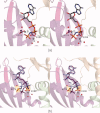
The binding of MgAMPPNP to the S. aureus biotin carboxylase. Shown in (a) is a close-up view of the region surrounding the MgAMPPNP ligand when bound to the enzyme (PDB accession no. 2VPQ). Both magnesium ions, depicted as gray spheres, display octahedral coordination geometry. Water molecules are shown as red spheres. The differences in AMPPNP binding, with or without magnesium ions, is shown in (b). The nucleotide, with its accompanying metal ions, is displayed in violet. Superimposed on this is the conformation of the nucleotide when it binds into the active site without its accompanying metals (light blue, PDB accession no. 2J9G).
Figure 7
Figure 7
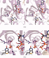
The binding of biotin, bicarbonate, and MgADP to the E. coli biotin carboxylase. A stereo-view of the biotin carboxylase active site with its bound ligands is shown in (a). Potential hydrogen bonds are indicated by the dashed lines. An overlay of the structure of biotin carboxylase solved in the presence of biotin, bicarbonate, and MgADP with that determined only in the presence of inorganic phosphate is shown in (b). The bicarbonate and phosphate are located in nearly identical positions in the active sites of the two models. X-ray coordinates were obtained from the Protein Data Bank (3G8C and 1BNC).
Figure 8
Figure 8
The binding of pyridopyrimidines and aminooxazoles to biotin carboxylase. The chemical structures for three pyridopyrimidine inhibitors of biotin carboxylase (1, 2, and 3) and one aminooxazole (4) are shown in (a). The manners in which the pyridopyrimidine inhibitors bind to biotin carboxylase are depicted in stereo in (b). Compounds 1, 2, and 3 are highlighted in light orange, light green, and light blue, respectively. For comparison purposes, the conformation of ATP, when bound to biotin carboxylase, is also shown. Compound 4 binds in a similar orientation to the pyridopyrimidine inhibitors as depicted in (c). X-ray coordinates were obtained from the Protein Data Bank (2V58, 2V59, 2V5A, 2W71, and 1DV2).
Figure 9
Figure 9
The binding of inhibitors to biotin carboxylase. The chemical structure of 2-(2-chlorobenzylamino)-1-(cyclo-hexylmethyl)-1H-benzo[d]imidazole-5-carboxamide is shown in (a). The manner in which this compound binds to biotin carboxylase is depicted in (b). On the basis of this initial structural analysis, a series of novel inhibitors was created. X-ray coordinates used were from the Protein Data Bank (3JZF). Soraphen A is a potent antifungal agent with the chemical structure shown in (c). It binds to yeast biotin carboxylase in a pocket located at the interface between the N- and C-terminal domains as indicated by the space-filling representation in (d). X-ray coordinates were from the Protein Data Bank (1W96).
Figure 10
Figure 10
The molecular architecture of the C-terminal domain of the E. coli BCCP. The protein is characterized by two layers of antiparallel β-sheet, highlighted in blue and green. Presumed hydrogen bonds between Thr 94 and biotin are indicated by the dashed lines. Subsequent NMR studies suggest that these hydrogen bonds do not occur. X-ray coordinates were from the Protein Data Bank (1BDO).
Figure 11
Figure 11
The structure of enoyl CoA hydratase. Members of the crotonase superfamily typically display either trimeric or hexameric quaternary structures with their active sites wedged between subunits. Shown in (a) is a ribbon representation of one subunit of enoyl CoA hydratase, an enzyme that catalyzes the second step in fatty acid oxidation. X-ray coordinates were from the Protein Data Bank (2DUB). The subunit can be envisioned in terms of two domains. The N-terminal region is dominated by two layers of β-sheet (displayed in blue and magenta), whereas the C-terminal motif contains three α-helical regions (all α-helices are shown in green). Members of the crotonase superfamily contain an oxyanion hole, which is formed by two peptidic nitrogen groups (b). Also, in these family members, it is thought that a helix dipole moment plays a role in activating the carbonyl carbons of the substrates for nucleophilic attacks.
Scheme 4
Scheme 4
Figure 12
Figure 12
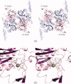
The structure of the E. coli carboxyltransferase. The quaternary structure of the enzyme is an α22 heterodimer. X-ray coordinates were from the Protein Data Bank (2F9Y). The α- and β-chains are colored in light blue and pink, respectively in (a). The view shown is looking down the twofold rotational axis that relates one α,β dimer to another. Each β-subunit contains a four-cysteine zinc finger as shown in (b).
Figure 13
Figure 13
The carboxyltransferase domain of yeast acetyl-CoA carboxylase and the α-subunit of glutaconyl-CoA decarboxylase demonstrate similar quaternary structures. Both function as homodimers. Shown in (a) is the carboxyltransferase of the yeast acetyl-CoA carboxylase. X-ray coordinates were from the Protein Data Bank (1OD2). Each subunit of the homodimer can be envisioned in terms of an N-terminal and a C-terminal domain, displayed in pink and blue, respectively. Likewise, the two α-subunits of the glutaconyl-CoA decarboxylase dimer adopt similar bilobal type architectures. The N- and C-terminal domains are depicted in pink and blue, respectively. X-ray coordinates were from the Protein Data Bank (1PIX).
Figure 14
Figure 14
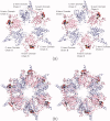
The transcarboxylase 12S subunit shows a hexameric quaternary structure. The quaternary structure of the enzyme can be envisioned as a dimer of trimers. X-ray coordinates were from the Protein Data Bank (1ON3). The trimer is shown in (a) with the methylmalonyl-CoA ligands displayed in sphere representations. Each monomer consists of an N- and a C-terminal domain, colored in pink and blue, respectively. The complete hexamer is shown in (b).
Figure 15
Figure 15
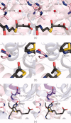
Oxyanion holes in carboxyltransferases. In the 12S subunit of transcarboxylase, the oxyanion hole is formed by the backbone amide group of Ala 143 and a water molecule as depicted in (a). X-ray coordinates were from the Protein Data Bank (1ON3). In contrast, in the β-subunit of propionyl-CoA carboxylase, one backbone amide group forms the oxyanion hole as shown in (b). X-ray coordinates were from the Protein Data Bank (1XNY). The oxyanion hole in propionyl-CoA carboxylase is formed by the backbone amide group of Ala 420 and the positive end of a helix dipole moment. X-ray coordinates were from the Protein Data Bank (1XNY).
Figure 16
Figure 16
Possible reaction mechanism for propionyl-CoA carboxylase.

Similar articles

Cited by

References

    1. Kögl F, Tönnis B. Uber das Bios-Problem. Darstellung von Krystallisierten Biotin aus Eigelb. Z Physiol Chem. 1936;242:43–73.
    1. Eakin RE, Snell EE, Williams RJ. A constituent of raw egg white capable of inactivating biotin in vitro. J Biol Chem. 1940;136:801–802.
    1. du Vigneaud V. The structure of biotin. Science. 1942;96:455–461. - PubMed
    1. Lardy HA, Peanasky R. Metabolic functions of biotin. Physiol Rev. 1953;33:560–565. - PubMed
    1. Wakil SJ, Titchener EB, Gibson DM. Evidence for the participation of biotin in the enzymic synthesis of fatty acids. Biochim Biophys Acta. 1958;29:225–226. - PubMed

LinkOut - more resources