

Pancreatico-biliary endoscopic ultrasound: a systematic review of the levels of evidence, performance and outcomes
- PMID: 22969187
- PMCID: PMC3436039
- DOI: 10.3748/wjg.v18.i32.4243
Pancreatico-biliary endoscopic ultrasound: a systematic review of the levels of evidence, performance and outcomes
Abstract
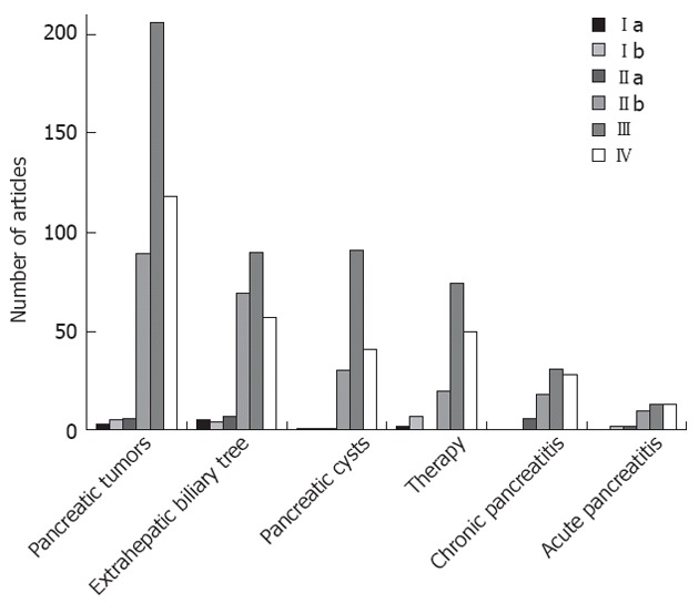
Our aim was to record pancreaticobiliary endoscopic ultrasound (EUS) literature of the past 3 decades and evaluate its role based on a critical appraisal of published studies according to levels of evidence (LE). Original research articles (randomized controlled trials, prospective and retrospective studies), meta-analyses, reviews and surveys pertinent to gastrointestinal EUS were included. All articles published until September 2011 were retrieved from PubMed and classified according to specific disease entities, anatomical subdivisions and therapeutic applications of EUS. The North of England evidence-based guidelines were used to determine LE. A total of 1089 pertinent articles were reviewed. Published research focused primarily on solid pancreatic neoplasms, followed by disorders of the extrahepatic biliary tree, pancreatic cystic lesions, therapeutic-interventional EUS, chronic and acute pancreatitis. A uniform observation in all six categories of articles was the predominance of LE III studies followed by LE IV, II b, II a, I b and I a, in descending order. EUS remains the most accurate method for detecting small (< 3 cm) pancreatic tumors, ampullary neoplasms and small (< 4 mm) bile duct stones, and the best test to define vascular invasion in pancreatic and peri-ampullary neoplasms. Detailed EUS imaging, along with biochemical and molecular cyst fluid analysis, improve the differentiation of pancreatic cysts and help predict their malignant potential. Early diagnosis of chronic pancreatitis appears feasible and reliable. Novel imaging techniques (contrast-enhanced EUS, elastography) seem promising for the evaluation of pancreatic cancer and autoimmune pancreatitis. Therapeutic applications currently involve pancreaticobiliary drainage and targeted fine needle injection-guided antitumor therapy. Despite the ongoing development of extra-corporeal imaging modalities, such as computed tomography, magnetic resonance imaging, and positron emission tomography, EUS still holds a leading role in the investigation of the pancreaticobiliary area. The major challenge of EUS evolution is its expanding therapeutic potential towards an effective and minimally invasive management of complex pancreaticobiliary disorders.
Keywords: Acute pancreatitis; Bile duct stones; Chronic pancreatitis; Contrast harmonic endoscopic ultrasound; Duct drainage; Endoscopic ultrasound; Fine needle aspiration; Pancreatic cysts; Pancreatic tumors.
Figures
References
-
- Classen M, Strohm WD, Kurtz W. Pancreatic pseudocysts and tumors in endosonography. Scand J Gastroenterol Suppl. 1984;94:77–84. - PubMed
-
- Strohm WD, Kurtz W, Classen M. Detection of biliary stones by means of endosonography. Scand J Gastroenterol Suppl. 1984;94:60–64. - PubMed
-
- Yasuda K, Tanaka Y, Fujimoto S, Nakajima M, Kawai K. Use of endoscopic ultrasonography in small pancreatic cancer. Scand J Gastroenterol Suppl. 1984;102:9–17. - PubMed
-
- Fusaroli P, Kypreos D, Alma Petrini CA, Caletti G. Scientific publications in endoscopic ultrasonography: changing trends in the third millennium. J Clin Gastroenterol. 2011;45:400–404. - PubMed
-
- Fusaroli P, Vallar R, Togliani T, Khodadadian E, Caletti G. Scientific publications in endoscopic ultrasonography: a 20-year global survey of the literature. Endoscopy. 2002;34:451–456. - PubMed
Publication types
MeSH terms
LinkOut - more resources
Full Text Sources
Miscellaneous
