

Epstein-Barr virus-positive ileal extraosseous plasmacytoma containing plasmablastic lymphoma components with CD20-positive lymph node involvement
- PMID: 22969303
- PMCID: PMC3437912
- DOI: 10.2147/IJGM.S33549
Epstein-Barr virus-positive ileal extraosseous plasmacytoma containing plasmablastic lymphoma components with CD20-positive lymph node involvement
Abstract
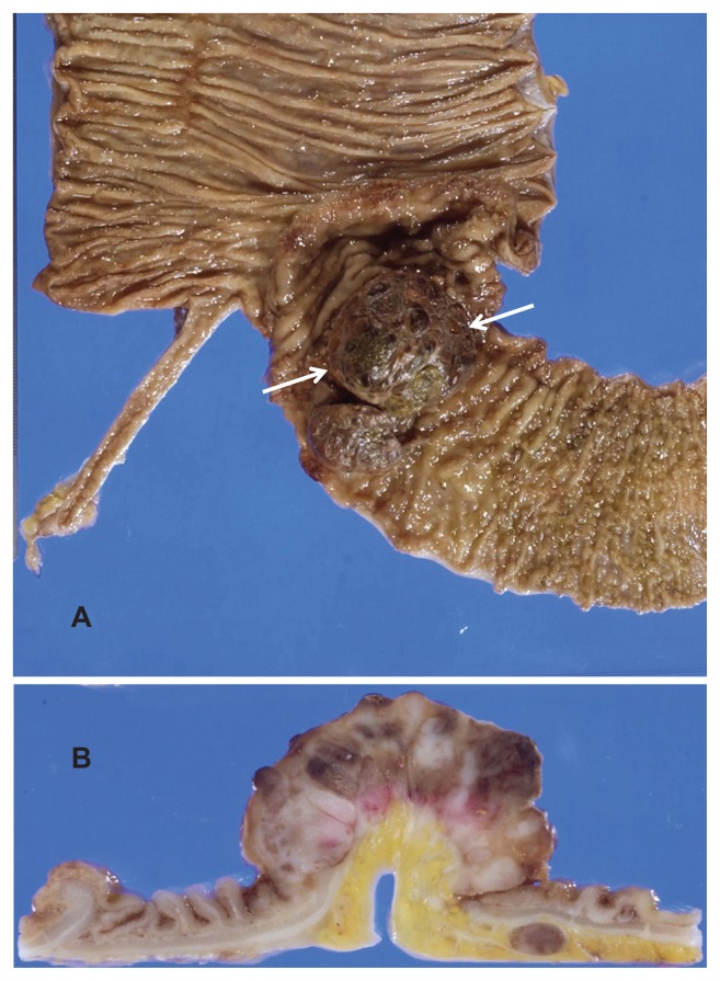
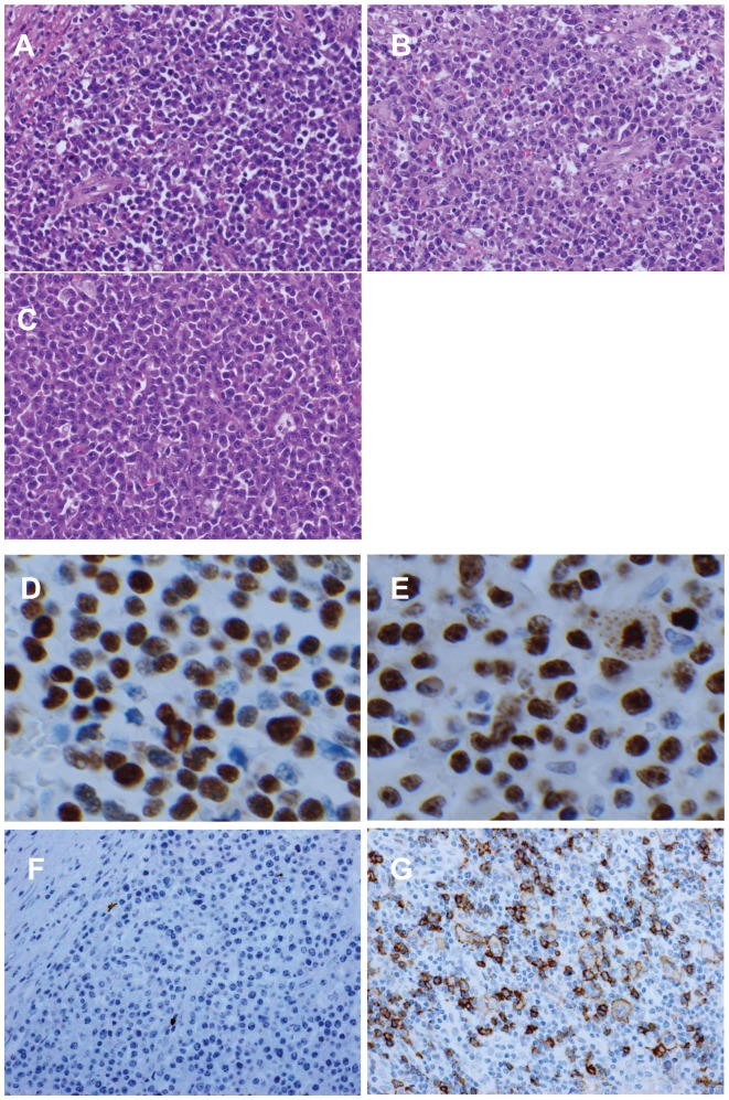
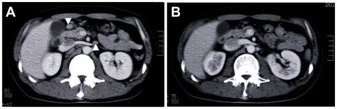
We report a case of Epstein-Barr virus (EBV)-positive ileal extraosseous plasmacytoma containing plasmablastic lymphoma components with CD20-positive lymph node involvement. A 34-year-old healthy Japanese male developed intussusception due to an ileal plasmacytoma. The lesion was positive for EBV-encoded small nuclear RNA in in situ hybridization, with the surrounding lymph nodes showing the expression of CD20. Tumor cells in the ileal and lymph node lesions contained high-grade malignant features compatible with plasmablastic lymphoma. Because his abdominal lymph nodes recurred 6 months after resection, he received six cycles of R-CHOP chemotherapy (rituximab, cyclophosphamide, doxorubicin, vincristine, and prednisolone), and had a complete remission. Although his case was complicated by acute promyelocytic leukemia, he has so far survived, recurrence-free, for more than 7.5 years after chemotherapy for extraosseous plasmacytoma.
Keywords: CD20; Epstein-Barr virus (EBV); R-CHOP; ileal extraosseous plasmacytoma; lymph node involvement; plasmablastic lymphoma.
Figures
Similar articles
-
CD3+ CD56+ EBER1+ atypical extraosseous plasmacytoma of the nasal cavity.Int J Hematol. 2018 Sep;108(3):344-347. doi: 10.1007/s12185-018-2438-y. Epub 2018 Mar 23. Int J Hematol. 2018. PMID: 29572766
-
Epstein-Barr virus-positive plasmacytoma in immunocompetent patients.Histopathology. 2015 Aug;67(2):225-34. doi: 10.1111/his.12640. Epub 2015 Feb 19. Histopathology. 2015. PMID: 25556356
-
Secondary cutaneous Epstein-Barr virus-associated diffuse large B-cell lymphoma in a patient with angioimmunoblastic T-cell lymphoma: a case report and review of literature.Diagn Pathol. 2012 Jan 19;7:7. doi: 10.1186/1746-1596-7-7. Diagn Pathol. 2012. PMID: 22260632 Free PMC article. Review.
-
Unusual course of generalized lymph node primary plasmacytoma in a patient with Sjögren's syndrome: a case report.J Med Case Rep. 2017 Apr 20;11(1):116. doi: 10.1186/s13256-017-1266-7. J Med Case Rep. 2017. PMID: 28424097 Free PMC article.
-
CD20-positive primary gastric T-cell lymphoma poorly responding to initial treatment with rituximab plus CHOP, and a literature review.Int J Hematol. 2015 Dec;102(6):702-8. doi: 10.1007/s12185-015-1841-x. Epub 2015 Aug 7. Int J Hematol. 2015. PMID: 26251099 Review.
Cited by
-
CD3+ CD56+ EBER1+ atypical extraosseous plasmacytoma of the nasal cavity.Int J Hematol. 2018 Sep;108(3):344-347. doi: 10.1007/s12185-018-2438-y. Epub 2018 Mar 23. Int J Hematol. 2018. PMID: 29572766
-
Plasmablastic Lymphoma Associated with Adjacent Mature Plasma Cell Population Exhibiting Opposite Light Chain Restriction.Case Rep Pathol. 2020 Dec 28;2020:8875547. doi: 10.1155/2020/8875547. eCollection 2020. Case Rep Pathol. 2020. PMID: 33489398 Free PMC article.
-
Expect the Unexpected: Report of a Case of Pediatric Pharyngeal Extraosseous Plasmacytoma with Tumefactive Amyloidosis ("Amyloidoma") and a Review of the Literature.Head Neck Pathol. 2015 Dec;9(4):431-5. doi: 10.1007/s12105-015-0614-4. Epub 2015 Feb 12. Head Neck Pathol. 2015. PMID: 25672253 Free PMC article. Review.
-
Plasmablastic transformation of a pre-existing plasmacytoma: a possible role for reactivation of Epstein Barr virus infection.Haematologica. 2014 Nov;99(11):e235-7. doi: 10.3324/haematol.2014.111872. Epub 2014 Sep 5. Haematologica. 2014. PMID: 25193957 Free PMC article. No abstract available.
-
Epstein-Barr virus-positive plasmacytoma in immunocompetent patients: a diagnostic dilemma.Int J Clin Exp Pathol. 2020 Mar 1;13(3):582-586. eCollection 2020. Int J Clin Exp Pathol. 2020. PMID: 32269699 Free PMC article.
References
-
- Alexiou C, Kau RJ, Dietzfelbinger H, et al. Extramedullary plasmacytoma: tumor occurrence and therapeutic concepts. Cancer. 1999;85:2305–2014. - PubMed
-
- McKenna RW, Kyle RA, Kuehl WM, Grogan TM, Harris NL, Coupland RW. Plasma cell neoplasms. In: Swerdlow SH, Campo E, Harris NL, Jaffe ES, Pileri SA, editors. WHO Classification of Tumours of Haematopoietic and Lymphoid Tissues. 4th ed. Lyon, France: IARC; 2008. pp. 200–213.
-
- Lin BT, Weiss LM. Primary plasmacytoma of lymph nodes. Hum Pathol. 1997;28:1083–1090. - PubMed
-
- Bachar G, Goldstein D, Brown D, et al. Solitary extramedullary plasmacytoma of the head and neck – long-term outcome analysis of 68 cases. Head Neck. 2008;30:1012–1019. - PubMed
Publication types
LinkOut - more resources
Full Text Sources
Research Materials
