

Direct measurement of mammographic X-ray spectra with a digital CdTe detection system
- PMID: 22969406
- PMCID: PMC3436035
- DOI: 10.3390/s120608390
Direct measurement of mammographic X-ray spectra with a digital CdTe detection system
Abstract
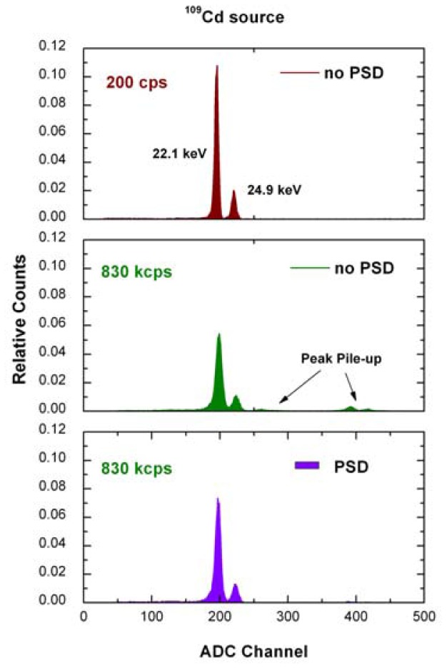
In this work we present a detection system, based on a CdTe detector and an innovative digital pulse processing (DPP) system, for high-rate X-ray spectroscopy in mammography (1-30 keV). The DPP system performs a height and shape analysis of the detector pulses, sampled and digitized by a 14-bit, 100 MHz ADC. We show the results of the characterization of the detection system both at low and high photon counting rates by using monoenergetic X-ray sources and a nonclinical X-ray tube. The detection system exhibits excellent performance up to 830 kcps with an energy resolution of 4.5% FWHM at 22.1 keV. Direct measurements of clinical molybdenum X-ray spectra were carried out by using a pinhole collimator and a custom alignment device. A comparison with the attenuation curves and the half value layer values, obtained from the measured and simulated spectra, from an ionization chamber and from a solid state dosimeter, also shows the accuracy of the measurements. These results make the proposed detection system a very attractive tool for both laboratory research, calibration of dosimeters and advanced quality controls in mammography.
Keywords: CdTe detectors; X-ray spectroscopy; digital pulse processing; high photon counting rate; mammography.
Figures


Similar articles
-
High-rate x-ray spectroscopy in mammography with a CdTe detector: a digital pulse processing approach.Med Phys. 2010 Dec;37(12):6147-56. doi: 10.1118/1.3512804. Med Phys. 2010. PMID: 21302771
-
Uncertainty estimation and statistical comparative methodology for mammography x-ray energy spectra.Biomed Phys Eng Express. 2020 Apr 21;6(3):035018. doi: 10.1088/2057-1976/ab817d. Biomed Phys Eng Express. 2020. PMID: 33438663
-
Comparison of two portable solid state detectors with an improved collimation and alignment device for mammographic x-ray spectroscopy.Med Phys. 2006 Sep;33(9):3469-77. doi: 10.1118/1.2229431. Med Phys. 2006. PMID: 17022243
-
Direct measurement of clinical mammographic x-ray spectra using a CdTe spectrometer.Med Phys. 2017 Jul;44(7):3504-3511. doi: 10.1002/mp.12287. Epub 2017 May 26. Med Phys. 2017. PMID: 28429382
-
Tutorial on X-ray photon counting detector characterization.J Xray Sci Technol. 2018;26(1):1-28. doi: 10.3233/XST-16210. J Xray Sci Technol. 2018. PMID: 29154310 Free PMC article. Review.
Cited by
-
A Novel Extraction Procedure of Contact Characteristic Parameters from Current-Voltage Curves in CdZnTe and CdTe Detectors.Sensors (Basel). 2023 Jul 1;23(13):6075. doi: 10.3390/s23136075. Sensors (Basel). 2023. PMID: 37447923 Free PMC article.
-
Ballistic Deficit Pulse Processing in Cadmium-Zinc-Telluride Pixel Detectors for High-Flux X-ray Measurements.Sensors (Basel). 2022 Apr 29;22(9):3409. doi: 10.3390/s22093409. Sensors (Basel). 2022. PMID: 35591099 Free PMC article.
-
High-rate dead-time corrections in a general purpose digital pulse processing system.J Synchrotron Radiat. 2015 Sep;22(5):1190-201. doi: 10.1107/S1600577515013776. Epub 2015 Aug 7. J Synchrotron Radiat. 2015. PMID: 26289270 Free PMC article.
References
-
- Sanborg M., Dance D.R., Alm Carlsson G., Persliden J. A Monte Carlo for the simulation of image quality and absorbed dose in diagnostic radiology. Comput. Methods Prog. Biomed. 1994;31:167–180. - PubMed
-
- Boone J.M., Fewell T.R., Jennings R.J. Molybdenum, rhodium, and tungsten anode spectral models using interpolating polynomials with application to mammography. Med. Phys. 1997;24:1863–1874. - PubMed
-
- Sidky E.Y., Yu L., Pan X., Zou Y., Vannier M. A robust method of X-ray source spectrum estimation from transmission measurements: Demonstrated on computer simulated, scatter-free transmission data. J. Appl. Phys. 2005;97:124701.
-
- Silva M.C., Herdade S.B., Lammoglia P., Costa P.R., Terini R.A. Determination of the voltage applied to X-ray tubes from the bremsstrahlung spectrum obtained with a silicon PIN photodiode. Med. Phys. 2000;27:2617–2623. - PubMed
-
- Assiamah M., Nam T.L., Keddy R.J. Comparison of mammography radiation dose values obtained from direct incident air kerma measurements with values from measured X-ray spectral data. Appl. Radiat. Isot. 2005;52:551–560. - PubMed
MeSH terms
Substances
LinkOut - more resources
Full Text Sources
Medical
