. 2012 Aug 30:6:249.
doi: 10.3389/fnhum.2012.00249. eCollection 2012.

Differential vulnerability of substantia nigra and corpus striatum to oxidative insult induced by reduced dietary levels of essential fatty acids

Affiliations

Differential vulnerability of substantia nigra and corpus striatum to oxidative insult induced by reduced dietary levels of essential fatty acids

Henriqueta D Cardoso et al. Front Hum Neurosci. .

Abstract

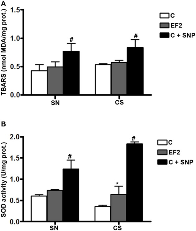
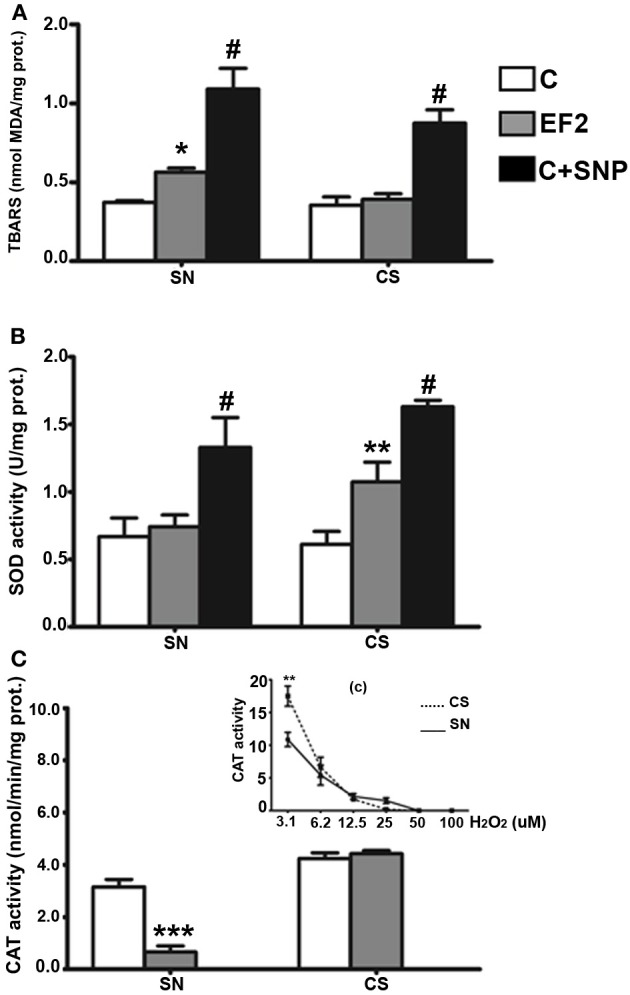
Oxidative stress (OS) has been implicated in the etiology of certain neurodegenerative disorders. Some of these disorders have been associated with unbalanced levels of essential fatty acids (EFA). The response of certain brain regions to OS, however, is not uniform and a selective vulnerability or resilience can occur. In our previous study on rat brains, we observed that a two-generation EFA dietary restriction reduced the number and size of dopaminergic neurons in the substantia nigra (SN) rostro-dorso-medial. To understand whether OS contributes to this effect, we assessed the status of lipid peroxidation (LP) and anti-oxidant markers in both SN and corpus striatum (CS) of rats submitted to this dietary treatment for one (F1) or two (F2) generations. Wistar rats were raised from conception on control or experimental diets containing adequate or reduced levels of linoleic and α-linolenic fatty acids, respectively. LP was measured using the thiobarbituric acid reaction method (TBARS) and the total superoxide dismutase (t-SOD) and catalase (CAT) enzymatic activities were assessed. The experimental diet significantly reduced the docosahexaenoic acid (DHA) levels of SN phospholipids in the F1 (~28%) and F2 (~50%) groups. In F1 adult animals of the experimental group there was no LP in both SN and CS. Consistently, there was a significant increase in the t-SOD activity (p < 0.01) in both regions. In EF2 young animals, degeneration in dopaminergic and non-dopaminergic neurons and a significant increase in LP (p < 0.01) and decrease in the CAT activity (p < 0.001) were detected in the SN, while no inter-group difference was found for these parameters in the CS. Conversely, a significant increase in t-SOD activity (p < 0.05) was detected in the CS of the experimental group compared to the control. The results show that unbalanced EFA dietary levels reduce the redox balance in the SN and reveal mechanisms of resilience in the CS under this stressful condition.

Keywords: DHA; catalase; corpus striatum; lipid peroxidation; neurodegeneration; oxidative stress; substantia nigra; superoxide dismutase.


Figures

Figure 1
Figure 1
Thiobarbituric acid-reactant substances (TBARS) levels (A) and total superoxide dismutase (t-SOD) activities (B) in the pool of Substantia Nigra and Corpus Striatum from first generation adult rats fed essential fatty acid restricted diet and controls (n = 12 per group). *P < 0.05 compared to control group. Treatment of control homogenates with sodium nitroprusside (SNP) was used as positive control in all the experiments. #P < 0.001 compared to control or EF1 groups.
Figure 2
Figure 2
Thiobarbituric acid-reactant substances (TBARS) levels (A), total superoxide dismutase (t-SOD) activities (B), and catalase (CAT) activities (C) in the pool of Substantia Nigra and Corpus Striatum from young rats fed an essential fatty acid restricted diet over two generations and respective controls (n = 12 per group). *P < 0.05; **P < 0.001 compared to control group. Treatment of control homogenates with sodium nitroprusside (SNP) was used as positive control for TBARS and t-SOD in all the experiments. The insert (c) in the panel (C) shows the H2O2 concentration-dependent manner of the CAT activity in the SN and CS control homogenates obtained as positive controls. #P < 0.0001 compared to control or EF2 groups.
Figure 3
Figure 3
Fluoro-Jade C staining in brain parasagittal sections of F1- (panel A) or F2- (panel B) generation groups at the level of Substantia nigra (SN) or Corpus Striatum (CS). No labeling was detected in cell bodies or processes of SN (Aa, Ab) and CS (Ac, Ad) in adult animals of F1 generation. However, FJC positive cell bodies and processes were detected in the SN pars reticulata (Bb) and pars compacta (Bd) of EF2 group while no labeling in these regions was seen in the controls (Ba, Bc). No FJC labeling was detected in cell bodies of the CS in the EF2 (Bf) or control (Be) groups. A slight and non-specific labeling was seen in regions rich in myelin such as cerebral peduncle (cp), corpus callosum (cc), or myelinated fibers crossing the CS.
Figure 4
Figure 4
Photographs of epifluorescence microscopy showing SN sections from a representative EF2 animal subjected to TH immunostaining followed by Fluoro-Jade C staining. Examples of single (FJC; yellow arrows) or double (TH + FJC; black arrows) labeled cells can be seen either in the SN pars reticulata (A,B, and C) or in the pars compacta (D,E, and F). High magnification of the region (d) is shown in the bottom panel. Scale bar of A = B,C,D,E, and F represents 60 μm while the scale bar of bottom panel represents 20 μm.
Figure 5
Figure 5
Photographs of epifluorescence microscopy showing CS sections from a representative EF2 animal subjected to TH immunostaining (A) followed by Fluoro-Jade C staining (B). Note the absence of FJC-positive cells surrounded by TH-positive neuronal terminals. cc = corpus calosum, Scale bar = 40 μm.

Similar articles

Cited by

References

    1. Aebi H. (1984). Catalase in vitro. Methods Enzymol. 105, 121–126 - PubMed
    1. Ahmad S. O., Park J. H., Radel J. D., Levant B. (2008). Reduced numbers of dopamine neurons in the substantia nigra pars compacta and ventral tegmental area of rats fed an n-3 polyunsaturated fatty acid-deficient diet: a stereological study. Neurosci. Lett. 438, 303–307 10.1016/j.neulet.2008.04.073 - DOI - PMC - PubMed
    1. Berry J. F., Cevallos W. H., Wade R. R. (1965). Lipid class and fatty acid composition of intact peripheral nerve and during Wallerian degeneration. J. Am. Oil Chem. Soc. 42, 492–500 - PubMed
    1. Bian G. L., Wei L. C., Shi M., Wang Y. Q., Cao R., Chen L. W. (2007). Fluoro-Jade C can specifically stain the degenerative neurons in the substantia nigra of the 1-methyl-4-phenyl-1 2 3, 6-tetrahydro pyridine-treated C57BL/6 mice. Brain Res. 30, 55–61 10.1016/j.brainres.2007.02.078 - DOI - PubMed
    1. Borba J. M. C., Rocha-de-Melo A. P., Amâncio dos Santos A., Andrade da Costa B. L. S., Silva R. P. (2010). Essential fatty acid deficiency reduces cortical spreading depression propagation in rats: a two-generation study. Nutr. Neurosci. 13, 144–150 10.1179/147683010X12611460763887 - DOI - PubMed