. 2012 Aug 27:3:295.
doi: 10.3389/fpsyg.2012.00295. eCollection 2012.

Resting state networks and consciousness: alterations of multiple resting state network connectivity in physiological, pharmacological, and pathological consciousness States

Affiliations

Resting state networks and consciousness: alterations of multiple resting state network connectivity in physiological, pharmacological, and pathological consciousness States

Lizette Heine et al. Front Psychol. .

Abstract

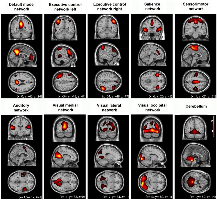
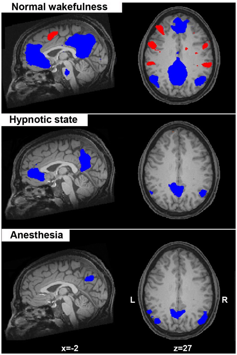
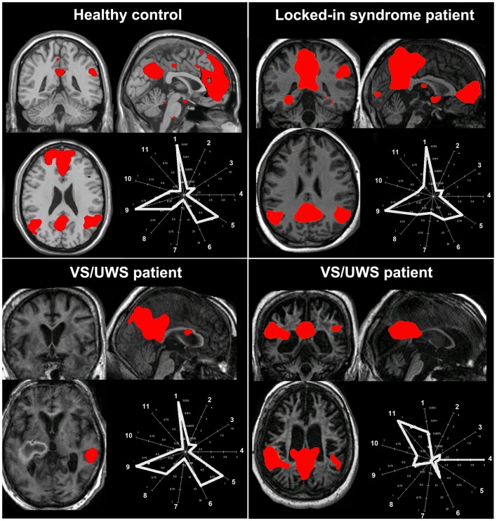
In order to better understand the functional contribution of resting state activity to conscious cognition, we aimed to review increases and decreases in functional magnetic resonance imaging (fMRI) functional connectivity under physiological (sleep), pharmacological (anesthesia), and pathological altered states of consciousness, such as brain death, coma, vegetative state/unresponsive wakefulness syndrome, and minimally conscious state. The reviewed resting state networks were the DMN, left and right executive control, salience, sensorimotor, auditory, and visual networks. We highlight some methodological issues concerning resting state analyses in severely injured brains mainly in terms of hypothesis-driven seed-based correlation analysis and data-driven independent components analysis approaches. Finally, we attempt to contextualize our discussion within theoretical frameworks of conscious processes. We think that this "lesion" approach allows us to better determine the necessary conditions under which normal conscious cognition takes place. At the clinical level, we acknowledge the technical merits of the resting state paradigm. Indeed, fast and easy acquisitions are preferable to activation paradigms in clinical populations. Finally, we emphasize the need to validate the diagnostic and prognostic value of fMRI resting state measurements in non-communicating brain damaged patients.

Keywords: anesthesia; coma; consciousness; default mode network; hypnosis; resting state networks; sleep.


Figures

Figure 1
Figure 1
Multiple cerebral networks can be identified with fMRI in healthy controls (n = 10) during normal wakeful resting state using independent component analysis. These networks reflect “higher-order” cognitive (i.e., default mode, left and right executive control, salience networks), and “lower-order” sensorimotor, and sensory (auditory, visual) function. For illustrative purposes, group-level spatial maps (z values) are rendered on a structural T1 magnetic resonance template and x, y, and z values indicate the Montreal Neurological Institute coordinates of the represented sections.
Figure 2
Figure 2
Spontaneous fMRI BOLD activity in the default mode network (in blue; considered to reflect self-related mentation) anticorrelates with the activity of a lateral frontoparietal system (in red; considered to mediate conscious perception of the external world). Here, this anticorrelated activity is shown for normal wakefulness, hypnotic state, and during deep anesthesia. Of note is the absence of the activity in the “extrinsic” frontoparietal system in the two conditions of altered sense of awareness (hypnosis, anesthesia) which is considered as suggestive of a diminished “external” awareness (i.e., the perception of the environment through the senses). Statistical maps are thresholded at a false discovery error rate p < 0.05 and rendered on a structural T1 magnetic resonance image of a healthy subject (x and z values indicate Talairach coordinates of the represented sections).
Figure 3
Figure 3
The challenge of selecting the “right” independent component as the resting state network of interest in pathological conditions. The figure illustrates the spatial pattern (brain maps, z values 0.8–10) and spatial-temporal properties (fingerprints: a representation of the component in a multidimensional space of parameters; De Martino et al., 2007) of the default mode network in healthy consciousness states (healthy subject, patient with locked-in syndrome; upper row) and in two patients with vegetative state/unresponsive wakefulness syndrome (VS/UWS; lower row). For the healthy control, the locked-in syndrome and the VS/UWS patient in the lower left corner, the default mode network shows the characteristic properties in both the spatial and the temporal domain (i.e., the fingerprints pick in the 0.02–0.05 Hz frequency band labeled with the number 9) even if for the VS/UWS patient the spatial pattern is only partially preserved. Of note is that the second VS/UWS patient exhibits the spatial pattern of the default mode network but importantly the time course of this component is characterized by high frequency fluctuations, in the 0.1–0.25 Hz frequency band and high spatial entropy (labeled, respectively, with the number 11 and 4 in the fingerprint). Therefore, such activity cannot be considered of neuronal origin. As a consequence, if the component selection was merely based on a spatial similarity test (e.g., with a predefined template), then this component could be erroneously selected and further statistically analyzed. A “compromise” in the selection of the appropriate network of interest in the space and time domain is needed to will eventually exclude non-neuronal contributions [Fingerprint labels: (1) degree of clustering, (2) skewness, (3) kurtosis, (4) spatial entropy, (5) autocorrelation, (6) temporal entropy, power: (7) 0–0.008 Hz, (8) 0.008–0.02 Hz, (9) 0.02–0.05 Hz, (10) 0.05–0.1 Hz, (11) 0.1–0.25 Hz].

Similar articles

Cited by

References

    1. Allen E. A., Erhardt E. B., Damaraju E., Gruner W., Segall J. M., Silva R. F., Havlicek M., Rachakonda S., Fries J., Kalyanam R., Michael A. M., Caprihan A., Turner J. A., Eichele T., Adelsheim S., Bryan A. D., Bustillo J., Clark V. P., Feldstein Ewing S. W., Filbey F., Ford C. C., Hutchison K., Jung R. E., Kiehl K. A., Kodituwakku P., Komesu Y. M., Mayer A. R., Pearlson G. D., Phillips J. P., Sadek J. R., Stevens M., Teuscher U., Thoma R. J., Calhoun V. D. (2011). A baseline for the multivariate comparison of resting-state networks. Front. Syst. Neurosci. 5:2.10.3389/fnsys.2011.00002 - DOI - PMC - PubMed
    1. American Society of Anesthesiologists Task Force on Intraoperative Awareness (2006). Practice advisory for intraoperative awareness and brain function monitoring a report by the American society of anesthesiologists task force on intraoperative awareness. Anesthesiology 104, 847–86410.1097/00000542-200604000-00031 - DOI - PubMed
    1. Anderson J. S., Druzgal T. J., Lopez-Larson M., Jeong E. K., Desai K., Yurgelun-Todd D. (2010). Network anticorrelations, global regression, and phase-shifted soft tissue correction. Hum. Brain Mapp. 32, 919–93410.1002/hbm.21079 - DOI - PMC - PubMed
    1. Bardin J. C., Fins J. J., Katz D. I., Hersh J., Heier L. A., Tabelow K., Dyke J. P., Ballon D. J., Schiff N. D., Voss H. U. (2011). Dissociations between behavioural and functional magnetic resonance imaging-based evaluations of cognitive function after brain injury. Brain 134, 769–78210.1093/brain/awr005 - DOI - PMC - PubMed
    1. Beckmann C. F., Deluca M., Devlin J. T., Smith S. M. (2005). Investigations into resting-state connectivity using independent component analysis. Philos. Trans. R. Soc. Lond. B Biol. Sci. 360, 1001–101310.1098/rstb.2005.1634 - DOI - PMC - PubMed