. 2012 Aug 27:3:304.
doi: 10.3389/fpsyg.2012.00304. eCollection 2012.

From objects to landmarks: the function of visual location information in spatial navigation

Affiliations

From objects to landmarks: the function of visual location information in spatial navigation

Edgar Chan et al. Front Psychol. .

Abstract

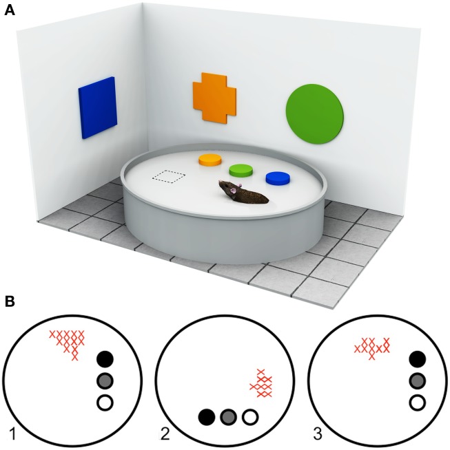
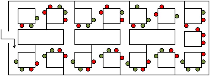
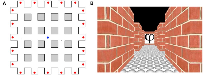
Landmarks play an important role in guiding navigational behavior. A host of studies in the last 15 years has demonstrated that environmental objects can act as landmarks for navigation in different ways. In this review, we propose a parsimonious four-part taxonomy for conceptualizing object location information during navigation. We begin by outlining object properties that appear to be important for a landmark to attain salience. We then systematically examine the different functions of objects as navigational landmarks based on previous behavioral and neuroanatomical findings in rodents and humans. Evidence is presented showing that single environmental objects can function as navigational beacons, or act as associative or orientation cues. In addition, we argue that extended surfaces or boundaries can act as landmarks by providing a frame of reference for encoding spatial information. The present review provides a concise taxonomy of the use of visual objects as landmarks in navigation and should serve as a useful reference for future research into landmark-based spatial navigation.

Keywords: hippocampus; landmarks; navigation; parahippocampal gyrus; retrosplenial cortex; spatial memory; striatum; topographical disorientation.


Figures

Figure 1
Figure 1
Approaches to understanding landmark processing in rodents. (A) Example of a typical Morris Water Maze setup. The Morris Water Maze paradigm (Morris, 1981), originally developed for rodents, requires the animal to locate a hidden platform (dashed square) that is submerged below the surface of a large circular arena filled with opaque water, using available room cues (in this illustration, the colored geometric shapes). The start position is varied between trials so that animals cannot use proprioceptive or egocentric strategies, but must learn to use the configural relationships between objects within the room to locate the platform. By manipulating the available visual information, or by lesioning specific neural structures, it is possible to determine the types of cues and brain regions that are important for solving the task. (B) Firing rate maps of a hippocampal place cell adapted from Cressant et al. (1997). “Place cells” are spatially selective cells, found in the rodent hippocampus, that fire maximally when the rodent is within a well-defined region of the environment (i.e., within the cell’s “place field”) but not at any other location, independent of head and body direction. Place cell activity is thought to reflect a rodent’s internal spatial representation of the environment. As such, the navigational relevance of different types of environmental object can be inferred from the manner in which they influence place cell activity. The firing rates of a rodent hippocampal place cell within a circular arena are shown in the example. The filled circles represent three prominent objects within the arena. As the spatial locations of objects are rotated between trials (1–3), the place field of the place cell shifts accordingly (red crosses). In this example, the rodent’s internal spatial representation is anchored by the three objects.
Figure 2
Figure 2
The influence of spatial location on object processing adapted from Janzen and van Turennout (2004). An aerial schematic of the virtual museum used by Janzen and van Turennout (2004). Red dots indicate locations of decision point objects, and green dots indicate locations of non-decision point objects.
Figure 3
Figure 3
Heading direction selectivity in humans adapted from Baumann and Mattingley (2010). (A) Aerial schematic of the virtual environment used in the study by Baumann and Mattingley (2010). Red dots represent the locations of the abstract symbols that acted as orientation landmarks. The blue dot represents the starting location of each learning trial. (B) An example of a single image viewed by participants during the test phase.

Similar articles

Cited by

References

    1. Able K. P. (1991). Common themes and variations in animal orientation systems. Am. Zool. 31, 157–167
    1. Aguirre G. K., D’Esposito M. (1999). Topographical disorientation: a synthesis and taxonomy. Brain 122, 1613–162810.1093/brain/122.9.1613 - DOI - PubMed
    1. Alyan S. (1996). Evidence for resetting the directional component of path integration in the house mouse (Mus musculus). Ethology 102, 629–63810.1111/j.1439-0310.1996.tb01154.x - DOI
    1. Alyan S., Jander R. (1994). Short-range homing in the house mouse, Mus musculus: stages in the learning of directions. Anim. Behav. 48, 285–29810.1006/anbe.1994.1242 - DOI
    1. Aminoff E., Gronau N., Bar M. (2007). The parahippocampal cortex mediates spatial and nonspatial associations. Cereb. Cortex 17, 1493–150310.1093/cercor/bhl078 - DOI - PubMed

LinkOut - more resources