. 2012 Aug 28:3:260.
doi: 10.3389/fimmu.2012.00260. eCollection 2012.

Chromatinized Protein Kinase C-θ: Can It Escape the Clutches of NF-κB?

Affiliations

Chromatinized Protein Kinase C-θ: Can It Escape the Clutches of NF-κB?

Elissa L Sutcliffe et al. Front Immunol. .

Abstract

We recently provided the first description of a nuclear mechanism used by Protein Kinase C-theta (PKC-θ) to mediate T cell gene expression. In this mode, PKC-θ tethers to chromatin to form an active nuclear complex by interacting with proteins including RNA polymerase II, the histone kinase MSK-1, the demethylase LSD1, and the adaptor molecule 14-3-3ζ at regulatory regions of inducible immune response genes. Moreover, our genome-wide analysis identified many novel PKC-θ target genes and microRNAs implicated in T cell development, differentiation, apoptosis, and proliferation. We have expanded our ChIP-on-chip analysis and have now identified a transcription factor motif containing NF-κB binding sites that may facilitate recruitment of PKC-θ to chromatin at coding genes. Furthermore, NF-κB association with chromatin appears to be a prerequisite for the assembly of the PKC-θ active complex. In contrast, a distinct NF-κB-containing module appears to operate at PKC-θ targeted microRNA genes, and here NF-κB negatively regulates microRNA gene transcription. Our efforts are also focusing on distinguishing between the nuclear and cytoplasmic functions of PKCs to ascertain how these kinases may synergize their roles as both cytoplasmic signaling proteins and their functions on the chromatin template, together enabling rapid induction of eukaryotic genes. We have identified an alternative sequence within PKC-θ that appears to be important for nuclear translocation of this kinase. Understanding the molecular mechanisms used by signal transduction kinases to elicit specific and distinct transcriptional programs in T cells will enable scientists to refine current therapeutic strategies for autoimmune diseases and cancer.

Keywords: NF-κB; PKC-theta; T cells; chromatin; immune response gene; microRNA; nuclear PKC-theta; signaling kinase.


Figures
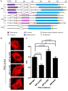
Figure 1
Figure 1
SPT243 is the minimum motif for nuclear localization of PKC-θ regulated by phosphorylation at S241 and T243. (A) SPT-like motifs and nuclear localization sequences (NLS) were displayed relative to the domain organization of PKC members (nPKC: δ, θ, η, ε; cPKC: α, βI, βII, γ; and aPKC: ζ, ℩, or λ). SPT-like motifs are depicted in red and the locations of NLS are shown in yellow. The C2-like domain (purple) is characteristic of nPKCs, while cPKCs share the calcium binding C2 domain (pink). Both nPKC and cPKC isoforms possess the C1 domain (white), that is composed of C1a and C1b. In comparison, aPKC isoforms only contain C1a of the C1 domain. All PKC family members contain a C-terminal catalytic domain (blue). (B) The full length PKC-θ wild type gene sequence and its derivatives wherein putative phosphorylation sites at S241 and T243 were mutated to either the non-phosphorylatable alanine (SPT to APA) or the phosphomimetic glutamine (SPT to EPE), were cloned into the pTracer-CMV vector in frame with a C-terminal HA-tag. The vector also codes for GFP, which is translated independent of the insert and serves as an internal marker for transfected cells. Subconfluent cultures of Cos-7 cells were transfected and subsequently the fixed cells were probed with rabbit antibody to HA-tag, followed by secondary antibody to rabbit immunoglobulins conjugated to Alexa-Fluor 568. Localization of expressed PKC-θ was studied with confocal laser scanning microscopy as detailed in the methods. Representative images for each construct are shown. (C) Fn/c values for each construct are shown in (C), with significant differences between datasets indicated. Data shown are mean ± SEM, n > 15 for each dataset.
Figure 2
Figure 2
PKC-θ activity in T lymphocytes. Jurkat T cells were either non-stimulated (NS) or stimulated (ST) with PMA/CaI for 4 h. The PKC-θ specific antibody was used to immunoprecipitate PKC-θ from either whole cells or nuclei from both NS and ST treated Jurkat T cells. PKC ELISA-based kinase assays were performed on these PKC-θ fractions and absorbance was measured at 450 nm. Data is plotted as relative kinase activity compared to the negative control that did not have any antibody. Data is representative of the mean ± SE of three independent experiments and statistical significance was determined by a two-tailed paired t-test using GraphPad Prism 5.03.
Figure 3
Figure 3
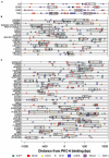
Motif superfamilies identified within promoters of PKC-θ bound genes. A greater proportion of the promoters of the PKC-bound genes had motifs for members of the SP1F, ZBPF, MAZF, MZF1, PAX5, EKLF, E2FF, NFKB, AHRR, and EBOX motif superfamilies, when compared to a background set of promoters. The representative members of the superfamilies shown are SP1_01 (SP1F), ZNF219_01 (ZBPF), MAZR_01 (MAZR), MZF1_01 (MZF1), PAX5_01 (PAX5), EKLF_01 (EKLF), NFKAPPAB_01 (NFKB), E2F_03 (E2FF), AHRARNT_02 (AHRR), and MYCMAX_03 (EBOX).
Figure 4
Figure 4
Transcription factor binding motifs in the PKC-bound sequences. Motifs for members of the NFKB (red), E2FF (green), PAX5 (orange), AHRR (blue), EBOX (purple), MZF (gray), and SP1F (black) superfamilies were identified by their Position Weight Matrices. PKC binds four inducible genes [+150 bp downstream from the transcription start site (A)], in promoter sequences (B) or within genes (C). All motifs within a superfamily are shown for (A) while only NFKAPPAB_01 (NFKB), E2F_03 (E2FF), AHRARNT_02 (AHRR), PAX5_01 (PAX5), and SP1_01 (SP1F) are shown for (B) and (C). Commonly co-occurring motifs are boxed together. NFKB_SP1F_E2FF commonly co-occur in the promoter regions (B), while the NFKB_E2FF_AHRR and NFKB_AHRR_PAX5 combinations are significantly over-represented in the within-gene PKC binding regions (C).
Figure 5
Figure 5
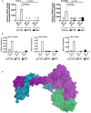
Binding of the PKC-θ active transcription complex to the CD69 promoter appears dependent on NF-κB. (A) Resting Jurkat T cells were pre-incubated with either Pentoxifylline (PTX), Bay, or left untreated (control) for 1 h. Cells were then left non-stimulated (NS) or activated with PMA/CaI (ST) for 4 h. Total mRNA was isolated for cDNA reverse transcription. Gene specific Taqman® expression assay was used to detect the transcript levels of human IL-2 and CD69. Data is expressed as arbitrary mRNA copies normalized to GAPDH. Data represent the mean ± SD of four independent experiments. Statistical significance between the activated control and inhibited samples was determined by a two-tailed paired t-test using GraphPad Prism 5.03. PKC-θ, Pol II, and LSD1 ChIP experiments were performed on the samples described in (A). (B) Sybr-green real-time PCR was used to detect the relative enrichment of these proteins across the CD69 promoter (−214 to −52 relative to the transcription start site). (C) The catalytic domain of PKC-θ (green, pdb code 2JED) is modeled interacting with the LSD1-CoREST complex (purple and blue, respectively, pdb code 2IW5). In this model the peptide-binding groove of PKC-θ contacts the SWIRM and amine oxidase domains of LSD1.
Figure 6
Figure 6
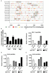
PKC-θ binding to the miR-200c promoter is not impaired by the absence of NF-κB in T cells. (A) Transcription factor binding motifs in the microRNA PKC-θ bound sequences. Motifs for NFKAPPAB_01 (NFKB, red), E2F_03 (E2FF, green), and PAX5_01 (PAX5, orange) were identified by their Position Weight Matrices. The three motifs commonly occur within 300 bp of each other near the PKC binding sites in microRNA genes. (B) TaqMan miR-200c microRNA cDNA was isolated from resting (NS) and 4 h PMA/CaI stimulated (ST) Jurkat T cells in the presence or absence of Pentoxifylline (PTX) or Bay. Data expressed as fold change in miR-200c relative to NS set to 1 and normalized to RNU6B. Data representative of the mean ± SE of three independent experiments. (C) PKC-θ, (D) Pol II, and (E) LSD1 ChIP were carried out on samples as described for (B). Sybr-green real-time PCR was used to detect the relative enrichment of these proteins across the miR-200c promoter. ChIP data are expressed as relative enrichment and plotted as the mean ± SE of two independent experiments performed in duplicate.
Figure 7
Figure 7
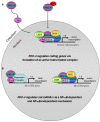
Putative model for PKC-θ tethering to coding vs. microRNA genes in T cells. (A) T cell activation induces a complex signaling cascade that involves PKC-θ, which provokes the activation and nuclear translocation of the transcription factor c-Rel. This event ultimately results in the binding of c-Rel to a unique transcription factor module at gene regulatory regions. We propose that c-Rel then initiates the recruitment of PKC-θ that may enter the nucleus via a phosphorylatable SPT sequence and bind to chromatin, thereby allowing the formation of the active transcription complex on coding genes to enable chromatin accessibility and gene transcription. (B) Following T cell stimulation, there is a reduction in PKC-θ binding to chromatin at microRNA genes compared to resting T cells. Instead, NF-κB is recruited via a distinct transcription factor motif and may form part of a repressive complex on these genes to dampen gene transcription. When NF-κB is removed from this system, more of the active PKC-θ complex assembles on microRNA gene promoters thereby overcoming transcriptional repression.

Similar articles

Cited by

References

    1. Anest V., Hanson J. L., Cogswell P. C., Steinbrecher K. A., Strahl B. D., Baldwin A. S. (2003). A nucleosomal function for IkappaB kinase-alpha in NF-kappaB-dependent gene expression. Nature 423, 659–66310.1038/nature01648 - DOI - PubMed
    1. Beausoleil S. A., Villen J., Gerber S. A., Rush J., Gygi S. P. (2006). A probability-based approach for high-throughput protein phosphorylation analysis and site localization. Nat. Biotechnol. 24, 1285–129210.1038/nbt1240 - DOI - PubMed
    1. Birbach A., Gold P., Binder B. R., Hofer E., de Martin R., Schmid J. A. (2002). Signaling molecules of the NF-kappa B pathway shuttle constitutively between cytoplasm and nucleus. J. Biol. Chem. 277, 10842–1085110.1074/jbc.M112475200 - DOI - PubMed
    1. Caamano J., Hunter C. A. (2002). NF-kappaB family of transcription factors: central regulators of innate and adaptive immune functions. Clin. Microbiol. Rev. 15, 414–42910.1128/CMR.15.3.414-429.2002 - DOI - PMC - PubMed
    1. Carlotti F., Dower S. K., Qwarnstrom E. E. (2000). Dynamic shuttling of nuclear factor kappa B between the nucleus and cytoplasm as a consequence of inhibitor dissociation. J. Biol. Chem. 275, 41028–4103410.1074/jbc.M006179200 - DOI - PubMed

LinkOut - more resources