. 2012 Aug 27:3:265.
doi: 10.3389/fimmu.2012.00265. eCollection 2012.

Autoimmune dysregulation and purine metabolism in adenosine deaminase deficiency

Affiliations

Autoimmune dysregulation and purine metabolism in adenosine deaminase deficiency

Aisha Vanessa Sauer et al. Front Immunol. .

Abstract

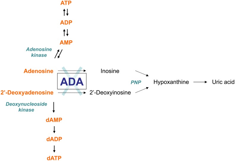
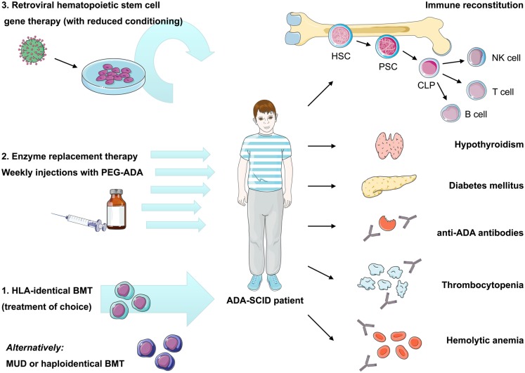
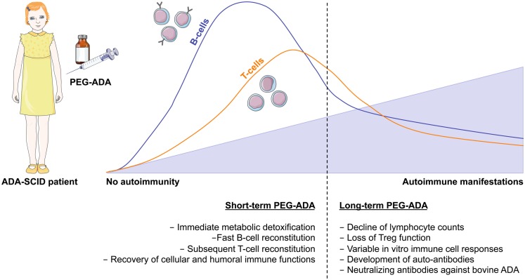
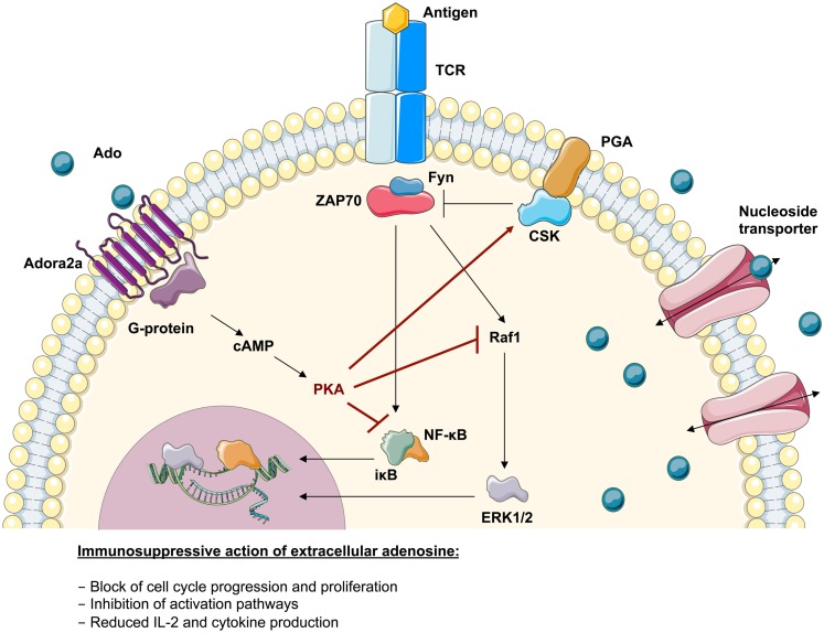
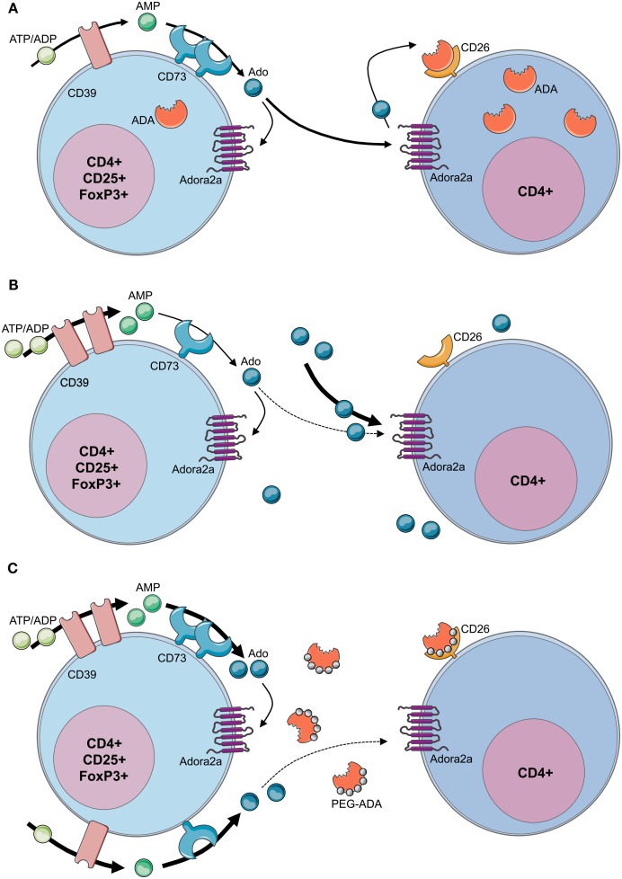
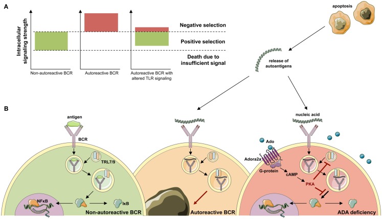
Genetic defects in the adenosine deaminase (ADA) gene are among the most common causes for severe combined immunodeficiency (SCID). ADA-SCID patients suffer from lymphopenia, severely impaired cellular and humoral immunity, failure to thrive, and recurrent infections. Currently available therapeutic options for this otherwise fatal disorder include bone marrow transplantation (BMT), enzyme replacement therapy with bovine ADA (PEG-ADA), or hematopoietic stem cell gene therapy (HSC-GT). Although varying degrees of immune reconstitution can be achieved by these treatments, breakdown of tolerance is a major concern in ADA-SCID. Immune dysregulation such as autoimmune hypothyroidism, diabetes mellitus, hemolytic anemia, and immune thrombocytopenia are frequently observed in milder forms of the disease. However, several reports document similar complications also in patients on long-term PEG-ADA and after BMT or GT treatment. A skewed repertoire and decreased immune functions have been implicated in autoimmunity observed in certain B-cell and/or T-cell immunodeficiencies, but it remains unclear to what extent specific mechanisms of tolerance are affected in ADA deficiency. Herein we provide an overview about ADA-SCID and the autoimmune manifestations reported in these patients before and after treatment. We also assess the value of the ADA-deficient mouse model as a useful tool to study both immune and metabolic disease mechanisms. With focus on regulatory T- and B-cells we discuss the lymphocyte subpopulations particularly prone to contribute to the loss of self-tolerance and onset of autoimmunity in ADA deficiency. Moreover we address which aspects of immune dysregulation are specifically related to alterations in purine metabolism caused by the lack of ADA and the subsequent accumulation of metabolites with immunomodulatory properties.

Keywords: ADA-SCID; adenosine deaminase; autoimmunity; gene therapy; severe combined immunodeficiency.


Figures

Figure 1
Figure 1
The adenosine deaminase (ADA) metabolism. ADA is an enzyme of the purine salvage pathway, which catalyzes the irreversible deamination of adenosine and 2′-deoxyadenosine into inosine and 2′-deoxyinosine, respectively. Most adenosine derives from endogenous breakdown of ATP and degradation of RNA, or is taken up exogenously by ubiquitously expressed nucleoside transporters. Unlike adenosine, 2′-deoxyadenosine is formed by DNA degradation is predominantly catabolized by ADA. Further conversion of inosine nucleoside leads to hypoxanthine, which can either enter a non-reversible pathway to uric acid or salvaged back into other mononucleosides. In the absence of ADA, the presence of these alternative “bypass” pathways results in normal concentrations of the catabolic products of the enzyme reaction in patients with ADA-SCID. Conversely, the levels of ADA substrates, adenosine and 2′-deoxyadenosine, are not only found in increased amounts in extracellular body fluids, but they also “spill over” into additional pathways normally only minimally utilized, thus contributing to the pathogenic mechanisms of the disease.
Figure 2
Figure 2
Current therapeutic options in ADA-SCID and reported autoimmune manifestations after treatment. Immune reconstitution in ADA deficiency can be achieved by bone marrow transplantation, enzyme replacement, or gene therapy, nonetheless recovery of immune functions may vary depending on the applied treatment and patient’s characteristics. Treatment of choice remains bone marrow transplantation from an HLA-identical sibling donor, while transplants from alternative donors are associated with high morbidity and mortality. Enzyme replacement therapy using pegylated bovine ADA is a non-curative treatment requiring weekly intramuscular injections with PEG-ADA. ADA-SCID has been the pioneer disease for the development of human gene therapy. It is based on the reinfusion of autologous HSC transduced with a retroviral vector containing the ADA cDNA. Variable degrees of immune reconstitution can be achieved by these treatments, but onset of autoimmunity is of concern in post-treatment ADA-SCID patients. Reported autoimmune manifestations include: autoimmune hypothyroidism, diabetes mellitus, thrombocytopenia, hemolytic anemia, and development of anti-ADA antibodies. HLA, human leukocyte antigen; BMT, bone marrow transplantation; MUD, matched unrelated donor; PEG-ADA, pegylated bovine ADA; HSC, hematopoietic stem cell; PSC, pluripotent stem cell; CLP, committed lymphocyte precursor; NK, natural killer cell.
Figure 3
Figure 3
Immune reconstitution and development of autoimmunity after PEG-ADA treatment. Enzyme replacement therapy with pegylated bovine ADA is a lifesaving but non-curative treatment for ADA-SCID patients. It provides metabolic detoxification and protective immune function with patients remaining clinically well, but immune reconstitution is often suboptimal and may not be long-lived. Shortly after initiation of PEG-ADA treatment, patients show recovery of B-cell counts, followed by a gradual increase in T-cell numbers and reconstitution of immune cell functions. However, the long-term consequences of PEG-ADA treatment are unknown. Immune recovery in B and T- cells is below normal levels. Major concerns are the susceptibility to opportunistic infections and the development of autoimmunity due to lymphopenia with gradual decline of immune functions and perturbation of T- and B-cell tolerance.
Figure 4
Figure 4
Immunosuppressive action of adenosine in lymphoid cells. Upon TCR-antigen-binding and T-cell activation, the translocation of activated transcription factors and the expression of anti-apoptotic factors supports their survival, proliferation, and differentiation. The adenosine 2a receptor (Adora2a) present on T-lymphocytes interacts with coupled G-proteins to stimulate cAMP formation. The effects of cAMP in T cells are almost entirely mediated by the cAMP-dependent protein kinase A (PKA). It has been shown that the inhibitory properties of PKA are mediated via activation of Csk or inhibition of Raf1. Consistently it has been shown that the severely compromised effector functions in ADA-deficient T cells associates with an intrinsically reduced ERK1/2 signaling, a decreased phosphorylation of IκB and an altered nuclear recruitment of NF-κB. This suggests that the accumulating adenosine levels in an ADA-deficient environment can directly antagonize biochemical events arising from TCR engagement.
Figure 5
Figure 5
Loss of regulatory T-cell function in ADA-SCID. (A) By concomitant expression of CD39 and CD73, Tregs have the enzymatic machinery to generate and maintain high levels of extracellular adenosine. Contrarily to T effector cells, Tregs express low levels of ADA and CD26. Extracellular ATP or ADP is converted by the ectonucleotidase CD39 into AMP, which is further converted into adenosine by the CD73 ectoenzyme. The produced adenosine binds to the Adora2A receptor expressed on activated effector T cells, which are enriched in ADA and the surface-bound glycoprotein CD26. The coordinated expression of CD39 and CD73 on Tregs and Adora2a on T effector cells enables adenosine-mediated immunosuppression. (B) In the absence of ADA, deficient Tregs express high levels of CD39, increasing their capacity for ATP hydrolysis, but reduced levels of CD73 on their surface. Although both CD39 and CD73 are rate limiting for extracellular adenosine generation, CD73 is the last component of the ectoenzymatic chain. With accumulating adenosine levels, possibly to avoid a further increase of extracellular concentrations, CD73 is reduced and ADA−/− Tregs display a decreased suppressive activity toward T effector cells. Nevertheless no apparent autoimmune manifestations can be observed at onset, likely due to the severely reduced effector T- and B-cell populations. Moreover, the accumulating extracellular adenosine is likely to maintain an anti-inflammatory environment in these mice. (C) After PEG-ADA treatment, the adenosinergic machinery of CD39 and CD73 are upregulated, indicating an increased requirement for ATP hydrolysis and enhanced adenosine production. Despite the initial rescue of suppressive activity by upregulation of CD73 for elevated adenosine production, long-term PEG-ADA treatment interferes with Treg function by augmenting adenosine turnover. PEG-ADA present in the extracellular space eliminates adenosine produced by the ectoenzymatic chain and hinders adenosine-mediated suppression by interfering between adenosine and Adora2a expressed on T effector cells.
Figure 6
Figure 6
Development of ANA-expressing B-cell clones in ADA deficiency. (A) During B-cell development in the bone marrow, BCR signals provide a cell-intrinsic measure for negative or positive selection. By excessively weak or strong BCR signals, two thresholds identify functionally unfit or autoreactive B-cell clones. B cells that fail to rearrange and signal through their BCR die of neglect. Negative selection against autoreactive BCR specificities occurs following high avidity BCR interactions with antigen. While positive selection of B-cells requires persistent intermediate BCR signaling in both developing and mature B-cells. (B) Upon antigen-binding the BCR is internalized and transported to the cytoplasmic compartments containing Toll-like receptor 9 (TLR9) or TLR7. Physiological engagement of immunoreceptors leads to the phosphorylation and proteasomal degradation of IκB, thereby releasing NF-κB into the cytoplasm. Subsequently, NF-κB translocates to the nucleus and initiates the transcription of NF-κB target genes required for immune cell development and function. During the bone marrow differentiation of B cells, a major source of autoantigen is developing lymphocytes, which undergo apoptosis. Autoantigens are released in the extracellular space and form immune complexes. Binding of nucleic acid-containing immune complexes to an autoreactive BCR produces a strong activation signal through simultaneous activation of TLRs and leads to the depletion or receptor editing of the B cell. It can be hypothesized that in ADA deficiency this negative selection is dampened by adenosine present in the extracellular place and engagement of the Adora2a. Activation of Adora2a elevates intracellular cAMP through activation of adenylyl cyclase. In turn, cAMP activates PKA that blocks the BCR-induced phosphorylation of IκB to inhibit immunoreceptor-mediated NF-κB activation in the cytoplasm. The strong depletion signal coming from BCR and TLR coengagement is thereby lowered and ADA-deficient B cells carrying an autoreactive BCR egress into the periphery.

Similar articles

Cited by

References

    1. Abuchowski A., van Es T., Palczuk N. C., Davis F. F. (1977). Alteration of immunological properties of bovine serum albumin by covalent attachment of polyethylene glycol. J. Biol. Chem. 252, 3578–3581 - PubMed
    1. Aiuti A. (2004). Gene therapy for adenosine-deaminase-deficient severe combined immunodeficiency. Best Pract. Res. Clin. Haematol. 17, 505–51610.1016/j.beha.2004.05.012 - DOI - PubMed
    1. Aiuti A., Cassani B., Andolfi G., Mirolo M., Biasco L., Recchia A., Urbinati F., Valacca C., Scaramuzza S., Aker M., Slavin S., Cazzola M., Sartori D., Ambrosi A., Di Serio C., Roncarolo M. G., Mavilio F., Bordignon C. (2007). Multilineage hematopoietic reconstitution without clonal selection in ADA-SCID patients treated with stem cell gene therapy. J. Clin. Invest. 117, 2233–224010.1172/JCI31666 - DOI - PMC - PubMed
    1. Aiuti A., Cattaneo F., Galimberti F., Benninghoff U., Cassani B., Callegaro L., Scaramuzza S., Andolfi G., Mirolo M., Brigida I., Tabucchi A., Carlucci F., Eibl M., Aker M., Slavin S., Al-Mousa H., Al Ghonaium A., Ferster A., Duppenthaler A., Notarangelo L., Wintergerst U., Buckley R., Bregni M., Valsecchi M., Rossi P., Ciceri F., Miniero R., Bordignon C., Roncarolo M. (2009). Long-term safety and efficacy of gene therapy for adenosine deaminase (ADA)-deficient severe combined immunodeficiency. N. Engl. J. Med. 360, 447–45810.1056/NEJMoa0805817 - DOI - PubMed
    1. Aiuti A., Ficara F., Cattaneo F., Bordignon C., Roncarolo M. G. (2003). Gene therapy for adenosine deaminase deficiency. Curr. Opin. Allergy Clin. Immunol. 3, 461–46610.1097/00130832-200312000-00007 - DOI - PubMed