. 2012 Sep 4:3:278.
doi: 10.3389/fimmu.2012.00278. eCollection 2012.

TNF receptor family signaling in the development and functions of medullary thymic epithelial cells

Affiliations

TNF receptor family signaling in the development and functions of medullary thymic epithelial cells

Taishin Akiyama et al. Front Immunol. .

Abstract

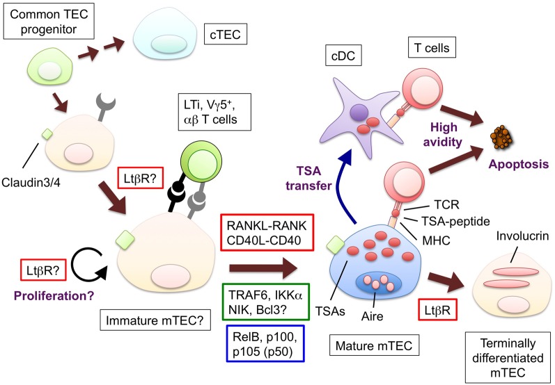
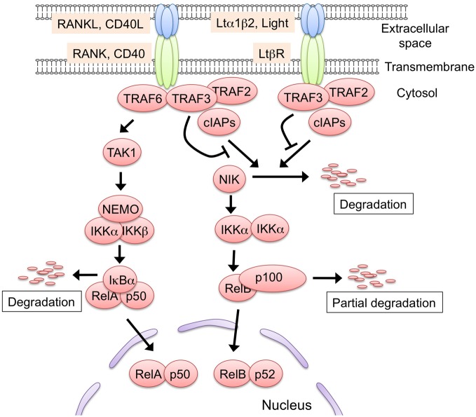
Thymic epithelial cells (TECs) provide the microenvironment required for the development of T cells in the thymus. A unique property of medullary thymic epithelial cells (mTECs) is their expression of a wide range of tissue-restricted self-antigens, critically regulated by the nuclear protein AIRE, which contributes to the selection of the self-tolerant T cell repertoire, thereby suppressing the onset of autoimmune diseases. The TNF receptor family (TNFRF) protein receptor activator of NF-κB (RANK), CD40 and lymphotoxin β receptor (LtβR) regulate the development and functions of mTECs. The engagement of these receptors with their specific ligands results in the activation of the NF-κB family of transcription factors. Two NF-κB activation pathways, the classical and non-classical pathways, promote the development of mature mTECs induced by these receptors. Consistently, TNF receptor-associated factor (TRAF6), the signal transducer of the classical pathway, and NF-κB inducing kinase (NIK), the signal transducer of the non-classical pathway, are essential for the development of mature mTECs. This review summarizes the current understanding of how the signaling by the TNF receptor family controls the development and functions of mTEC.

Keywords: NF-κB; TNF receptor family; autoimmune disease; medullary thymic epithelial cells; self-tolerance; signal transduction.


Figures

Figure 1
Figure 1
The hypothetical model for the development and functions of mTECs and molecules to promote the mTEC development. Bipotent common progenitors give rise to the mTEC progenitors expressing claudin-3 and -4. Lymphoid tissue inducer (LTi), Vγ5+ DETC progenitor (Vγ5+) or positively selected α β T cells produce RANKL; CD40L is supplied only by α β T cells. The interaction between RANKL and RANK promotes the differentiation and/or proliferation of the mTECs. The similar contribution of the interaction of CD40L and CD40 begins at the neonatal stage. RANK and CD40 signaling results in the translocation of the NF-κB family member RelB through the activation of signal transducers TRAF6 and NIK. The mature mTECs express a wide variety of tissue-specific antigens (TSAs) and AIRE. The TSAs are in part transferred to conventional DCs; as a result, both the mTECs and DCs would display the TSA-peptides to the developing T cells. The T cells expressing TCR bound to the complex of the TSA-peptide and MHC molecules with high avidity undergo apoptosis. Mature mTECs might further differentiate into involucrin-expressing mTECs in the LtβR and AIRE-dependent process. The LtβR signaling might regulate the up-regulation of RANK in the TECs and/or enhance the proliferation of the RANK-expressing TECs. Ligands and receptors involved in each process are surrounded by red line rectangles, signal transducers are by green line rectangles, transcription factors are by blue line rectangles.
Figure 2
Figure 2
The two NF-κB activation pathways induced by RANK, CD40, and Ltβ R signaling. Either RANK, CD40, or LtβR signaling is capable of activating the non-classical NF-κB pathway. In the steady state, the protein complex consisting of TRAF2, TRAF3, and cIAP1/2 binds to and poly-ubiquitinates NF-κB inducing kinase (NIK). The poly-ubiquitinated NIK is immediately degraded by the 26S-proteasome machinery. The engagement of the receptors with their ligands recruits the TRAF2, TRAF3, and cIAP1/2 complex, leading to the release of NIK from the complex. The stabilized NIK activates the IKK α complex by phosphorylation, which in turn phosphorylates p100, thus sequestering RelB in the cytoplasm. Subsequently, p100 is poly-ubiquitinated and converted to p52 by partial degradation, an event that leads to the nuclear translocation of the active RelB/p52 complex. The RANKL-RANK and CD40L-CD40 interaction recruits TRAF6 to their cytoplasmic tails. TRAF6 activates the downstream serine/threonine kinase, typically TGF-β activating kinase 1 (TAK1), and activated TAK1 causes the activation of the IKK complex, which consists of IKK α, IKK β, and NEMO. Thereafter, the activated IKK complex phosphorylates IkB α, thereby inducing the degradation of IkB α to lead to the nuclear translocation of the RelA complex. The TRAF6-mediated NF-κB activation might induce the expression of RelB. Although LtβR also can activate the classical pathway, there is no supporting evidence to date that the LtβR-mediated classical pathway regulates mTEC development. Such protein modifications as phosphorylation and ubiquitination are omitted in this figure for simplicity.

References

    1. Aggarwal B. (2003). Signalling pathways of the TNF superfamily: a double-edged sword. Nat. Rev. Immunol. 3, 745–756 10.1038/nri1184 - DOI - PubMed
    1. Aggarwal B. B., Gupta S. C., Kim J. H. (2012). Historical perspectives on tumor necrosis factor and its superfamily: 25 years later, a golden journey. Blood 119, 651–665 10.1182/blood-2011-04-325225 - DOI - PMC - PubMed
    1. Akiyama T., Maeda S., Yamane S., Ogino K., Kasai M., Kajiura F., Matsumoto M., Inoue J. (2005). Dependence of self-tolerance on TRAF6-directed development of thymic stroma. Science 308, 248–251 10.1126/science.1105677 - DOI - PubMed
    1. Akiyama T., Shimo Y., Yanai H., Qin J., Ohshima D., Maruyama Y., Asaumi Y., Kitazawa J., Takayanagi H., Penninger J., Matsumoto M., Nitta T., Takahama Y., Inoue J. (2008). The tumor necrosis factor family receptors RANK and CD40 cooperatively establish the thymic medullary microenvironment and self-tolerance. Immunity 29, 423–437 10.1016/j.immuni.2008.06.015 - DOI - PubMed
    1. Anderson D. M., Maraskovsky E., Billingsley W. L., Dougall W. C., Tometsko M. E., Roux E. R., Teepe M. C., DuBose R. F., Cosman D., Galibert L. (1997). A homologue of the TNF receptor and its ligand enhance T-cell growth and dendritic-cell function. Nature 390, 175–179 10.1038/36593 - DOI - PubMed