

Posterior semicircular canal benign paroxysmal positional vertigo presenting with torsional downbeating nystagmus: an apogeotropic variant
- PMID: 22969807
- PMCID: PMC3434409
- DOI: 10.1155/2012/413603
Posterior semicircular canal benign paroxysmal positional vertigo presenting with torsional downbeating nystagmus: an apogeotropic variant
Abstract
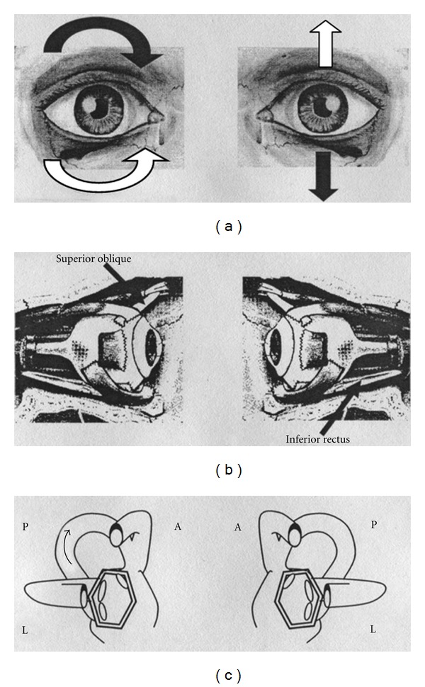
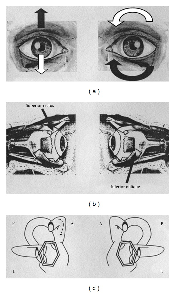
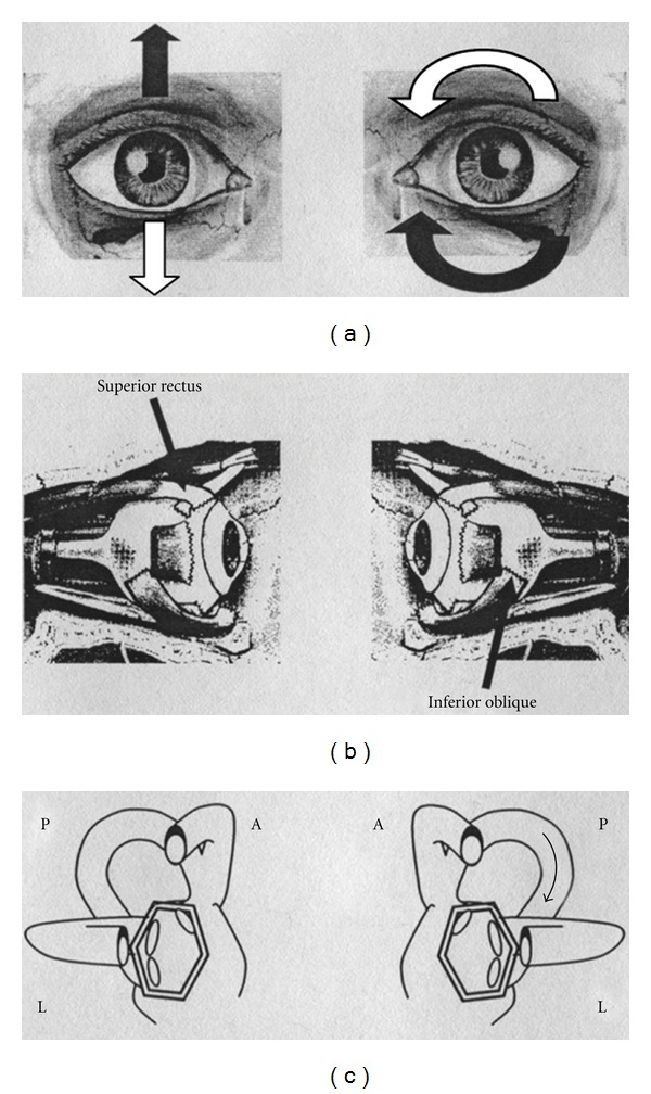
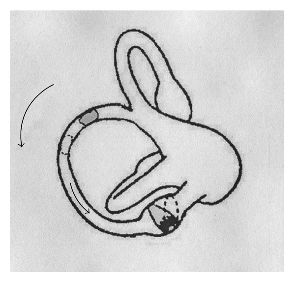
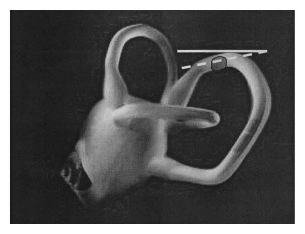
The aim of this study is to verify the hypothesis that free-floating particles could sometimes localize into the distal portion of the non ampullary arm of the posterior semicircular canal (PSC) so that assuming the Dix-Hallpike's positions, the clot could move towards the ampulla eliciting a inhibitory torsional-down beating paroxysmal positional nystagmus (PPNy), instead of typical excitatory torsional-up beating PPNy. Among 45 patients with vestibular signs suggesting anterior semicircular canal paroxysmal positional vertigo (PPV), collected from February 2003 to August 2006, we detected a group of 6 subjects whose clinical findings showed a singular behaviour during follow-up. At the first check-up, all patients were submitted to different types of physical manoeuvres for ASC canalolithiasis. Patients were controlled during the same session and after one week. When we found that nystagmus was qualitatively changed we adopted the appropriate physical therapies for that sign. At a next check-up, after having performed some physical therapies, all patients had a typical PSC PPNy of the opposite side, with respect to that of the ASC initially diagnosed. Basing on these observations we conclude that PSC PPV, similarly to lateral semicircular canal PPV, could manifests in a apogeotropic variant.
Figures
Similar articles
-
Congruous Torsional Down Beating Nystagmus in the Third Position of the Semont's Maneuver in Patients Treated for Canalithiasis of Posterior Semicircular Canal Benign Paroxysmal Positional Vertigo: Its Significance and Prognostic Value.Front Neurol. 2020 Sep 15;11:949. doi: 10.3389/fneur.2020.00949. eCollection 2020. Front Neurol. 2020. PMID: 33071926 Free PMC article.
-
Apogeotropic Posterior Semicircular Canal Benign Paroxysmal Positional Vertigo: Some Clinical and Therapeutic Considerations.Audiol Res. 2015 Mar 31;5(1):130. doi: 10.4081/audiores.2015.130. eCollection 2015 Jan 21. Audiol Res. 2015. PMID: 26557364 Free PMC article.
-
Apogeotropic Posterior Semicircular Canal BPPV-A Case Series from South Rajasthan.Ann Indian Acad Neurol. 2023 Nov-Dec;26(6):989-993. doi: 10.4103/aian.aian_706_23. Epub 2023 Sep 27. Ann Indian Acad Neurol. 2023. PMID: 38229625 Free PMC article.
-
Classification, diagnostic criteria and management of benign paroxysmal positional vertigo.Auris Nasus Larynx. 2017 Feb;44(1):1-6. doi: 10.1016/j.anl.2016.03.013. Epub 2016 May 9. Auris Nasus Larynx. 2017. PMID: 27174206 Review.
-
Benign paroxysmal positional vertigo and its variants.Handb Clin Neurol. 2016;137:241-56. doi: 10.1016/B978-0-444-63437-5.00018-2. Handb Clin Neurol. 2016. PMID: 27638076 Review.
Cited by
-
Case report: Atypical patterns of nystagmus suggest posterior canal cupulolithiasis and short-arm canalithiasis.Front Neurol. 2022 Oct 10;13:982191. doi: 10.3389/fneur.2022.982191. eCollection 2022. Front Neurol. 2022. PMID: 36299265 Free PMC article.
-
Incidence and Clinical Significance of Positional Downbeat Nystagmus in Posterior Canal Benign Paroxysmal Positional Vertigo.J Clin Neurol. 2019 Apr;15(2):143-148. doi: 10.3988/jcn.2019.15.2.143. Epub 2018 May 31. J Clin Neurol. 2019. PMID: 29856161 Free PMC article.
-
Downbeat nystagmus: a clinical and pathophysiological review.Front Neurol. 2024 May 24;15:1394859. doi: 10.3389/fneur.2024.1394859. eCollection 2024. Front Neurol. 2024. PMID: 38854962 Free PMC article. Review.
-
Natural course of positional down-beating nystagmus of peripheral origin.J Neurol. 2013 Jun;260(6):1489-96. doi: 10.1007/s00415-012-6815-9. Epub 2013 Jan 5. J Neurol. 2013. PMID: 23292207 Clinical Trial.
-
A New Variant of Posterior Canal Benign Paroxysmal Positional Vertigo: A Nonampullary or Common Crus Canalolithiasis.Case Rep Otolaryngol. 2015;2015:816081. doi: 10.1155/2015/816081. Epub 2015 May 31. Case Rep Otolaryngol. 2015. PMID: 26114003 Free PMC article.
References
-
- Hall SF, Ruby RRF, McClure JA. The mechanics of benign paroxysmal vertigo. Journal of Otolaryngology. 1979;8(2):151–158. - PubMed
-
- Epley JM. New dimensions of benign paroxysmal positional vertigo. Otolaryngology—Head and Neck Surgery. 1980;88(5):599–605. - PubMed
-
- Brandt T, Steddin S. Current view of the mechanism of benign paroxysmal positioning vertigo: cupulolithiasis or canalolithiasis? Journal of Vestibular Research. 1993;3(4):373–382. - PubMed
-
- Mauri S, De Bernardi F, Scotti E, Quaglieri S. La storia della vertigine parossistica posizionale. XVI Giornata Italiana di Otoneurologia, Revisione critica di venti anni di vertigine parossistica posizionale benigna (VPPB '99); March 1999; Sorrento, Italy. pp. 43–54.
-
- Brandt T. Positional and positioning vertigo and nystagmus. Journal of the Neurological Sciences. 1990;95(1):3–28. - PubMed
LinkOut - more resources
Full Text Sources
Miscellaneous
