. 2012 Feb;3(2):165-170.
doi: 10.3892/etm.2011.393. Epub 2011 Nov 28.

Osteogenic differences in cultured rat periosteal cells under hypoxic and normal conditions

Affiliations

Osteogenic differences in cultured rat periosteal cells under hypoxic and normal conditions

Takehiro Ichijima et al. Exp Ther Med. 2012 Feb.

Abstract

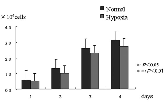
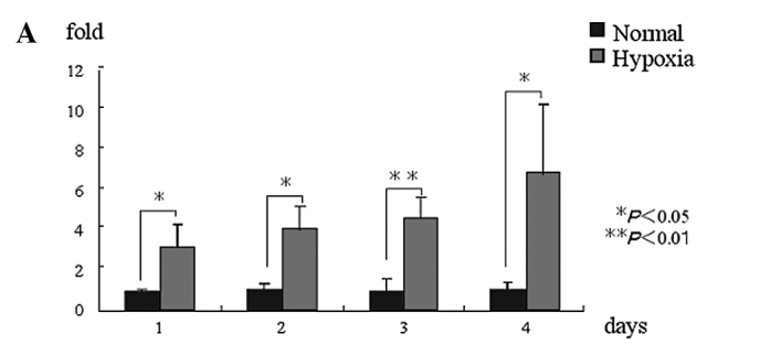
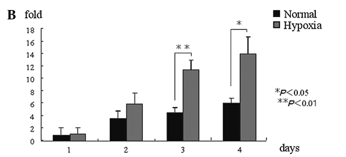
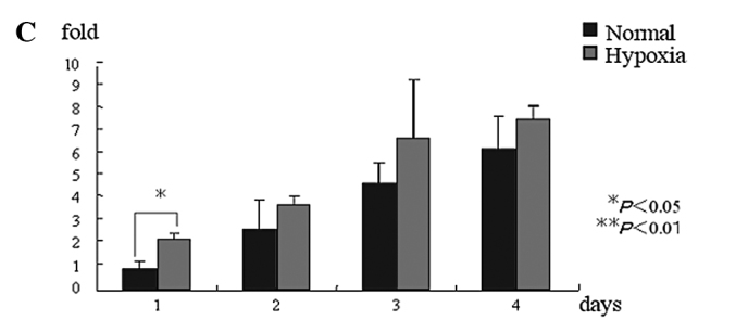
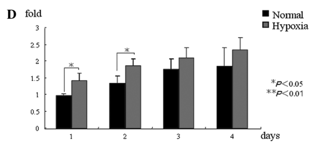
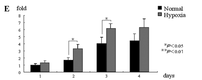
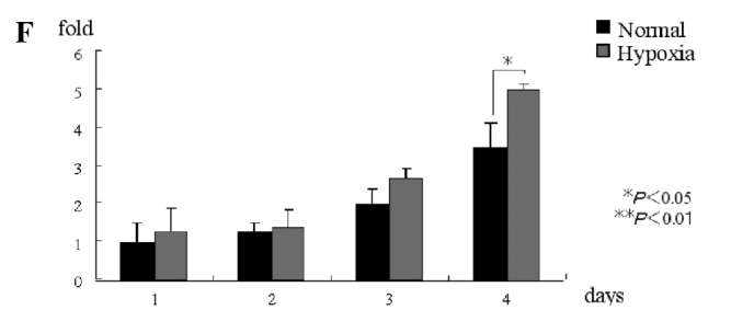
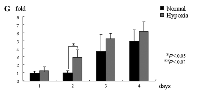
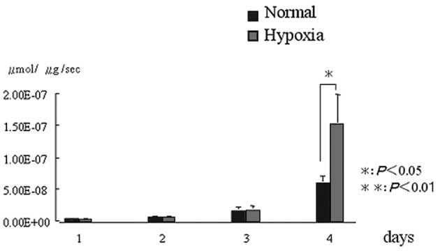
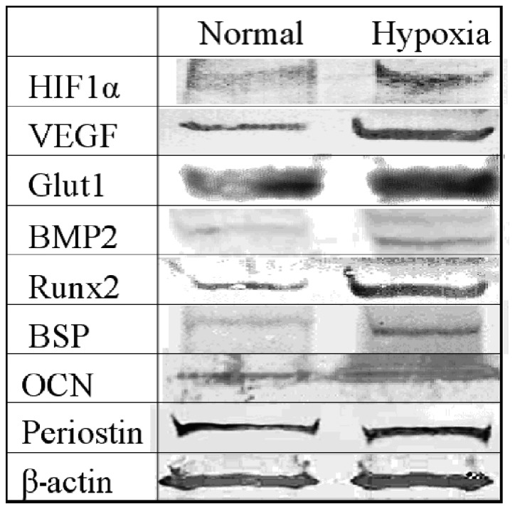
The aim of the present study was to investigate the osteogenic capability of rat calvarial periosteal cells in hypoxic conditions in vitro. Periosteum was obtained from the calvarial bone of Sprague-Dawley rats. Following primary tissue culture, subcultured cells were used in hypoxic or normal conditions. On days 1, 2, 3 and 4 following the cell culture, cell proliferation and mRNA and protein expression levels were evaluated. No significant difference in the cell proliferation rate was found between the normal and hypoxic condition groups. The hypoxic condition group exhibited a stronger expression of hypoxia-inducible factor (HIF)1α, vascular endothelial growth factor (VEGF), Runx2, alkaline phosphatase (ALP), bone sialoprotein (BSP), osteocalcin (OCN) and periostin at the mRNA level compared to that of the normal condition group. The hypoxic condition group also exhibited a stronger expression of HIF1α, VEGF, bone morphogenetic protein (BMP)2, Runx2, ALP and BSP at the protein level compared to that of the normal condition group. In conclusion, periosteal cells cultured in hypoxic conditions demonstrated activated osteogenic capability in vitro.


Figures

Figure 1.
Figure 1.
Cell proliferation rate. No difference was noted between the hypoxic and normal condition groups at any of the time-periods.
Figure 2.
Figure 2.
mRNA expression. (A) HIF1α mRNA expression in the hypoxic condition group was significantly higher than that in the normal condition group at all of the time-periods. (B) VEGF mRNA expression in the hypoxic condition group was significantly higher than that in the normal condition group on days 2 and 3, but was not significantly different at other time-periods. (C) Runx2 mRNA expression in the hypoxic condition group was significantly higher than that in the normal condition group on day 1, but was not significantly different at other time-periods. (D) ALP mRNA expression in the hypoxic condition group was significantly higher than that in the normal condition group on days 1 and 2, but was not significantly different at other time-periods. (E) BSP mRNA expression in the hypoxic condition group was significantly higher than that in the normal condition group on days 2 and 3, but was not significantly different at other time-periods. (F) OCN mRNA expression in the hypoxic condition group was significantly higher than that in the normal condition group on day 4, but was not significantly different at other time-periods. (G) Periostin mRNA expression in the hypoxic condition group was significantly higher than that in the normal condition group on day 2, but was not significantly different at other time-periods. *P<0.05, **P<0.01. HIF1α, hypoxia-inducible factor; VEGF, vascular endothelial growth factor; ALP, alkaline phosphatase; BSP, bone sialoprotein; OCN, osteocalcin.
Figure 2.
Figure 2.
mRNA expression. (A) HIF1α mRNA expression in the hypoxic condition group was significantly higher than that in the normal condition group at all of the time-periods. (B) VEGF mRNA expression in the hypoxic condition group was significantly higher than that in the normal condition group on days 2 and 3, but was not significantly different at other time-periods. (C) Runx2 mRNA expression in the hypoxic condition group was significantly higher than that in the normal condition group on day 1, but was not significantly different at other time-periods. (D) ALP mRNA expression in the hypoxic condition group was significantly higher than that in the normal condition group on days 1 and 2, but was not significantly different at other time-periods. (E) BSP mRNA expression in the hypoxic condition group was significantly higher than that in the normal condition group on days 2 and 3, but was not significantly different at other time-periods. (F) OCN mRNA expression in the hypoxic condition group was significantly higher than that in the normal condition group on day 4, but was not significantly different at other time-periods. (G) Periostin mRNA expression in the hypoxic condition group was significantly higher than that in the normal condition group on day 2, but was not significantly different at other time-periods. *P<0.05, **P<0.01. HIF1α, hypoxia-inducible factor; VEGF, vascular endothelial growth factor; ALP, alkaline phosphatase; BSP, bone sialoprotein; OCN, osteocalcin.
Figure 3.
Figure 3.
Alkaline phosphatase (ALP) activity. Compared to the normal condition group, the hypoxic condition group demonstrated a higher expression of ALP activity on day 4. *P<0.05, **P<0.01
Figure 4.
Figure 4.
Protein expression. We carried out an evaluation of the normal and the hypoxic condition group on day 4 after the incubation. HIF1α, VEGF, BMP2, Runx2 and BSP were all expressed more strongly in the hypoxic condition cell group than in the normal condition group. Glut1, OCN and periostin were all expressed in both the normal and hypoxic condition groups. HIF1α, hypoxia-inducible factor; VEGF, vascular endothelial growth factor; BMP2, bone morphogenetic protein 2; ALP, alkaline phosphatase; BSP, bone sialoprotein; OCN, osteocalcin.

Similar articles

Cited by

References

    1. Zheng Y, Ringe J, Liang Z, et al. Osteogenic potential of human periosteum-derived progenitor cells in PLGA scaffold using allogeneic serum. J Zhejiang Univ. 2006;7:817–824. - PMC - PubMed
    1. Hayashi O, Katsube Y, Hirose M, et al. Comparison of osteogenic ability of rat mesenchymal stem cells from bone marrow, periosteum, and adipose tissue. Calcif Tissue Int. 2008;82:238–247. - PubMed
    1. Gallie WE, Robertson DE. The periosteum. Can Med Assoc J. 1914;4:33–36. - PMC - PubMed
    1. Okuda K, Tanabe H, Suzuki K, et al. Platelet-rich plasma combined with a porous hydroxyapatite graft for the treatment of intrabony periodontal defects in humans:a comparative controlled clinical study. J Periodontal Res. 2005;76:890–898. - PubMed
    1. Yamamiya K, Okuda K, Kawase T, et al. Tissue-engineered cultured periosteum sheets combined with platelet-rich plasma and porous hydroxyapatite graft in treating human osseous defects. J Periodontol. 2008;79:811–818. - PubMed

LinkOut - more resources