. 2012;7(8):e44285.
doi: 10.1371/journal.pone.0044285. Epub 2012 Aug 27.

Stromal-to-epithelial transition during postpartum endometrial regeneration

Affiliations

Stromal-to-epithelial transition during postpartum endometrial regeneration

Cheng-Chiu Huang et al. PLoS One. 2012.

Abstract

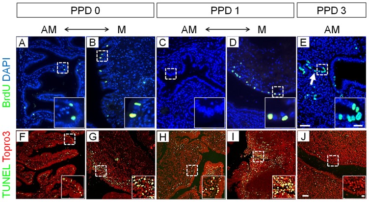
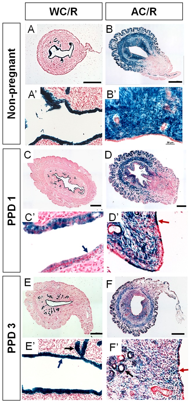
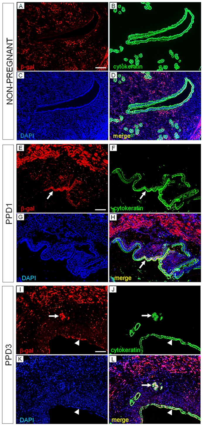
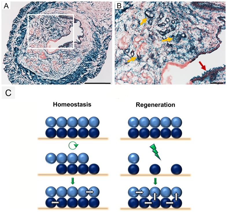
Endometrium is the inner lining of the uterus which is composed of epithelial and stromal tissue compartments enclosed by the two smooth muscle layers of the myometrium. In women, much of the endometrium is shed and regenerated each month during the menstrual cycle. Endometrial regeneration also occurs after parturition. The cellular mechanisms that regulate endometrial regeneration are still poorly understood. Using genetic fate-mapping in the mouse, we found that the epithelial compartment of the endometrium maintains its epithelial identity during the estrous cycle and postpartum regeneration. However, whereas the stromal compartment maintains its identity during homeostatic cycling, after parturition a subset of stromal cells differentiates into epithelium that is subsequently maintained. These findings identify potential progenitor cells within the endometrial stromal compartment that produce long-term epithelial tissue during postpartum endometrial regeneration.


Conflict of interest statement

Competing Interests: The authors have declared that no competing interests exist.

Figures

Figure 1
Figure 1. Cellular dynamics during endometrial regeneration after parturition.
Cell proliferation assayed by BrdU labeling (A–E) and cell death assayed by TUNEL (F–J) in the postpartum endometrium. AM: anti-mesometrial region of uterus; M: mesometrial region; PPD 0, 6 hours after parturition; PPD 1, 30 hours after parturition; PPD 3, 72 hours after parturition. Boxed areas are high-magnification images from highlighted zones in A to J. Scale bars  = 50 µm in A–J; 10 µm in all boxed areas.
Figure 2
Figure 2. Stromal-to-epithelial transition in the postpartum endometrium.
β-gal stained uterine cross sections from non-pregnant and postpartum (PPD 1 and 3) Wnt7a-Cre; R26R-lacZ (WC/R) and Amhr2-Cre; R26R-lacZ (AC/R) mice. (A and A′) Non-prenant WC/R female (n = 5). All luminal and glandular epithelia are β-gal positive but the stroma and myometrium are negative. (B and B′) Non-pregnant AC/R female (n = 5). The stroma is β-gal positive but the epithelia are negative. (C and C′), PPD 1 in WC/R mice (n = 4) is similar to the non-pregnant WC/R female except that there are regions of the luminal epithelium that are β-gal negative (blue arrows). (D and D′), PPD 1 in AC/R mice (n = 5) is similar to the non-pregnant female except there are regions of the luminal epithelium that are β-gal positive (red arrow). (E and E′), PPD 3 in WC/R mice (n = 4) appears the same as non-pregnant, only luminal and glandular epithelial tissues are β-gal positive. (F and F′), PPD 3 in AC/R mice (n = 5) appears similar to PPD1 with stroma-derived luminal epithelium (red arrow) except that there are also stroma-derived glandular epithelium (black arrow). Scale bars  = 500 µm in A–F; 50 µm in A′–F′.
Figure 3
Figure 3. Stroma-derived luminal and glandular epithelium express the cytokeratin epithelial marker.
β-gal and cytokeratin double immunofluorescence labeling of Amhr2-Cre; R26R virgin and postpartum endometrium. (A–D) In non-pregnant uteri, β-gal immunoreactivity is specifically detected in the endometrial stroma and muscle cells. Luminal epithelial (LE) and glandular epithelial (GE) cells are labeled with cytokeratin immunofluorescence (E–H) In PPD 1 uteri, strong β-gal immunosignal is detected in the LE (white arrows) and co-localize with cytokeratin staining (I–L) At PPD3, β-gal-marked stroma-derived GE and LE cells express cytokeratin (white arrows: GE; white arrow heads: LE). Scale bars  = 50 µm.
Figure 4
Figure 4. Persistence of stroma-derived epithelial cells and current working models of uterine homeostasis and regeneration.
(A) Stroma-derived epithelial cells are detected in the endometrium of Amhr2-Cre; R26R mice more than two months after parturition. Scale bar  = 100 µm. (B) Higher magnification of Figure 4A, showing stroma-derived luminal (red arrow) and glandular (yellow arrows) epithelium. Scale bar  = 100 µm. (C) Models of uterine homeostasis and postpartum regeneration. A surface epithelial sheet (light blue) and stroma (dark blue) represent the endometrial tissue compartments. Cells lost during estrous cycles are replaced by new cells generated from the same cell compartments. Endometrial damage caused by parturition causes loss of both epithelium and stroma. In addition to stromal cells generating more stroma, some stromal cells sense and respond to the damage and initiate the regeneration process by differentiating into epithelium.

Similar articles

Cited by

References

    1. Roy A, Matzuk MM (2011) Reproductive tract function and dysfunction in women. Nat Rev Endocrinol 7: 517–525. - PubMed
    1. Bulun SE (2010) Endometriosis. N Engl J Med 360: 268–279. - PubMed
    1. Amant F, Moerman P, Neven P, Timmerman D, Van Limbergen E, et al. (2005) Endometrial cancer. Lancet 366: 491–505. - PubMed
    1. Dharma SJ, Kholkute SD, Nandedkar TD (2001) Apoptosis in endometrium of mouse during estrous cycle. Indian J Exp Biol 39: 218–222. - PubMed
    1. Wood GA, Fata JE, Watson KL, Khokha R (2007) Circulating hormones and estrous stage predict cellular and stromal remodeling in murine uterus. Reproduction 133: 1035–1044. - PubMed

Publication types

MeSH terms

Substances