. 2012;7(9):e43454.
doi: 10.1371/journal.pone.0043454. Epub 2012 Sep 6.

A fluorescent, genetically-encoded voltage probe capable of resolving action potentials

Affiliations

A fluorescent, genetically-encoded voltage probe capable of resolving action potentials

Lauren Barnett et al. PLoS One. 2012.

Abstract

There is a pressing need in neuroscience for genetically-encoded, fluorescent voltage probes that can be targeted to specific neurons and circuits to allow study of neural activity using fluorescent imaging. We created 90 constructs in which the voltage sensing portion (S1-S4) of Ciona intestinalis voltage sensitive phosphatase (CiVSP) was fused to circularly permuted eGFP. This led to ElectricPk, a probe that is an order of magnitude faster (taus ~1-2 ms) than any currently published fluorescent protein-based voltage probe. ElectricPk can follow the rise and fall of neuronal action potentials with a modest decrease in fluorescence intensity (~0.7% ΔF/F). The probe has a nearly linear fluorescence/membrane potential response to both hyperpolarizing and depolarizing steps. This is the first probe based on CiVSP that captures the rapid movements of the voltage sensor, suggesting that voltage probes designed with circularly permuted fluorescent proteins may have some advantages.


Conflict of interest statement

Competing Interests: The authors have the following conflicts: Funding for this study came in part from The John B. Pierce Laboratory, Inc. Authors Jelena Platisa and Vincent A. Pieribone are employees of The John B. Pierce Laboratory, Inc. There are no other relevant declarations relating to employment, consultancy, patents, products in development or marketed products. This does not alter the authors' adherence to all the PLoS ONE policies on sharing data and materials.

Figures

Figure 1
Figure 1. Probe design, expression level and ΔF/ΔV sensitivity of CiVSP::cpEGFP constructs.
Two variables were systematically explored: 1) the amino acid position within CiVSP in which the cpEGFP was fused (horizontal axis, e.g.1.x), and 2) the size and position (vertical axis, e.g. x.1) of the hole in the cpEGFP. Gray numbers are construct number. The green shaded boxes within the plot indicate probes that produced fluorescence in HEK 293 cells. Bold numbers in boxes represent average values of peak %ΔF/F ± SEM for +100 mV/200 ms steps from a holding potential of −70 mV; NS = no detectable ΔF/F signal; Red numbers in boxes indicate reversed responses (increase in fluorescence) to depolarizing steps. The response kinetics of constructs 9.1,10.1,5.5,6.5,1.6,2.6,8.6,6.9,7.9 was either non existent or too small to accurately measure. Constructs 7.7 and 8.7 exhibited relatively slow (τ∼69 ms) on and off kinetics. Constructs 10.5,1.7,2.7, 4.7,5.7,1.8,10.8,2.9,4.9,5.9,8.9,9.9,10.9 had on and off rates that were dominated by extremely fast components (τ∼2 ms).
Figure 2
Figure 2. Schematic diagram of CiVSP::cpEGFP based construct design.
The S1–4 domain of Ciona intestinalis voltage sensitive phosphatase (yellow) is fused to circularly permuted EGFP (green).
Figure 3
Figure 3. The protein sequence of the various fusion sites of cpEGFP with CiVSP described in this report.
From top: CiVSP sequence includes S4 domain (bold cyan), linker domain (cyan and black) and phosphatase domain (red). The fusion sites and linker sequences of VSFP 2.1, 3.1 (17, 18) and Mermaid (4) probes. The ten different fusion sites and linkers described in this report (pLB1.x-10.x; see Figure 1). The fusion site is identified by the last CiVSP amino acid present in the probe (bold red). In all cases, this residue is followed by a five amino acid linker (purple) and the cpEGFP (green). The amino acid at the beginning of the cpEGFP depends on the hole size and position of the cpEGFP (See Figure 1).
Figure 4
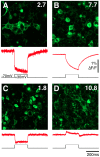
Figure 4. Examples of CiVSP::cpEGFP constructs expression in HEK293 cells and voltage sensitivity.
In all cases the upper panel is a confocal image of HEK293 cells transiently expressing the construct and the lower panel is the average fluorescence response (red trace) to ten +100 mV/200 ms voltage steps applied under whole-cell patch clamp configuration. Correction for FP photobleaching has been removed by division of a double exponential fit to the portions of the trace outside the effects of the voltage step. A) The ElectricPk (pLB 2.7) probe is localized both in the membrane and intracellularly. The probe exhibits a rapid decrease in fluorescence with a relatively low signal to noise ratio due to relatively weak basal fluorescence. B) The high expression levels of construct pLB7.7 produces a relatively high signal to noise ratio, however much of the fluorescent protein is localized intracellularly, and its fluorescence response is dominated by a slow component. C) Construct pLB1.8 has mixed membrane and intracellular distribution that produces a small and rapid negative response with a moderate signal to noise ratio. D) pLB10.8 has predominantly intracellular localization and a small positive fluorescence response with a moderate signal to noise ratio.
Figure 5
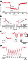
Figure 5. Voltage sensitivity and response kinetics of ElectricPk.
A) Voltage-dependent fluorescence sensitivity of an HEK293 cell when transiently expressing ElectricPK (red trace) verses mUKG in Mermaid (upper black trace) tested with voltage steps of +100 mV/200 ms from a −70 mV holding potential (lower black trace). Fluorescence, in all cases, is normalized to initial and peak level. B) Single exponential fits to the dominant on and off response rates of ElectricPk and mUKG of Mermaid in cells tested as in A. C) and D) Response of HEK 293 cell expressing ElectricPk (red trace) to a trains of voltage steps of +100 mV from a −70 mV holding potential with a variable duration; 100 ms, 3 ms and 1 ms (black trace).
Figure 6
Figure 6. Linear ΔF/ΔV response of ElectricPK.
A) Fluorescence response (upper traces) of HEK293 cells transiently expressing ElectricPk to depolarizing and hyperpolarizing voltage steps (−170 to +30 mV from a −70 mV holding potential, lower traces). B) ΔF/ΔV curve of ElectricPk derived from data presented in A.
Figure 7
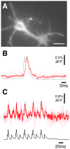
Figure 7. Detection of action potentials in hippocampal neurons in vitro using ElectricPK.
A) Wide field image of an in vitro hippocampal neuron expressing ElectricPk. Bar = 15 µm. B) Single (light red trace) and averaged (red trace) optical response to action potentials evoked in the neuron seen in (A) taken using wide field microscopy and a RedShirtImaging NeuroCCD camera. The red trace is an average of 32 action potentials. All responses captured at 2000 fps. C) Fluorescence change (light red trace-unfiltered, red trace-filtered) to a single train of evoked action potentials recorded from an in vitro hippocampal neuron expressing ElectricPk. Lower black trace is the voltage recording made from the patch electrode. All fluorescence traces are bleach subtracted and where indicated, low pass filtered (Bessel) at 350 Hz.

Similar articles

Cited by

References

    1. Siegel MS, Isacoff EY (1997) A genetically encoded optical probe of membrane voltage. Neuron 19: 735–741. - PubMed
    1. Sakai R, Repunte-Canonigo V, Raj CD, Knopfel T (2001) Design and characterization of a DNA-encoded, voltage-sensitive fluorescent protein. Eur J Neurosci 13: 2314–2318. - PubMed
    1. Ataka K, Pieribone VA (2002) A genetically targetable fluorescent probe of channel gating with rapid kinetics. Biophys J 82: 509–516. - PMC - PubMed
    1. Tsutsui H, Karasawa S, Okamura Y, Miyawaki A (2008) Improving membrane voltage measurements using FRET with new fluorescent proteins. Nat Methods 5: 683–685. - PubMed
    1. Baker BJ, Mutoh H, Dimitrov D, Akemann W, Perron A, et al. (2008) Genetically encoded fluorescent sensors of membrane potential. Brain Cell Biol 36: 53–67. - PMC - PubMed

Publication types

Substances