. 2012;7(9):e43722.
doi: 10.1371/journal.pone.0043722. Epub 2012 Sep 7.

Porphyromonas gingivalis FimA fimbriae: fimbrial assembly by fimA alone in the fim gene cluster and differential antigenicity among fimA genotypes

Affiliations

Porphyromonas gingivalis FimA fimbriae: fimbrial assembly by fimA alone in the fim gene cluster and differential antigenicity among fimA genotypes

Keiji Nagano et al. PLoS One. 2012.

Abstract

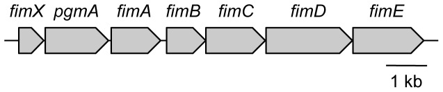
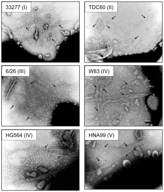
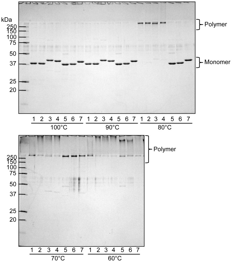
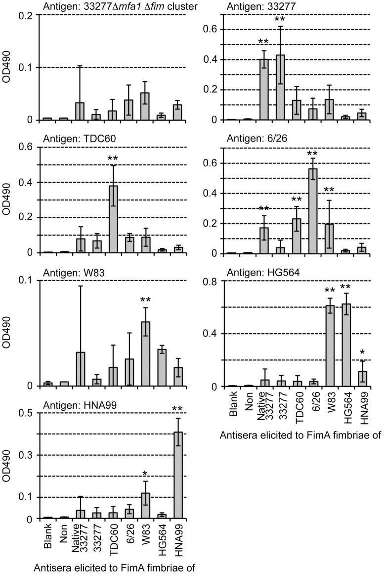
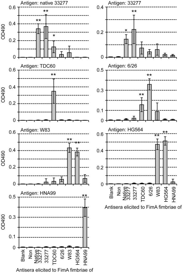
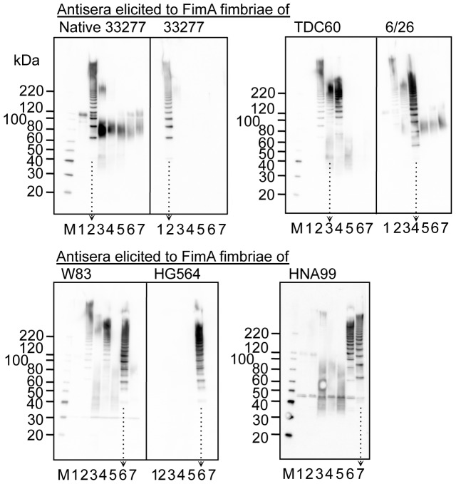
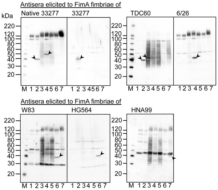
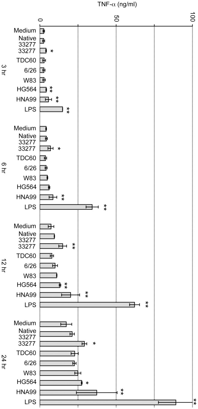
The periodontal pathogen Porphyromonas gingivalis colonizes largely through FimA fimbriae, composed of polymerized FimA encoded by fimA. fimA exists as a single copy within the fim gene cluster (fim cluster), which consists of seven genes: fimX, pgmA and fimA-E. Using an expression vector, fimA alone was inserted into a mutant from which the whole fim cluster was deleted, and the resultant complement exhibited a fimbrial structure. Thus, the genes of the fim cluster other than fimA were not essential for the assembly of FimA fimbriae, although they were reported to influence FimA protein expression. It is known that there are various genotypes for fimA, and it was indicated that the genotype was related to the morphological features of FimA fimbriae, especially the length, and to the pathogenicity of the bacterium. We next complemented the fim cluster-deletion mutant with fimA genes cloned from P. gingivalis strains including genotypes I to V. All genotypes showed a long fimbrial structure, indicating that FimA itself had nothing to do with regulation of the fimbrial length. In FimA fimbriae purified from the complemented strains, types I, II, and III showed slightly higher thermostability than types IV and V. Antisera of mice immunized with each purified fimbria principally recognized the polymeric, structural conformation of the fimbriae, and showed low cross-reactivity among genotypes, indicating that FimA fimbriae of each genotype were antigenically different. Additionally, the activity of a macrophage cell line stimulated with the purified fimbriae was much lower than that induced by Escherichia coli lipopolysaccharide.


Conflict of interest statement

Competing Interests: The authors have declared that no competing interests exist.

Figures

Figure 1
Figure 1. P. gingivalis fim gene cluster.
We constructed a mutant with the whole region of the fim gene cluster from fimX through fimE deleted. The schema was drawn on the basis of ATCC 33277. However, genome-analyzed strains of W83 and TDC60 show that they are the same as that of ATCC 33277 in the gene arrangement.
Figure 2
Figure 2. Immunoblot analysis for FimA using whole-cell sonicates.
Whole-cell sonicates were denatured in an SDS-containing buffer with 2-mercaptoethanol by heating at 100°C (upper) and 80°C (lower) for 10 min, then subjected to SDS-PAGE and immunoblot analysis. A mixture of specific antisera to the FimA polymer and monomer was used. Antigen samples were as follows: P. gingivalis ATCC 33277 Δmfa1 (expresses native FimA fimbriae, lane 1), P. gingivalis ATCC 33277 Δmfa1 Δfim cluster (FimA deficient, lane 2) with fimX-pgmA-fimA (lane 3), pgmA-fimA (lane 4), fimA (lane 5), fimX & fimA (lane 6) complementarily introduced, and P. gingivalis W83 Δfim cluster with fimA introduced (lane 7). All introduced genes originated from P. gingivalis ATCC 33277. Incomplete dissociation of FimA polymers produces a ladder-like band indicating oligomers in the lower panel. Note that bands slightly higher than 60 kDa (the lower panel) are dimers although monomers appear to be about 40 kDa (the upper panel).
Figure 3
Figure 3. Transmission electron microscopic observation of FimA fimbriae on the bacterial cell surface.
P. gingivalis ATCC 33277 Δmfa1 Δfim cluster cells with fimA from 33277, TDC60, 6/26, W83, HG564 and HNA99 introduced by using an expression vector. Samples were negatively stained with 1% ammonium molybdate. Arrows indicate fimbrial structure. Some fimbriae appear to be bundled. Bars show 0.2 µm.
Figure 4
Figure 4. Immunoblot analysis for PgmA using whole-cell sonicates.
Whole-cell sonicates (W) were fractionated into soluble (Sol), envelope (Env), inner membrane (IM) and outer membrane (OM) fractions. Samples were denatured in an SDS-containing buffer with 2-mercaptoethanol by heating at 100°C for 10 min, then subjected to SDS-PAGE and immunoblot analysis. Emp denotes 33277 Δmfa1 Δfim cluster/pT-COW::ragAP, carrying empty vector, used as a negative control; 33277 denotes the wild-type strain; Complement denotes 33277 Δmfa1 Δfim cluster carrying pT-COW::ragAP::fimX-pgmA-fimA. An arrow indicates PgmA as a 60-kDa protein. Degradation bands (below the 60-kDa) were also visualized because PgmA was highly sensitive to intrinsic proteases of this bacterium .
Figure 5
Figure 5. SDS-PAGE and CBB staining using purified FimA fimbriae.
Purified FimA fimbriae were denatured in an SDS-containing buffer with 2-mercaptoethanol by heating at 60 to 100°C for 10 min, then subjected to SDS-PAGE and CBB staining. Samples were as follows: purified from P. gingivalis ATCC 33277 Δmfa1 (native 33277 FimA fimbriae, lane 1), P. gingivalis ATCC 33277 Δmfa1 Δfim cluster with fimA of ATCC 33277 (I) (lane 2), TDC60 (II) (lane 3), 6/26 (III) (lane 4), W83 (IV) (lane 5), HG564 (IV) (lane 6), and HNA99 (V) (lane 7) introduced. Note that CBB staining did not visualize a ladder band as seen in immunoblot analysis in Fig. 2.
Figure 6
Figure 6. ELISA using absorbed antisera and whole-cell sonicates as antigen.
Whole-cell sonicates were coated on ELISA plates as antigens. Antisera from mice immunized with each pure genotype fimbriae were used after absorption with the fimbria-deficient mutant 33277 Δmfa1 Δfim cluster. “Non” indicates non-immunized mouse sera. W83 rarely produces FimA protein and fimbriae. Data show mean ± SD. Asterisks indicate statistical significance compared with Non (* p<0.05, ** p<0.01). Note that scales of Y axes are adjusted in order to compare titers clearly.
Figure 7
Figure 7. ELISA using absorbed antisera and purified FimA fimbriae as antigen.
Pure FimA fimbriae, derived from each fimA gene, were coated on ELISA plates as antigens. Antisera from mice immunized with each pure genotype fimbriae were used after absorption with the fimbria-deficient mutant 33277 Δmfa1 Δfim cluster. “Non” indicates non-immunized mouse sera. Data show mean ± SD. Asterisks indicate statistical significance compared with Non (* p<0.05, ** p<0.01). Note that scales of Y axes are adjusted as in Fig. 6.
Figure 8
Figure 8. Immunoblot analysis using whole-cell sonicates partially denatured.
Whole-cell sonicates were denatured in an SDS-containing buffer with 2-mercaptoethanol by heating at 70°C for 10 min, and subjected to SDS-PAGE and immunoblot analysis by using antisera, 1,000-fold dilution, from mice immunized with purified FimA fimbriae. Antigen samples were as follows: P. gingivalis ATCC 33277 Δmfa1 Δfim cluster (FimA deficient, lane 1), and the wild-type strains of ATCC 33277 (lane 2), TDC60 (lane 3), 6/26 (lane 4), W83 (lane 5), HG564 (lane 6), and HNA99 (lane 7). M denotes a standard marker. W83 rarely produces FimA protein and fimbriae. Note that ladder bands are specific for FimA fimbriae whereas smear bands between 40–80 kDa are nonspecific. Arrows with dotted lines are placed in order to clearly discriminate each lane.
Figure 9
Figure 9. Immunoblot analysis using whole-cell sonicates completely denatured.
Whole-cell sonicates were denatured in an SDS-containing buffer with 2-mercaptoethanol by heating at 100°C for 10 min, and subjected to SDS-PAGE and immunoblot analysis using antisera, 1,000-fold dilution, from mice immunized with purified FimA fimbriae. Antigen samples were as follows: P. gingivalis ATCC 33277 Δmfa1 Δfim cluster (FimA deficient, lane 1), and the wild-type strains of ATCC 33277 (lane 2), TDC60 (lane 3), 6/26 (lane 4), W83 (lane 5), HG564 (lane 6), and HNA99 (lane 7). M denotes standard marker. Arrowheads show distinguishable bands corresponding to FimA monomers. Note that W83 rarely produces FimA protein and fimbriae.
Figure 10
Figure 10. TNF-α induction in mouse macrophage-like J774-1 cells.
Purified FimA fimbriae at 1 µg/ml and LPS at 10 EU/ml (corresponding to 2.6 ng/ml) were incubated with J774-1 cells for 3 to 24 hours. TNF-α in the medium was measured by ELISA. Medium denotes no addition; Native 33277 denotes purified FimA fimbriae from 33277 Δmfa1; 33277, TDC60, 6/26, W83, HG564 and HNA99 denote that pure FimA fimbriae, derived from the corresponding fimA in P. gingivalis ATCC 33277 Δmfa1 Δfim cluster, were used stimulants; LPS was E. coli LPS. Data show mean ± SD from two experiments with duplicate. Asterisks indicate statistical significance compared with Medium for each time (* p<0.05, ** p<0.01).

References

    1. Lamont RJ, Jenkinson HF (1998) Life below the gum line: pathogenic mechanisms of Porphyromonas gingivalis . Microbiol Mol Biol Rev 62: 1244–1263. - PMC - PubMed
    1. Yoshimura F, Murakami Y, Nishikawa K, Hasegawa Y, Kawaminami S (2009) Surface components of Porphyromonas gingivalis . J Periodont Res 44: 1–12. - PubMed
    1. Dickinson DP, Kubiniec MA, Yoshimura F, Genco RJ (1988) Molecular cloning and sequencing of the gene encoding the fimbrial subunit protein of Bacteroides gingivalis . J Bacteriol 170: 1658–1665. - PMC - PubMed
    1. Watanabe K, Onoe T, Ozeki M, Shimizu Y, Sakayori T, et al. (1996) Sequence and product analyses of the four genes downstream from the fimbrilin gene (fimA) of the oral anaerobe Porphyromonas gingivalis . Microbiol Immunol 40: 725–734. - PubMed
    1. Nelson KE, Fleischmann RD, DeBoy RT, Paulsen IT, Fouts DE, et al. (2003) Complete genome sequence of the oral pathogenic bacterium Porphyromonas gingivalis strain W83. J Bacteriol 185: 5591–5601. - PMC - PubMed

Publication types

MeSH terms

LinkOut - more resources