. 2012;7(9):e44093.
doi: 10.1371/journal.pone.0044093. Epub 2012 Sep 6.

α-Tomatine-mediated anti-cancer activity in vitro and in vivo through cell cycle- and caspase-independent pathways

Affiliations

α-Tomatine-mediated anti-cancer activity in vitro and in vivo through cell cycle- and caspase-independent pathways

Min-Wu Chao et al. PLoS One. 2012.

Abstract

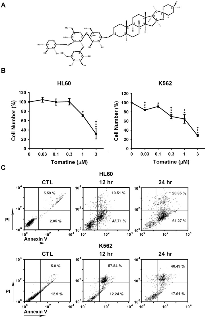
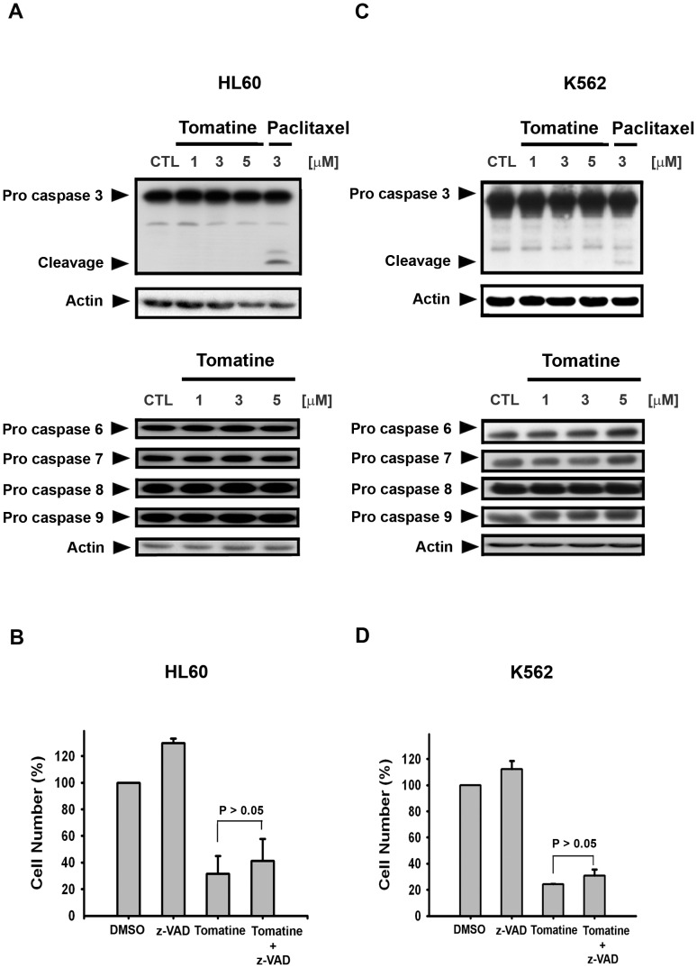
α-Tomatine, a tomato glycoalkaloid, has been reported to possess antibiotic properties against human pathogens. However, the mechanism of its action against leukemia remains unclear. In this study, the therapeutic potential of α-tomatine against leukemic cells was evaluated in vitro and in vivo. Cell viability experiments showed that α-tomatine had significant cytotoxic effects on the human leukemia cancer cell lines HL60 and K562, and the cells were found to be in the Annexin V-positive/propidium iodide-negative phase of cell death. In addition, α-tomatine induced both HL60 and K562 cell apoptosis in a cell cycle- and caspase-independent manner. α-Tomatine exposure led to a loss of the mitochrondrial membrane potential, and this finding was consistent with that observed on activation of the Bak and Mcl-1 short form (Mcl-1s) proteins. Exposure to α-tomatine also triggered the release of the apoptosis-inducing factor (AIF) from the mitochondria into the nucleus and down-regulated survivin expression. Furthermore, α-tomatine significantly inhibited HL60 xenograft tumor growth without causing loss of body weight in severe combined immunodeficiency (SCID) mice. Immunohistochemical test showed that the reduced tumor growth in the α-tomatine-treated mice was a result of increased apoptosis, which was associated with increased translocation of AIF in the nucleus and decreased survivin expression ex vivo. These results suggest that α-tomatine may be a candidate for leukemia treatment.


Conflict of interest statement

Competing Interests: The authors have declared that no competing interests exist.

Figures

Figure 1
Figure 1. α-Tomatine-induced apoptosis in human leukemia cell lines.
(A) The chemical structure of α-tomatine. (B) Cells were treated with or without α-tomatine for 24 hr, and cell viability was measured by using the mitochrondrial MTT reduction activity assay. Data are expressed as the means ± SEM of at least three determinations. * P<0.05, ** P<0.01, and *** P<0.001 compared with the control. (C) Flow cytometry analysis of plasma membranes with Annexin V-FITC/PI double staining. Cells were incubated with DMSO for 12 hr or in the presence of 5 µM α-tomatine for 12 and 24 hr. In the following experiments, 0.1% DMSO was used as control. Undamaged cells were stained negative by Annexin V-FITC/PI (bottom left quadrant). After incubation with 5 µM of α-tomatine for 12 hr, there were a significant number of apoptotic cells that stained positive with Annexin V-FITC and negative with PI (bottom right quadrant). Data are expressed from at least three separate determinations.
Figure 2
Figure 2. Cell cycle distribution of α-tomatine in HL60 and K562 cell lines.
(A) The upper lane shows HL60 cells that were treated with α-tomatine (10 µM) for the indicated time; the cell cycle distribution was assessed by FACScan flow cytometric analysis. The bottom lane, HL60 cells treated with paclitaxel (10 µM) for the indicated time, served as a positive control. (B) K562 cells were treated with α-tomatine (10 µM) for the indicated time. Data are expressed from at least three separate determinations.
Figure 3
Figure 3. α-Tomatine induced cell death independent of caspase activation in both K562 and HL-60 cell lines.
(A) HL60 cells were treated with α-tomatine (5 µM) or paclitaxel (3 µM) for 24 hr and caspase-3, -6, -7, -8, and -9 activations were detected. The proteins were separated and evaluated using Western blot analysis. Paclitaxel (3 µM) was used as a positive control. (B) HL60 cells were pretreated with 100 µM z-VAD-fmk for 30 min and then treated with α-tomatine (5 µM) for 24 hr. The cytotoxicity was determined by MTT assay. (C) K562 cells were treated with α-tomatine (5 µM) and caspase-3, -6, -7, -8, and -9 activations were detected. (D) K562 cells were pretreated with 100 µM z-VAD-fmk for 30 min and then treated with α-tomatine (5 µM) for 24 hr.
Figure 4
Figure 4. Effects of α-tomatine on the mitochondrial membrane potential in both HL60 and K562 cell lines.
The mitochondrial membrane potential was quantitated by flow cytometric analysis with rhodamine 123. The (A) HL60 and (B) K562 cell lines were treated with 10 µM rhodamine 123 and incubated at 37°C for 30 min in the presence of 5 µM α-tomatine. The horizontal axis shows the relative fluorescence intensity, and the vertical axis indicates the cell number. The green curve indicates the control. The blue curve indicates the α-tomatine-treated cells. A shift from the green curve to the blue curve indicates a loss of mitochondrial membrane potential. Data are expressed from at least three separate determinations.
Figure 5
Figure 5. α-Tomatine affected mitochondrial apoptotic or anti-apoptotic protein levels.
(A) α-Tomatine induced Mcl-1s and Bak up-regulations (pro-apoptotic) but did not affect Bcl-2 and Bid protein levels in the HL60 cells. (B) In the K562 cells, α-tomatine significantly enhanced the activation of Bak and up-regulated Mcl-1s; however α-tomatine did not influence Bcl-2 and Bid protein expressions. Both cell lines were treated with 5 µM α-tomatine for the indicated intervals. Data are expressed from at least three separate determinations.
Figure 6
Figure 6. α-Tomatine induced nuclear translocation of AIF and survivin down-regulation in both HL60 and K562 cell lines.
(A) HL60 and (B) K562 cells were treated with and without α-tomatine (5 µM) for 12 hr, 18 hr, 24 hr, and 30 hr. Cells were then fractionated into nuclear components, and the protein expressions of AIF and nucleolin (nuclear loading control) were evaluated by Western blot analysis. (C) HL60 and (D) K562 cells were treated with α-tomatine at the indicated concentrations and time. Survivin and actin protein levels were detected by Western blot analysis. Data are expressed from at least three separate determinations.
Figure 7
Figure 7. α-Tomatine significantly inhibited HL60 xenograft tumor growth and affected AIF and survivin expression in vivo.
HL-60 cells were ectopically implanted into SCID mice and when the tumor size reached 100 mm3, the mice were injected with 5 mg/kg (q2d, i.p.) doses of α-tomatine. (A) Effects of α-tomatine on tumor volume and the body weights of mice were studied. The growth curves are the means of the tumor sizes measured for each group (n = 5). (B) The tumors were then excised and processed for immunohistochemical staining. The upper lanes (a.c.e.g and i) are the control, and the down lanes (b.d.f.h and j) are the treated group, with α-tomatine (5 mg/kg). a,b: Hematoxylin and eosin staining; c,d,e and f staining for AIF (brown) ; g,h,i and j staining for survivin (brown). c,d,g and h are under 200× magnification; a,b,e,f and j are under 1000× magnification. (C) Western blot analysis was performed for AIF and survivin expressions together with actin as a loading control from randomly selected tumor in each of the control and 5 mg/kg α-tomatine treatment groups.

Similar articles

Cited by

References

    1. Fontaine TD, Irving GW Jr, et al. (1948) Isolation and partial characterization of crystalline tomatine, an antibiotic agent from the tomato plant. Arch Biochem 18: 467–475. - PubMed
    1. Friedman M (2002) Tomato glycoalkaloids: role in the plant and in the diet. J Agric Food Chem 50: 5751–5780. - PubMed
    1. Sandrock RW, Vanetten HD (1998) Fungal Sensitivity to and Enzymatic Degradation of the Phytoanticipin alpha-Tomatine. Phytopathology 88: 137–143. - PubMed
    1. Yang YW, Sheikh NA, Morrow WJ (2002) The ultrastructure of tomatine adjuvant. Biomaterials 23: 4677–4686. - PubMed
    1. Lee KR, Kozukue N, Han JS, Park JH, Chang EY, et al. (2004) Glycoalkaloids and metabolites inhibit the growth of human colon (HT29) and liver (HepG2) cancer cells. J Agric Food Chem 52: 2832–2839. - PubMed

Publication types

MeSH terms