. 2012;7(9):e44108.
doi: 10.1371/journal.pone.0044108. Epub 2012 Sep 10.

Expression of MUC17 is regulated by HIF1α-mediated hypoxic responses and requires a methylation-free hypoxia responsible element in pancreatic cancer

Affiliations

Expression of MUC17 is regulated by HIF1α-mediated hypoxic responses and requires a methylation-free hypoxia responsible element in pancreatic cancer

Sho Kitamoto et al. PLoS One. 2012.

Abstract

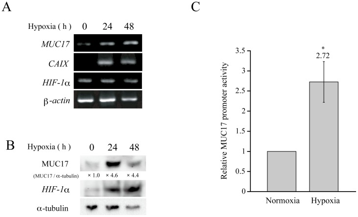
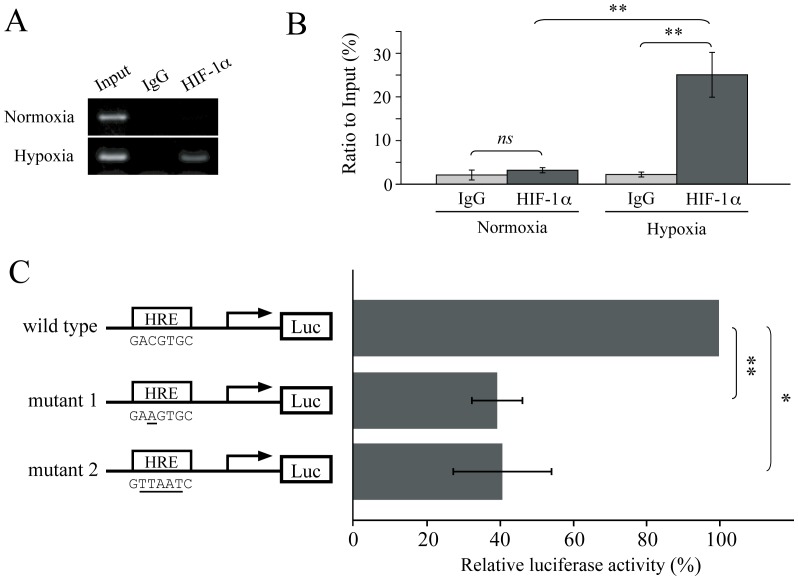
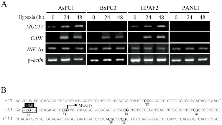
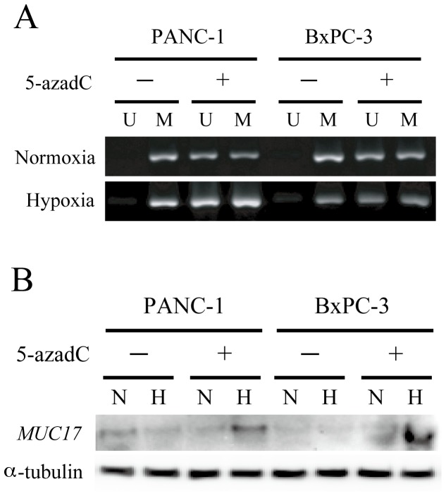
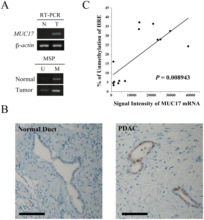
MUC17 is a type 1 membrane-bound glycoprotein that is mainly expressed in the digestive tract. Recent studies have demonstrated that the aberrant overexpression of MUC17 is correlated with the malignant potential of pancreatic ductal adenocarcinomas (PDACs); however, the exact regulatory mechanism of MUC17 expression has yet to be identified. Here, we provide the first report of the MUC17 regulatory mechanism under hypoxia, an essential feature of the tumor microenvironment and a driving force of cancer progression. Our data revealed that MUC17 was significantly induced by hypoxic stimulation through a hypoxia-inducible factor 1α (HIF1α)-dependent pathway in some pancreatic cancer cells (e.g., AsPC1), whereas other pancreatic cancer cells (e.g., BxPC3) exhibited little response to hypoxia. Interestingly, these low-responsive cells have highly methylated CpG motifs within the hypoxia responsive element (HRE, 5'-RCGTG-3'), a binding site for HIF1α. Thus, we investigated the demethylation effects of CpG at HRE on the hypoxic induction of MUC17. Treatment of low-responsive cells with 5-aza-2'-deoxycytidine followed by additional hypoxic incubation resulted in the restoration of hypoxic MUC17 induction. Furthermore, DNA methylation of HRE in pancreatic tissues from patients with PDACs showed higher hypomethylation status as compared to those from non-cancerous tissues, and hypomethylation was also correlated with MUC17 mRNA expression. Taken together, these findings suggested that the HIF1α-mediated hypoxic signal pathway contributes to MUC17 expression, and DNA methylation of HRE could be a determinant of the hypoxic inducibility of MUC17 in pancreatic cancer cells.


Conflict of interest statement

Competing Interests: The authors have declared that no competing interests exist.

Figures

Figure 1
Figure 1. MUC17 expression is enhanced by hypoxia.
(A) AsPC1 cells were cultured under hypoxic conditions (1% O2) for the indicated times. MUC17 mRNA expression was examined by RT-PCR at each time point. (B) AsPC1 cells were cultured under normoxic or hypoxic conditions for the indicated times. Cell lysates were probed with anti-MUC17, HIF1α, and α-tubulin antibodies by Western blot analysis. The intensities of the bands were quantitated by densitometric scanning, and the ratio of MUC17 to α-tubulin expression is shown under each band as the relative intensity compared with that obtained in normoxic AsPC1 cells. (C) MUC17 promoter activity was measured by a Dual-Luciferase Reporter Assay. After transfection of the MUC17 reporter plasmid, AsPC1 cells were incubated under normoxic or hypoxic conditions for 24 h. Cell lysates were assayed using a luciferase assay kit in a Tristar multimode microplate reader LB941 (Berthold Technologies). Transformation efficiency was normalized on the basis of Renilla luciferase activity. The promoter activity under normoxic conditions was given a value of 1. P values were determined using the Student's t-test. * P<0.05.
Figure 2
Figure 2. Hypoxic induction of MUC17 is dependent on HIF1α in AsPC1 cells.
(A) After the transfection of HIF1A siRNAs, AsPC1 cells were cultured under normoxia (N) or hypoxia (H) for 24 h. The level of mRNA was measured by RT-PCR. (B) Cell lysates from AsPC1 cells treated with HIF1A siRNAs were immunoblotted with the indicated antibodies. α-tubulin served as a loading control. (C) The densities of the acquired bands from Western blotting analysis were quantified and expressed as relative fold increases compared with that obtained from mock cells under hypoxic culture conditions. ns, not significant. * P<0.05, ** P<0.005.
Figure 3
Figure 3. Hypoxia enhances the recruitment of HIF1α to HRE and activates MUC17 transcription.
(A) Binding of HIF1α to chromatin was confirmed by a ChIP assay. AsPC1 cells were cultured under normoxia or hypoxia for 24 h. PCR was performed with specific primers covering HRE. (B) The densities of the acquired bands in panel (A) were quantified using Image J (NIH) and normalized to Input included in each experiment. (C) To evaluate the transactivation activity of HIF1α through HRE, a dual luciferase assay was conducted. AsPC1 cells were transfected with wild-type or HRE mutant MUC17 promoter constructs under hypoxic conditions for 24 h. P values were determined using Student's t-test. ns, not significant. * P<0.01, ** P<0.001.
Figure 4
Figure 4. Methylation status of HRE within the MUC17 proximal promoter region.
(A) Differential expression pattern of MUC17 induced by hypoxic exposure in pancreatic cancer cell lines. Each pancreatic cell line was cultured under normoxic (N) or hypoxic (H) conditions. Level of mRNA was determined by RT-PCR at each time point. (B) The human MUC17 gene promoter sequence, which spans positions −687 to +302 with respect to the transcription start site. The numbers of the CpG sites are underlined. The HRE site contains the CpG site (No. 13).
Figure 5
Figure 5. Methylation status of the HRE site determines the sensitivity of MUC17 to HIF1α-induced transactivation.
(A) Demethylation of CpG sites in the MUC17 promoter in BxPC3 and PANC1 cells after 1 µM 5-azadC treatment. MUC17-negative/low cell lines were treated with or without 5-azadC for 7 days. The methylation status of the MUC17 promoter harboring HRE was examined by MSP. The PCR products labeled M (methylated) were amplified by methylation-specific primers, and those labeled U (unmethylated) were amplified by primers specific for unmethylated DNA. (B) Restoration of MUC17 expression in pancreatic cancer cell lines by 5-azadC treatment. Cells were treated with or without 5-azadC for 7 days. During the last 24 h, each group was cultured under normoxic (N) or hypoxic (H) conditions. MUC17 expression was examined by Western blotting.
Figure 6
Figure 6. MUC17 expression is correlated with the hypomethylation of HRE within the MUC17 promoter in tissues from patients with PDAC.
(A) MUC17 mRNA expression and the HRE methylation status in the normal pancreas (N) and pancreatic tumor tissues (T) was examined by RT-PCR and MSP, respectively. (B) Representative immunohistochemical staining data for MUC17 in a patient with PDAC. Scale bar, 100 µm. (C) Correlation of MUC17 mRNA expression and the HRE methylation status was analyzed by Spearman's test. The densities of the acquired bands were quantified using Image J, and the relative amount of unmethylation in each sample was calculated as an index of the aberrant unmethylation status using the equation (%) = U/(U+M).

Similar articles

Cited by

References

    1. Jemal A, Siegel R, Ward E, Hao Y, Xu J, et al. (2009) Cancer statistics, 2009. CA: a cancer journal for clinicians 59: 225–249. - PubMed
    1. Bertout JA, Patel SA, Simon MC (2008) The impact of O2 availability on human cancer. Nature reviews Cancer 8: 967–975. - PMC - PubMed
    1. Semenza GL (2003) Targeting HIF-1 for cancer therapy. Nature reviews Cancer 3: 721–732. - PubMed
    1. Bhaskar KR, Garik P, Turner BS, Bradley JD, Bansil R, et al. (1992) Viscous fingering of HCl through gastric mucin. Nature 360: 458–461. - PubMed
    1. Ho SB, Dvorak LA, Moor RE, Jacobson AC, Frey MR, et al. (2006) Cysteine-rich domains of muc3 intestinal mucin promote cell migration, inhibit apoptosis, and accelerate wound healing. Gastroenterology 131: 1501–1517. - PubMed

Publication types

MeSH terms