

The role of coral-associated bacterial communities in Australian Subtropical White Syndrome of Turbinaria mesenterina
- PMID: 22970188
- PMCID: PMC3435329
- DOI: 10.1371/journal.pone.0044243
The role of coral-associated bacterial communities in Australian Subtropical White Syndrome of Turbinaria mesenterina
Abstract
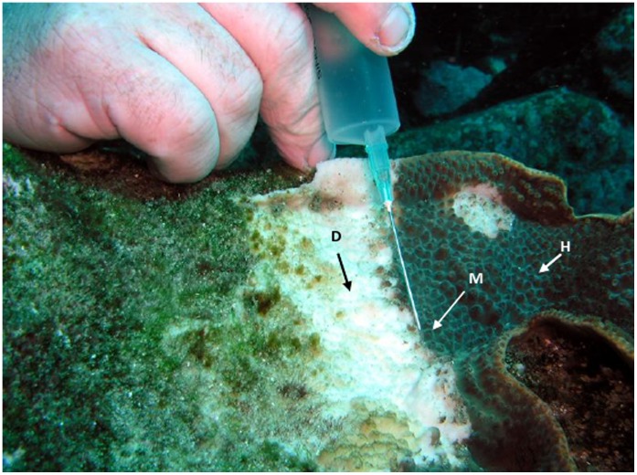
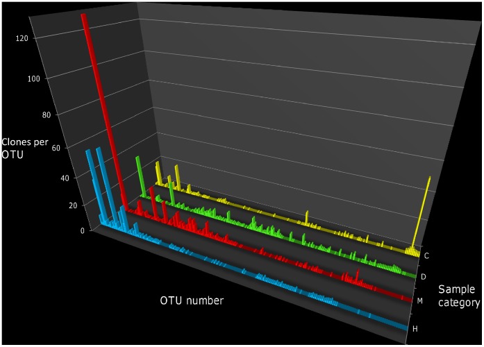
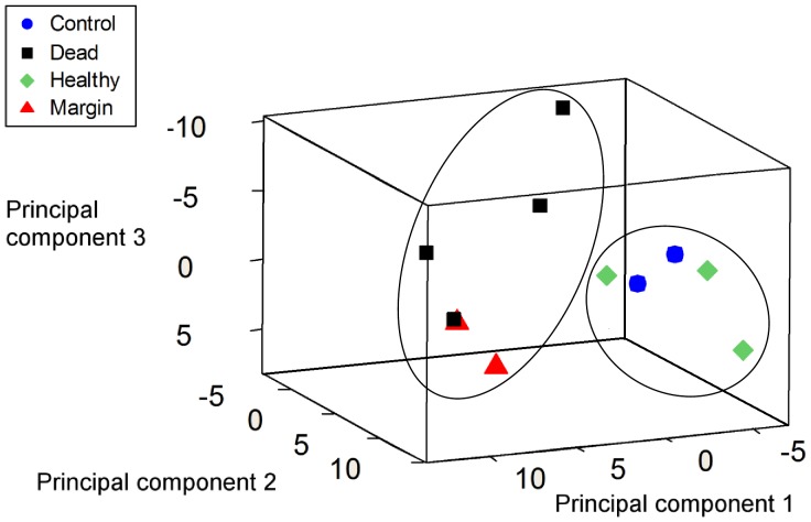
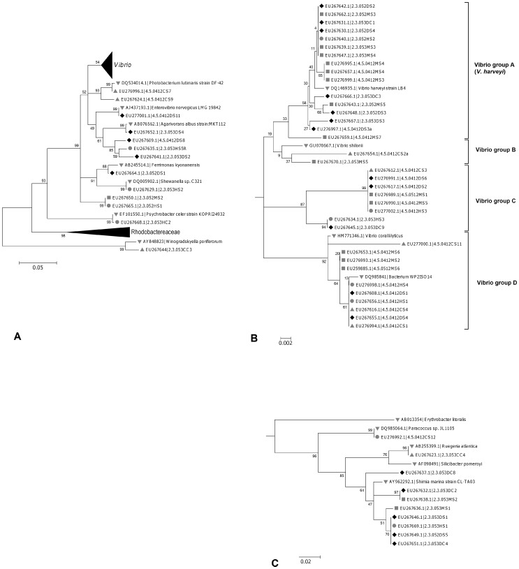
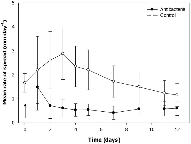
Australian Subtropical White Syndrome (ASWS) is an infectious, temperature dependent disease of the subtropical coral Turbinaria mesenterina involving a hitherto unknown transmissible causative agent. This report describes significant changes in the coral associated bacterial community as the disease progresses from the apparently healthy tissue of ASWS affected coral colonies, to areas of the colony affected by ASWS lesions, to the dead coral skeleton exposed by ASWS. In an effort to better understand the potential roles of bacteria in the formation of disease lesions, the effect of antibacterials on the rate of lesion progression was tested, and both culture based and culture independent techniques were used to investigate the bacterial communities associated with colonies of T. mesenterina. Culture-independent analysis was performed using the Oligonucleotide Fingerprinting of Ribosomal Genes (OFRG) technique, which allowed a library of 8094 cloned bacterial 16S ribosomal genes to be analysed. Interestingly, the bacterial communities associated with both healthy and disease affected corals were very diverse and ASWS associated communities were not characterized by a single dominant organism. Treatment with antibacterials had a significant effect on the rate of progress of disease lesions (p = 0.006), suggesting that bacteria may play direct roles as the causative agents of ASWS. A number of potential aetiological agents of ASWS were identified in both the culture-based and culture-independent studies. In the culture-independent study an Alphaproteobacterium closely related to Roseovarius crassostreae, the apparent aetiological agent of juvenile oyster disease, was found to be significantly associated with disease lesions. In the culture-based study Vibrio harveyi was consistently associated with ASWS affected coral colonies and was not isolated from any healthy colonies. The differing results of the culture based and culture-independent studies highlight the importance of using both approaches in the investigation of microbial communities.
Conflict of interest statement
Figures

References
-
- Sweatman H, Delean S, Syms C (2011) Assessing loss of coral cover on Australia’s Great Barrier Reef over two decades, with implications for longer-term trends. Coral Reefs 30: 521–531.
-
- Sutherland KP, Porter JW, Torres C (2004) Disease and immunity in Caribbean and Indo-Pacific zooxanthellate corals. Marine Ecology-Progress Series 266: 273–302.
-
- Willis BL, Page CA, Dinsdale EA (2004) Coral disease on the Great Barrier Reef. In: Rosenberg E, Loya Y, editors. Coral health and disease. Berlin: Springer.
-
- Weil E, Smith G, Gil-Agudelo DL (2006) Status and progress in coral reef disease research. Dis Aquat Organ 69: 1–7. - PubMed
Publication types
MeSH terms
Substances
Associated data
- Actions
- Actions
- Actions
- Actions
- Actions
- Actions
- Actions
- Actions
- Actions
- Actions
- Actions
- Actions
- Actions
- Actions
- Actions
- Actions
- Actions
- Actions
- Actions
- Actions
- Actions
- Actions
- Actions
- Actions
- Actions
- Actions
- Actions
- Actions
- Actions
- Actions
- Actions
- Actions
- Actions
- Actions
- Actions
- Actions
- Actions
- Actions
- Actions
- Actions
- Actions
- Actions
- Actions
- Actions
- Actions
- Actions
- Actions
- Actions
- Actions
- Actions
- Actions
- Actions
- Actions
- Actions
- Actions
- Actions
- Actions
- Actions
- Actions
- Actions
- Actions
- Actions
- Actions
- Actions
- Actions
- Actions
- Actions
- Actions
- Actions
- Actions
- Actions
- Actions
- Actions
- Actions
- Actions
- Actions
- Actions
- Actions
- Actions
- Actions
- Actions
- Actions
- Actions
- Actions
- Actions
- Actions
- Actions
- Actions
- Actions
- Actions
- Actions
- Actions
- Actions
- Actions
- Actions
- Actions
- Actions
- Actions
- Actions
- Actions
- Actions
- Actions
- Actions
- Actions
- Actions
- Actions
- Actions
- Actions
- Actions
- Actions
- Actions
- Actions
- Actions
- Actions
- Actions
- Actions
- Actions
- Actions
- Actions
- Actions
- Actions
- Actions
- Actions
- Actions
- Actions
- Actions
- Actions
- Actions
- Actions
- Actions
- Actions
- Actions
- Actions
- Actions
- Actions
- Actions
- Actions
- Actions
- Actions
- Actions
- Actions
- Actions
- Actions
- Actions
- Actions
- Actions
- Actions
- Actions
- Actions
- Actions
- Actions
- Actions
- Actions
- Actions
- Actions
- Actions
- Actions
- Actions
- Actions
- Actions
- Actions
- Actions
- Actions
- Actions
- Actions
- Actions
- Actions
- Actions
- Actions
- Actions
- Actions
- Actions
- Actions
- Actions
- Actions
- Actions
- Actions
- Actions
- Actions
- Actions
- Actions
- Actions
- Actions
- Actions
- Actions
- Actions
- Actions
- Actions
- Actions
- Actions
- Actions
- Actions
- Actions
- Actions
- Actions
- Actions
- Actions
- Actions
LinkOut - more resources
Full Text Sources
Medical
