. 2012;7(9):e44257.
doi: 10.1371/journal.pone.0044257. Epub 2012 Sep 6.

A chick model of retinal detachment: cone rich and novel

Affiliations

A chick model of retinal detachment: cone rich and novel

Colleen M Cebulla et al. PLoS One. 2012.

Abstract

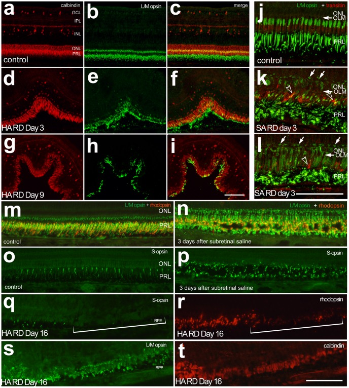
Background: Development of retinal detachment models in small animals can be difficult and expensive. Here we create and characterize a novel, cone-rich retinal detachment (RD) model in the chick.

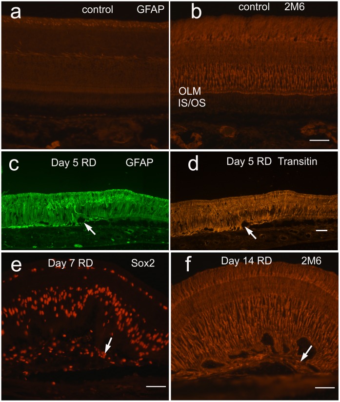
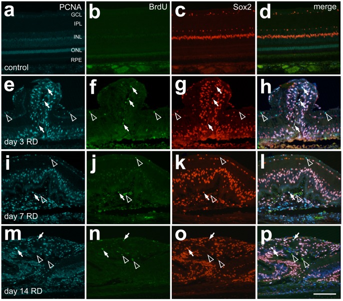
Methodology/principal findings: Retinal detachments were created in chicks between postnatal days 7 and 21 by subretinal injections of either saline (SA) or hyaluronic acid (HA). Injections were performed through a dilated pupil with observation via surgical microscope, using the fellow eye as a control. Immunohistochemical analyses were performed at days 1, 3, 7, 10 and 14 after retinal detachment to evaluate the cellular responses of photoreceptors, Müller glia, microglia and nonastrocytic inner retinal glia (NIRG). Cell proliferation was detected with bromodeoxyuridine (BrdU)-incorporation and by the expression of proliferating cell nuclear antigen (PCNA). Cell death was detected with terminal deoxynucleotidyl transferase dUTP nick end labeling (TUNEL). As in mammalian models of RD, there is shortening of photoreceptor outer segments and mis-trafficking of photoreceptor opsins in areas of RD. Photoreceptor cell death was maximal 1 day after RD, but continued until 14 days after RD. Müller glia up-regulated glial fibriliary acidic protein (GFAP), proliferated, showed interkinetic nuclear migration, and migrated to the subretinal space in areas of detachment. Microglia became reactive; they up-regulated CD45, acquired amoeboid morphology, and migrated toward outer retina in areas of RD. Reactive NIRG cells accumulated in detached areas.

Conclusions/significance: Subretinal injections of SA or HA in the chick eye successfully produced retinal detachments and cellular responses similar to those seen in standard mammalian models. Given the relatively large eye size, and considering the low cost, the chick model of RD offers advantages for high-throughput studies.


Conflict of interest statement

Competing Interests: The authors have declared that no competing interests exist.

Figures

Figure 1
Figure 1. Altered outer retinal morphology in chick retinal detachment.
Toluidine blue staining timecourse of retinal detachment frozen sections days 1, 3, 9, 10, and 14, showing the typical degenerative changes after HA RD as well as variability of the detachments generated. (a) Normal retina with details of outer retina, including oil droplets (predominantly orange-colored spheres) between the photoreceptor inner and outer segments and RPE apical processes (inset). (b–f) There is shortening of the outer segments and flattening of the RPE in areas of RD. Later there is a decrease in cell bodies in the outer nuclear layer (f). RPE hyperplasia is noted in some sections (C). ONL (outer nuclear layer), OPL (outer plexiform layer), PR (photoreceptor), IS (inner segments), OS (outer segments), RBC (red blood cells). Scale bar (50 microns).
Figure 2
Figure 2. Mistrafficking of photoreceptor opsins in RD.
Calbindin (red) and L/M opsin (green) immunostaining in control retina of the untreated fellow eye (a–c) and hyaluronic acid RD at day 3 (d–f) and 9 (g–i). Note decreased intensity of L/M opsin staining at day 9 HA RDs. Confocal microscopy demonstrates early mistrafficking of L/M opsin (green) with staining of the photoreceptor cell body and cone pedicles (arrows, k, l) in SA RDs at day 3 compared to untreated controls. The L/M opsin staining is present in areas consistent with the cone IS/OS as well as the cone ellipsoids in normal chick retina (j). Some transitin-positive glial processes are seen extending beneath the outer limiting membrane (OLM, arrowhead k, l). Similar to day 3 HA RDs, day 3 SA RDs show mistrafficking of rhodopsin (red) and L/M opsin (green, m, n) and S opsin (green, o, p), despite resolution of subretinal fluid. In advanced HA RD (Day 16 RD, q–t) there is redistribution of L/M opsin and calbindin to the cell body with loss of S opsin and rhodopsin (bracket). ONL (outer nuclear layer), OPL (outer plexiform layer), PRL (photoreceptor layer). Scale bar 50 microns.
Figure 3
Figure 3. Photoreceptor cell death in chick RD.
(a–f) Representative photographs of TUNEL-positive cells in control retina and day 1, 3, 7, 9 and 14 HA RDs. TUNEL positive cells are more prevalent in the outer nuclear layer early after detachment. The yellow lines mark the inner and outer limiting membranes. (g) Quantification of TUNEL: a bracket with an asterisk marks statistically significant differences between control and day 1 (p<0.0001) as well as day 1 and day 3 (p<0.0001), day 7 (p<0.0001), and day 14 (p = 0.0004). (h) Quantification of outer nuclear layer thickness: a bracket with an asterisk marks statistically significant differences between day 1 and day 7 (p = 0.0005) and day 14 (p = 0.0033). Error bars represent standard deviation.
Figure 4
Figure 4. Müller glia activated in chick RD: Expression of intermediate filaments GFAP (untreated (a), day 5 HA RD (c)) and transitin, the chick homologue of nestin, (day 5 HA RD (d), untreated not shown) are essentially negative in normal control retina and increase after RD by day 3 (not shown).
GFAP- and transitin-positive Müller glial processes extend beneath the outer limiting membrane (OLM) in HA RDs (arrows c, d) to the subretinal space. The Müller marker TOPAP (antibody 2M6) is present primarily in a vertical distribution throughout the untreated retina in Müller glia (b) and increases particularly in the outer retina below the OLM in HA RDs, with accompanying undulation of the outer retina (f). Cells in subretinal scars are positive for Müller progenitor marker Sox2 (arrow, e) and 2M6 (arrow, f).
Figure 5
Figure 5. Müller glia proliferate after HA RD.
Immunostaining to detect proliferating cells demonstrated more PCNA (blue) than BrdU (green) positive cells at the timepoints tested after RD (day 3, 7, 14). Sox2 (red) labels nuclei of Müller glia and NIRG cells. Merged images of PCNA, Sox2, and BrdU demonstrate a good correlation of PCNA and Sox2, including in subretinal scar. Cells labeling with all three markers are marked with arrows, while Sox2 negative cells labeling with PCNA and BrdU are marked with open arrowheads.
Figure 6
Figure 6. Microglia are up-regulated in chick SA and HA RD.
SA RDs (b) and HA RDs (c) show up-regulation of microglial marker CD45 (green) in areas of RD compared to control retina (a). The SA RD is resolved by day 3. The microglia acquire amoeboid morphology and migrate to the outer retina/subretinal space. Quantitation of CD45 staining in HA RD over time (d, density sum and e, mean area).
Figure 7
Figure 7. NIRG cells are mildly up-regulated after chick HA RD.
NIRG cells (arrows) are positive for Nkx2.2 (green, a–f) and Sox9 (orange, c, d) and are increased after HA RD compared to controls. The up-regulation of NIRGs is less than that of Müller glia (Sox9+/Nkx- cells in the inner nuclear layer and outer retina). NIRGs were identified in subretinal scars (f, arrow; green = Nkx, blue = DAPI).

References

    1. Agrawal RN, He S, Spee C, Cui JZ, Ryan SJ, et al. (2007) In vivo models of proliferative vitreoretinopathy. Nat Protoc 2: 67–77. - PubMed
    1. Mandava N, Blackburn P, Paul DB, Wilson MW, Read SB, et al. (2002) Ribozyme to proliferating cell nuclear antigen to treat proliferative vitreoretinopathy. Invest Ophthalmol Vis Sci 43: 3338–3348. - PubMed
    1. Schiff WM, Hwang JC, Ober MD, Olson JL, Dhrami-Gavazi E, et al. (2007) Safety and efficacy assessment of chimeric ribozyme to proliferating cell nuclear antigen to prevent recurrence of proliferative vitreoretinopathy. Arch Ophthalmol 125: 1161–1167. - PubMed
    1. Iribarne M, Ogawa L, Torbidoni V, Dodds CM, Dodds RA, et al. (2008) Blockade of endothelinergic receptors prevents development of proliferative vitreoretinopathy in mice. Am J Pathol 172: 1030–1042. - PMC - PubMed
    1. Luna G, Kjellstrom S, Verardo MR, Lewis GP, Byun J, et al. (2009) The effects of transient retinal detachment on cavity size and glial and neural remodeling in a mouse model of X-linked retinoschisis. Invest Ophthalmol Vis Sci 50: 3977–3984. - PMC - PubMed

Publication types