. 2012;7(9):e44452.
doi: 10.1371/journal.pone.0044452. Epub 2012 Sep 6.

The B. subtilis MgtE magnesium transporter can functionally compensate TRPM7-deficiency in vertebrate B-cells

Affiliations

The B. subtilis MgtE magnesium transporter can functionally compensate TRPM7-deficiency in vertebrate B-cells

Jaya Sahni et al. PLoS One. 2012.

Abstract

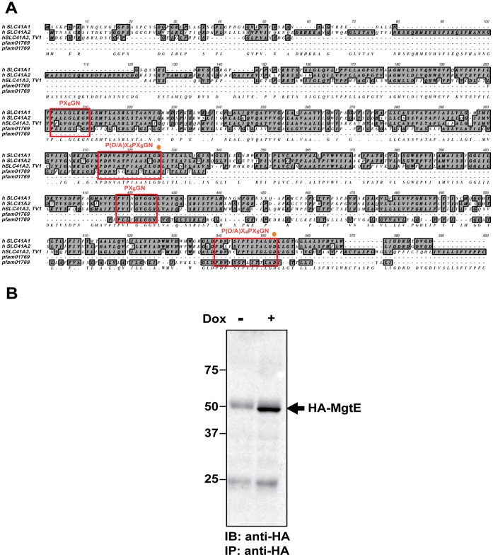
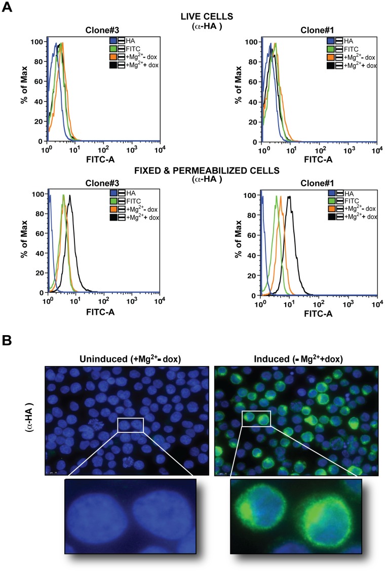
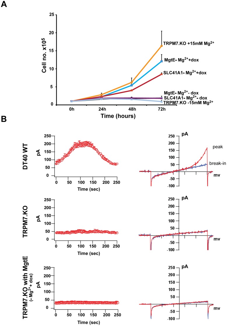
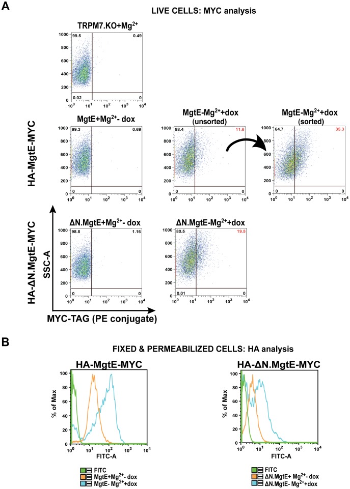
Recent studies have shown that the vertebrate magnesium transporters Solute carrier family 41, members 1 and 2 (SLC41A1, SLC41A2) and Magnesium transporter subtype 1 (MagT1) can endow vertebrate B-cells lacking the ion-channel kinase Transient receptor potential cation channel, subfamily M, member 7 (TRPM7) with a capacity to grow and proliferate. SLC41A1 and SLC41A2 display distant homology to the prokaryotic family of Mg(2+) transporters, MgtE, first characterized in Bacillus subtilis. These sequence similarities prompted us to investigate whether MgtE could potentially compensate for the lack of TRPM7 in the vertebrate TRPM7-deficient DT40 B-cell model system. Here, we report that overexpression of MgtE is able to rescue the growth of TRPM7-KO DT40 B-cells. However, contrary to a previous report that describes regulation of MgtE channel gating by Mg(2+) in a bacterial spheroplast model system, whole cell patch clamp analysis revealed no detectable current development in TRPM7-deficient cells expressing MgtE. In addition, we observed that MgtE expression is strongly downregulated at high magnesium concentrations, similar to what has been described for its vertebrate homolog, SLC41A1. We also show that the N-terminal cytoplasmic domain of MgtE is required for normal MgtE channel function, functionally confirming the predicted importance of this domain in regulation of MgtE-mediated Mg(2+) entry. Overall, our findings show that consistent with its proposed function, Mg(2+) uptake mediated by MgtE is able to restore cell growth and proliferation of TRPM7-deficient cells and supports the concept of functional homology between MgtE and its vertebrate homologs.


Conflict of interest statement

Competing Interests: The authors have declared that no competing interests exist.

Figures

Figure 1
Figure 1. Sequence alignment of the human SLC41 transporter family with MgtE pfam 01769 and MgtE expression analysis.
(A) ClustalW alignment of the amino acid sequences representing the three members of the human SLC41 transporters – A1, A2 and A3 transcript variant 1 (TV1) (includes both SLC41 D1 and D2 domains homologous to pfam01769) with the prokaryotic MgtE consensus sequence, pfam01769 from the conserved protein family database (pfam; http://pfam.sanger.ac.uk). The red boxes show the conserved motifs PX6GN and P (D/A)X4PX6D of which the former is considered to be the putative ion-conducting pore region and the last “D” (D432; aspartic acid residue) in the second motif has been established as an ion-selective site essential for Mg2+ uptake (denoted as an orange dot). (B) TRPM7-KO DT40 cells transfected with the N-terminal HA-tagged MgtE were doxycycline-induced and analyzed for MgtE protein expression by immunoprecipitation and immunoblotting with anti-HA. A clear band representing HA-MgtE was observed in the induced (dox +) cells. Data is representative of blots from at least two independent experiments.
Figure 2
Figure 2. Membrane orientation and cellular localization analysis of HA-MgtE in TRPM7-KO cells.
(A) Two clones of TRPM7-KO cells expressing HA-tagged MgtE (#3 and #1) were doxycycline-induced and either stained live with anti-HA (upper panel) or fixed & permeabilized and labeled with anti-HA (lower panel). Following staining with species-specific FITC, cells were acquired on BD LSRII. Data analysis by FlowJo demonstrated that the N-terminal HA-tag was only accessible intracellularly. (B) Both uninduced (left panel) and doxycycline inducible TRPM7-KO cells expressing HA-tagged MgtE (clone#3; right panel) were fixed & permeabilized followed by anti-HA and FITC staining for immunolocalization analysis of MgtE. The induced cells expressing the protein were clearly discernible from the uninduced cells on the basis of fluorescence. The upper panel shows staining of multiple cells in a single field and the lower panel shows a magnified view of two cells from the upper panel. All data are representative of results obtained from at least two independent experiments.
Figure 3
Figure 3. Growth and whole-cell patch clamp analysis of HA-tagged MgtE.
(A) TRPM7-KO cells, TRPM7-KO cells overexpressing HA-tagged MgtE and TRPM7-KO cells overexpressing HA-SLC41A1-MYC were induced with doxycycline and their growth was analyzed. Growth curves represent the average of three independent experiments and the error bars represent mean ± standard error of the mean (SEM). TRPM7-KO cells with 15 mM supplemental Mg2+, TRPM7-KO cells inducibly expressing either tagged MgtE or SLC41A1 displayed growth complementation over a period of 72 hours while the uninduced cells and TRPM7-KO cells in regular cell culture media did not grow. (B) Whole cell patch clamp analysis of DT40 WT, TRPM7-KO and TRPM7-KO cells overexpressing MgtE. Left panel shows graphical representation of current development over time and right panel shows I/V curves at peak (in red) and break-in (in blue). Although a TRPM7 mediated MIC current was observed in DT40 WT cells, no MIC current was observed either in TRPM7-KO cells or TRPM7-KO cells expressing HA-MgtE.
Figure 4
Figure 4. Downregulation of both MgtE expression and growth occurs in presence of high magnesium.
(A) TRPM7-KO cells expressing HA-MgtE were induced either in presence or absence of 15 mM supplemental Mg2+ and analyzed for expression after staining with anti-HA and species-specific FITC on BD LSRII at 24 and 48 hours. Cells induced in regular cell culture media had higher MgtE expression compared to cells induced in presence of 15 mM Mg2+. (B) Cells from similar conditions as mentioned in (A) were analyzed for expression at 5 days and 4 weeks post-induction and displayed fluorescence shifts similar to what was observed in (A) with significantly lower MgtE expression in cells induced in presence of 15 mM Mg2+ at 4 weeks. All data are representative of results obtained from at least three independent experiments. (C) Left panel: TRPM7-KO cells expressing MgtE were induced either in presence or absence of 15 mM supplemental Mg2+ and their growth was determined for a period of 98 h. Growth curves represent the average of three independent experiments and the error bars correspond to mean ± SEM. Cells induced in presence of 15 mM Mg2+ exhibited diminished growth compared to cells induced in regular cell culture media. Right panel: Cell size analysis of samples in the left panel at 98 h. TRPM7-KO cells expressing MgtE were kept either uninduced or doxycycline-induced in absence/presence of 15 mM Mg2+ and acquired on BD LSRII. Cells induced in presence of 15 mM Mg2+ were bigger in size compared to the uninduced and induced cells. Results are representative of three independent experiments.
Figure 5
Figure 5. Expression of the N domain deletion mutant results in stunted growth and reduced cell size.
(A) Left panel: TRPM7-KO cells expressing the HA-tagged N domain MgtE deletion (Δ1–137) mutant were either kept uninduced, doxycycline - induced or induced in presence of 15 mM supplemental Mg2+ following which their growth was monitored at 24, 48, 72 and 96 hours, respectively. TRPM7-KO cells expressing the HA-tagged WT MgtE were used as controls under similar growth conditions. Growth curves represent the average of three independent experiments and the error bars represent standard error of the mean. Right panel: Cell size analysis of TRPM7-KO cells expressing the HA-tagged N domain deletion mutant at 96 hours. Cells were either kept uninduced or induced in absence/presence of 15 mM magnesium and acquired on BD LSRII. Data is representative of at least three independent experiments. (B) TRPM7-KO cells expressing the HA-tagged N domain deletion mutant were analyzed for total protein expression by staining with anti-HA and FITC, post fixation and permeabilization. Single stained cells were used as controls and cells were acquired on BD LSRII followed by data analysis using FlowJo software. Comparable expression of the mutant was observed in cells induced in presence/absence of Mg2+. Results are representative of three independent experiments. (C) TRPM7-KO cells expressing the N domain deletion mutant were maintained under similar conditions as mentioned in (B) and analyzed for total protein expression (anti-HA/FITC) at four weeks. While the cells induced in presence of supplemental Mg2+ exhibited slightly increased expression (left panel), cells induced in regular cell culture media for up to 4 weeks displayed significantly lower expression levels compared to the ones induced for only 48 hours (right panel). In both (B) and (C), TRPM7-KO cells expressing the WT MgtE had highest expression at all time points. Results are representative of at least two independent sets of experiments.
Figure 6
Figure 6. C-terminal MYC-tagged WT MgtE and ΔN domain mutant display surface expression.
(A) Representative flow plots showing surface expression of the MYC-tagged WT MgtE and the N domain deletion mutant (HA-tagged at the N-terminus and MYC-tagged at the C-terminus) in stably transfected TRPM7-KO cells stained with MYC directly conjugated to PE. While the untransfected TRPM7-KO cells and the uninduced cells did not display any MYC-PE fluorescence, both the WT MgtE and the N domain deletion mutant demonstrated increased mean fluorescence intensities post staining. Sorting and analysis of WT MgtE cells based on the surface expression of MYC showed a significant shift to higher fluorescence intensity. (B) Uninduced and doxycycline induced cells in (A) were fixed & permeabilized and probed with anti-HA/anti-mouse FITC for analysis of the N-terminal HA-tag. A strong shift in fluorescence was observed in both induced WT MgtE and N-terminal mutant expressing cells relative to the uninduced controls. A small change in the fluorescence intensity of the uninduced cells was also observed in both the cases indicating a leaky promoter. Plots are representative of at least two independent experiments.

Similar articles

Cited by

References

    1. Maguire ME (2006) Magnesium transporters: properties, regulation and structure. Front Biosci 11: 3149–3163. - PubMed
    1. Moomaw AS, Maguire ME (2008) The unique nature of mg2+ channels. Physiology (Bethesda) 23: 275–285. - PMC - PubMed
    1. Runnels LW (2011) TRPM6 and TRPM7: A Mul-TRP-PLIK-cation of channel functions. Curr Pharm Biotechnol 12: 42–53. - PMC - PubMed
    1. Schmitz C, Perraud AL, Johnson CO, Inabe K, Smith MK, et al. (2003) Regulation of vertebrate cellular Mg2+ homeostasis by TRPM7. Cell 114: 191–200. - PubMed
    1. Mandt T, Song Y, Scharenberg AM, Sahni J (2011) SLC41A1 Mg2+ transport is regulated via Mg2+-dependent endosomal recycling through its N-terminal cytoplasmic domain. Biochem J 439: 129–139. - PubMed

Publication types

MeSH terms

LinkOut - more resources