. 2012;7(9):e44493.
doi: 10.1371/journal.pone.0044493. Epub 2012 Sep 6.

Protein disulfide isomerase-like protein 1-1 controls endosperm development through regulation of the amount and composition of seed proteins in rice

Affiliations

Protein disulfide isomerase-like protein 1-1 controls endosperm development through regulation of the amount and composition of seed proteins in rice

Yeon Jeong Kim et al. PLoS One. 2012.

Abstract

Protein disulfide isomerase (PDI) is a chaperone protein involved in oxidative protein folding by acting as a catalyst and assisting folding in the endoplasmic reticulum (ER). A genome database search showed that rice contains 19 PDI-like genes. However, their functions are not clearly identified. This paper shows possible functions of rice PDI-like protein 1-1 (PDIL1-1) during seed development. Seeds of the T-DNA insertion PDIL1-1 mutant, PDIL1-1Δ, identified by genomic DNA PCR and western blot analysis, display a chalky phenotype and a thick aleurone layer. Protein content per seed was significantly lower and free sugar content higher in PDIL1-1Δ mutant seeds than in the wild type. Proteomic analysis of PDIL1-1Δ mutant seeds showed that PDIL1-1 is post-translationally regulated, and its loss causes accumulation of many types of seed proteins including glucose/starch metabolism- and ROS (reactive oxygen species) scavenging-related proteins. In addition, PDIL1-1 strongly interacts with the cysteine protease OsCP1. Our data indicate that the opaque phenotype of PDIL1-1Δ mutant seeds results from production of irregular starch granules and protein body through loss of regulatory activity for various proteins involved in the synthesis of seed components.


Conflict of interest statement

Competing Interests: The authors have declared that no competing interests exist.

Figures

Figure 1
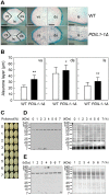
Figure 1. Isolation of the PDIL1-1Δ mutant and its seed phenotype.
(A) Identification of the T-DNA insertion site in the PDIL1-1 gene (locus number Os11g09280) by PCR. T-DNA insertion mutant PFG_1B-16041.R was provided by Dr. Gynheung An, POSTECH. Independent transgenic lines were analyzed by PCR using two sets of primers (described in Table S1); 1,229-bp fragments were amplified by PCR with LP and RP, but not by the primer set in transgenic homozygote lines. Approximately 700-bp fragments were amplified by PCR with BP and RP. (B) Identification of PDIL1-1 mutant rice by western blot. Total seed proteins extracted from the lines described in (A) were separated by SDS-PAGE and examined by western blot using an anti-PDIL1-1 antibody. (C) Grains of wild type and PDIL1-1Δ mutants were collected, and palea and lemma of the grains of wild type and two PDIL1-1Δ mutant alleles (PFG_1B-16041.R and PFG_2B-80111.R) were opened and removed. The mutants showed a chalky and uneven phenotype. Bar, 0.3 cm.
Figure 2
Figure 2. Comparison of seed proteins of WT and PDIL1-1Δ mutant.
(A) Total seed proteins were extracted from the WT and PDIL1-1Δ mutant and separated by 10–27% gradient SDS-PAGE. (B) Total proteins were extracted from mature seeds of the WT and PDIL1-1Δ mutant and then analyzed by 2-DE.
Figure 3
Figure 3. Aleurone layers of PDIL1-1Δ mutant seeds and analysis of PDIL1-1 protein level in differently polished seeds.
(A) Mature seeds of the WT and PDIL1-1Δ mutant were stained with methylene blue and then analyzed by light microscopy. Abbreviations: vs, ventral side; ds, dorsal side. ls, lateral side. Bar, 1 mm (left), and 0.5 mm (middle and right). (B) Thickness of aleurone layers was measured in both seeds of the WT and PDIL1-1Δ mutant. * and ** indicate a significant difference from the wild type at P<0.05 and P<0.01 by t-test, respectively. (C) Dried seeds were polished to different extents. (D The remaining seeds were ground thoroughly, and the PDIL1-1 level was examined by western blot with an anti-PDIL1-1 antibody (left). After detection, the membrane was stained with Coomassie brilliant blue (right). (E) PDIL1-1 levels were examined in the polished powder by western blot with an anti-PDIL1-1 antibody (left). After detection, the membrane was stained with Coomassie brilliant blue (right).
Figure 4
Figure 4. X-ray diffraction analysis of outer endosperm of the PDIL1-1Δ mutant seeds.
After polishing of the WT and the PDIL1-1Δ mutant seeds to a 10% weight loss, powders (the outer part of the endosperm), were collected and analyzed by an X-ray diffractometry. The two-theta angle (2θ) ranging from 4.0 to 40.0° was scanned to obtain values that were overlapping for comparison of the two samples.
Figure 5
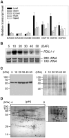
Figure 5. Expression pattern of PDIL1-1 gene.
(A) Expression profile of PDIL1-1 I gene during development. Total RNA was isolated from developing rice organs at the indicated time points and used for real-time RT-PCR. DAG, day after germination; DAF, days after flowering. (B) Total RNA was isolated from developing seeds at the indicated time points and separated on formaldehyde-agarose gels. After electrophoresis, total RNA was transferred onto a nylon membrane. The membrane was hybridized with 32P-labeled PDIL1-1 cDNA and exposed on X-ray film. DAF, days after flowering. (C) Examination of the level of PDIL1-1 protein during seed development. Total proteins were extracted from the wild type at the indicated time points. After 12% SDS-PAGE, proteins were transferred onto a nitrocellulose membrane and treated with an anti-PDIL1-1 antibody (left). After blotting, the membrane was stained with Coomassie brilliant blue (right). (D) Proteomic analysis of rice seed proteins. Total proteins were extracted from mature seeds of WT and then separated by 2-DE. Each protein spot was identified by MALDI-TOF MS (left). The boxed region was enlarged (right). Arrowheads indicate PDIL1-1 protein spots.
Figure 6
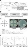
Figure 6. Physical interaction of PDIL1-1 with OsCP1.
(A) Phylogenetic tree of Arabidopsis cysteine protease 43 (AtCP43) amino acid sequences with its rice homologs. The sequences of the proteins were aligned using the CLUSTALW2 software program and a phylogenetic tree was generated using the MEGA5 software program. B) Map of conserved domains of OsCP1. The three conserved domains, inhibitor_I29, peptidase_C1A, and granulin, are indicated by boxes. (C) Partial OsCP1 cDNA encoding peptidase_C1A and granulin, and full-length PDIL1-1 cDNA were fused to sequences encoding the Gal4 activation domain (AD) and the Gal4 DNA-binding domain (BD) in pGADT7 and pGBKT7, respectively. Each number indicates yeast cells transformed with a combination of only pGADT7 or pGBKT7 vectors or recombinant plasmids. Combinations are described in the box. Transformants were plated onto minimal medium (Leu/Trp) and (Leu/Trp/His (5 mM 3-AT)), incubated for 4 days, and then photographed. (D) Expression analysis of OsCP1 and its homologs. Transcript levels of OsCP1 and its homologs, Os05g0108600 and Os01g0971400, were examined by real-time RT-PCR with gene-specific primers. These experiments were repeated three times independently. The reported values of transcript levels of OsCP1 homologs are normalized to numerical values relative to the transcript level of OsCP1 in DAF5, which is set at a value of 1.00±0.00. DAF, day after flowering.

Similar articles

Cited by

References

    1. Tsai C, Larkins B, Glover D (1978) Interaction of the opaque-2 gene with starch-forming mutant genes on the synthesis of zein in maize endosperm. Biochem Genet 16: 883–896. - PubMed
    1. Miles MJ, Morris VJ, Orford PD, Ring SG (1985) The roles of amylose and amylopectin in the gelation and retrogradation of starch. Carbohydr Res 135: 271–281.
    1. Gallant DJ, Bouchet B, Baldwin PM (1997) Microscopy of starch: evidence of a new level of granule organization. Carbohydr Polym 32: 177–191.
    1. Thompson DB (2000) On the non-random nature of amylopectin branching. Carbohydr. Polym 43: 223–239.
    1. Smith AM, Denyer K, Martin C (1997) The synthesis of the starch granule. Ann Rev Plant Biol 48: 67–87. - PubMed

Publication types

LinkOut - more resources