. 2012;7(9):e44523.
doi: 10.1371/journal.pone.0044523. Epub 2012 Sep 6.

Oral inoculation of young dairy calves with Mycoplasma bovis results in colonization of tonsils, development of otitis media and local immunity

Affiliations

Oral inoculation of young dairy calves with Mycoplasma bovis results in colonization of tonsils, development of otitis media and local immunity

Fiona Maunsell et al. PLoS One. 2012.

Abstract

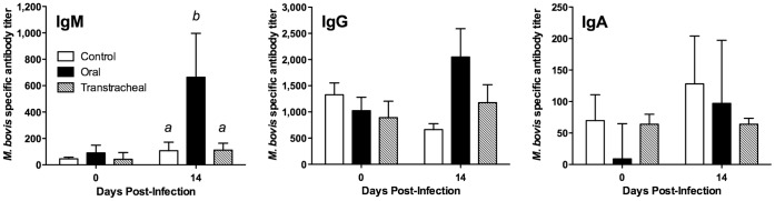
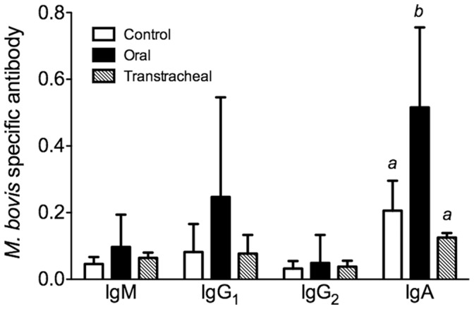
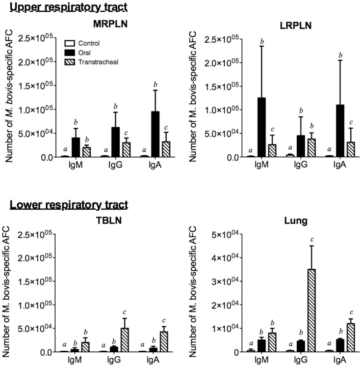
Because M. bovis otitis media is an economically important problem, there is a need to understand the pathogenesis of disease, not only to improve our understanding of the factors contributing to the development of this disease but also to inform the development of improved diagnostic tests and therapy. Oral ingestion of M. bovis-contaminated milk is linked, but not definitively proven, to development of otitis media. In the current study, we demonstrate that oral ingestion of M. bovis infected colostrum can result in an ascending infection and development of otitis media. Importantly, M. bovis was found to have a previously unrecognized tendency for colonization of the tonsils of calves, which most likely contributed to the subsequent development of otitis media. In contrast, transtracheal inoculation failed to produce clinically significant upper respiratory tract disease, although did induce lower respiratory tract disease. The upper respiratory tract was the major site of M. bovis-specific B cell and mucosal IgA responses in calves inoculated by the oral route. The oral inoculation route of infection presented here is particularly suited to the study of host-pathogen interactions during initial colonization of the tonsils, expansion of infection and dissemination to the lower respiratory tract and middle ear. In addition, it could be used to investigate potential new preventative or control strategies, especially those aimed at limiting colonization of the tonsils and/or spread to the middle ear.


Conflict of interest statement

Competing Interests: The authors have declared that no competing interests exist.

Figures

Figure 1
Figure 1. Oral inoculation of calves results in lesions in middle ear.
The severity of lesions in tympanic bulla was assessed using a subjective scoring system. No control or transtracheally-inoculated calf had lesion scores >1 in the tympanic bullae. In contrast, 3 of 8 orally inoculated calves developed significant lesions in the tympanic bullae (lesion score >1, P≤0.05). Two of these animals had bilateral lesion scores ≥3. Examples of the range of lesions and their associated scores are: a) Grade 1 shows normal bony trabecula, thin cuboidal to simple squamous mucosal epithelium and loose acellular lamina propria. 60× magnification. b) Grade 3 lesions demonstrate moderately dense collections of mixed inflammatory cells infiltrating the lamina propria and the mucosal epithelium. Moderate to large numbers of macrophages and neutrophils are present in the lumen. 40× magnification. c) Grade 4 lesions are characterized with a superficial epithelium that is moderately hyperplastic, with regions of squamous metaplasia. There are large numbers of mixed inflammatory cells infiltrating the lamina propria and epithelium. Prominent plump spindle cells and small capillaries lined by plump endothelia are present in the lamina propria. There are luminal collections of inflammatory cells and necrotic debris. 40× magnification. The insert shows large osteoclasts occupying prominent Howships lacunae (arrow) and osteoblasts lining the opposite side of the trabecula, indicative of bone remodeling. 40× magnification. d) Grade 5 lesions show full thickness mucosal necrosis with underlying osteonecrosis of trabecular bone. Note the superficial collections of inflammatory and necrotic debris. 20× magnification.
Figure 2
Figure 2. Lesions in the Eustachian tubes were common in calves infected by the oral route.
The severity of lesions was assessed using a subjective scoring system. Four of the 8 orally infected calves had lesion scores ≥2 (of a possible 3) in at least one Eustachian tube, with 3 calves having eustachitis in both ears. Two of the five transtracheally-inoculated calves however developed eustachitis, but were less severe than in orally-inoculated calves (lesion scores of 2 in one Eustachian tube). Examples of the range of lesions and their associated scores are: a) Grade 1 demonstrating curved dorso-medial cartilaginous support of the Eustachian tube. 2× magnification. b) Grade 1 lesion demonstrating columnar ciliated epithelium with loose collections of mononuclear cells in lamina propria. Cellular debris is visible in the lumen. 60× magnification. c) Grade 2 lesion demonstrating moderately dense collections of lymphocytes and plasma cells, with rare neutrophils. Low numbers of lymphocytes are present in the mucosal epithelium. 40× magnification. d) Grade 3 lesion demonstrating dense collections of lymphocytes and plasma cells intermixed with neutrophils in the lamina propria. The superficial mucosal epithelium is focally cuboidal and eroded, and the lumen contains collections of eosinophilic necrotic inflammatory debris. 40× magnification.
Figure 3
Figure 3. Ears with the highest lesions scores in tympanic bullae also had high lesion scores in corresponding Eustachian tubes.
Lesion scores for Eustachian tubes are plotted against with lesion scores for the corresponding tympanic bullae from calves experimentally infected with M. bovis by the oral route (N = 8; separate date points are shown for the left and right sides of each calf). For visual purposes, tissues with identical scores have been shown with values slightly offset.
Figure 4
Figure 4. Lung lesions were found in mycoplasma infected calves using either route of inoculation.
Histopathological findings in the lungs of control calves (N = 8) or calves inoculated with M. bovis by oral (N = 8) or transtracheal (N = 5) routes. Samples were collected at necropsy (14 days post-infection, except for one calf inoculated with M. bovis by the oral route which had to be euthanized at 10 days post-infection). a) Overall histopathological lesion scores. b) Subscores for lymphoid hyperplasia. Data are represented as scores for individual calves with the median value indicated by a horizontal line. Tissues were graded on a subjective scale from 1 (minimal or no lesions) to 5 (most severe lesions) and from 1 (no lymphoid hyperplasia) to 4 (marked lymphoid hyperplasia) for lesion scores and lymphoid hyperplasia subscores, respectively. abSuperscript letters indicate significant (P<0.05) differences between groups. c) Representative histopathologic appearance of a lung section with a lesion score of 1 (control calf). Magnification ×10. d) Representative histopathologic appearance of a lung section with a lesion score of 5 (orally-inoculated calf from which M. bovis was recovered from the lung). Magnification ×10.
Figure 5
Figure 5. Tonsils are a major site of M. bovis colonization in the upper respiratory tract.
At 14 days after oral inoculation of calves, mycoplasma numbers in nasal passages and tonsils were determined. Calves infected by oral ingestion of M. bovis had significantly higher (about 1 log higher) microbial loads in tonsils than did transtracheally-inoculated calves. Mycoplasmas were not recovered from any tissue of any control calf (N = 8). For visual purposes, tissues with identical scores have been shown with values slightly offset. Asterisks “*” denotes a significant difference between groups (P≤0.05).
Figure 6
Figure 6. The numbers of M. bovis recovered in the Eustachian tubes and tympanic bullae were dramatically higher in orally-inoculated than in transtracheally-inoculated calves.
A t 14 days after oral- (N = 8) or transtracheal-inoculation of calves (N = 5), mycoplasma numbers in corresponding a) Eustachian tubes and b) tympanic bullae were determined (left and right sides are shown separately for each inoculated calf). Mycoplasmas were not recovered from any tissue of any control calf (N = 8). (data not shown). High numbers of mycoplasmas were recovered from the ears of several animals in the mycoplasma-inoculated group. c) In all animals where mycoplasmas were recovered from the tympanic bullae, organisms were recovered from the corresponding Eustachian tube; however, there were several animals (outlined in dashed box) where organisms were recovered from the Eustachian tubes and not the corresponding tympanic bullae. Data represent all orally (N = 8) and transtracheally (N = 5) inoculated calves, with left and right sides shown separately for each calf. d) Ears with the highest lesions scores in tympanic bullae also had high lesion scores in corresponding Eustachian tubes. Data represent all orally (N = 8) and transtracheally (N = 5) inoculated calves, with left and right sides shown separately for each calf For visual purposes, tissues with identical scores have been shown with values slightly offset. Asterisks “*” denotes a significant difference between groups (P≤0.05).
Figure 7
Figure 7. The degree of tonsil colonization was associated with development of colonization of Eustachian tubes.
Relationship between the number of M. bovis recovered from pharyngeal tonsils and the number recovered from the left and right Eustachian tubes in calves inoculated with M. bovis by either the oral (N = 8) or transtracheal (N = 5) routes. When only the undiluted broth was positive, results were assigned a log10 value of 0.5. No mycoplasmas were recovered from carrier inoculated control calves (N = 8, data not shown).
Figure 8
Figure 8. The level of lower respiratory tract infection with M. bovis was similar between orally- and transtracheally-inoculated groups of calves.
At 14 days after oral- (N = 8) or transtracheal- (N = 5) inoculation of calves, mycoplasma numbers in a) trachea, b) bronchi and c) lung were determined. Control calves were orally inoculated with milk that did not contain mycoplasma, and mycoplasmas were not recovered from any tissue (N = 8) (Data not shown). There were no significant differences in the numbers of mycoplasma recovered from lower respiratory tracts of orally- and transtracheally-inoculated calves. For visual purposes, tissues with identical scores have been shown with values slightly offset. Data for the left and right primary bronchi are shown separately.
Figure 9
Figure 9. Serum IgM responses were higher in orally-inoculated calves.
Specific antibody levels were measured in serum obtained from calves inoculated with M. bovis. Serum samples were collected at on the day of infection and at necropsy (14 days post infection) from calves inoculated by the oral route (N = 8, except for one calf inoculated with M. bovis by the oral route which had to be euthanized at 10 days post-infection), the transtracheal route (N = 5; 14 days post-infection), and from control calves (N = 8; 14 days post-sham inoculation). Data are expressed as the mean ± SD of the antibody titer in the sample for each isotype. abSuperscript letters indicate significant (P<0.05) differences between groups.
Figure 10
Figure 10. Antibody responses in nasal lavages however were found in orally-, but not transtracheally-, inoculated calves.
Specific antibody levels were measured in nasal lavage fluids obtained from calves inoculated with M. bovis. Samples were collected at necropsy from calves inoculated by the oral route (N = 8; 14 days post-infection, except for one calf inoculated with M. bovis by the oral route which had to be euthanized at 10 days post-infection), the transtracheal route (N = 5; 14 days post-infection), and from control calves (N = 8; 14 days post-sham inoculation). Data are expressed as the mean ± SD of the optical density (O.D.) adjusted for the total amount of immunoglobulin (Ig), calculated as the optical density for M. bovis-specific Ig/total Ig (µg/ml) in the sample for each isotype. abSuperscript letters indicate significant (P<0.05) differences between groups.
Figure 11
Figure 11. The upper respiratory tract had a greater increase in the number of mycoplasma-specific antibody-forming cells (AFC) than did the lower respiratory tract.
ELISpot assays were performed on tissue collected at necropsy (14 days post infection except for one calf that was euthanized at 10 days post infection) from control calves (N = 5; white bars) and from calves inoculated by oral (N = 4; black bars) or transtracheal (N = 3; hatched bars) routes. Tissues from other calves in the study could not be analyzed due to low cell recovery. Cells were collected from Medial retropharyngeal lymph nodes (MRPLN), Lateral retropharyngeal lymph nodes (LRPLN), Tracheobronchial lymph nodes (TBLN) and Lung. The data for lymph nodes are represented as the mean ± SD of the total number of cells within that tissue. Data for the lungs are represented as the mean (± SD) of the number of cells/g of tissue. abcSuperscript letters indicate significant (P<0.05) differences in class-specific responses between groups.

References

    1. Hewicker-Trautwein M, Feldmann M, Kehler W, Schmidt R, Thiede S, et al. (2002) Outbreak of pneumonia and arthritis in beef calves associated with Mycoplasma bovis and Mycoplasma californicum . Vet Rec 151: 699–703. - PubMed
    1. Maeda T, Shibahara T, Kimura K, Wada Y, Sato K, et al. (2003) Mycoplasma bovis-associated suppurative otitis media and pneumonia in bull calves. J Comp Pathol 129: 100–110. - PubMed
    1. Francoz D, Fecteau G, Desrochers A, Fortin M (2004) Otitis media in dairy calves: a retrospective study of 15 cases (1987 to 2002). Can Vet J 45: 661–666. - PMC - PubMed
    1. Lamm CG, Munson L, Thurmond MC, Barr BC, George LW (2004) Mycoplasma otitis in California calves. J Vet Diagn Invest 16: 397–402. - PubMed
    1. Ayling RD, Bashiruddin SE, Nicholas RA (2004) Mycoplasma species and related organisms isolated from ruminants in Britain between 1990 and 2000. Vet Rec 155: 413–416. - PubMed

Publication types