. 2012;7(9):e44552.
doi: 10.1371/journal.pone.0044552. Epub 2012 Sep 10.

Estrogen deficiency induces the differentiation of IL-17 secreting Th17 cells: a new candidate in the pathogenesis of osteoporosis

Affiliations

Estrogen deficiency induces the differentiation of IL-17 secreting Th17 cells: a new candidate in the pathogenesis of osteoporosis

Abdul M Tyagi et al. PLoS One. 2012.

Abstract

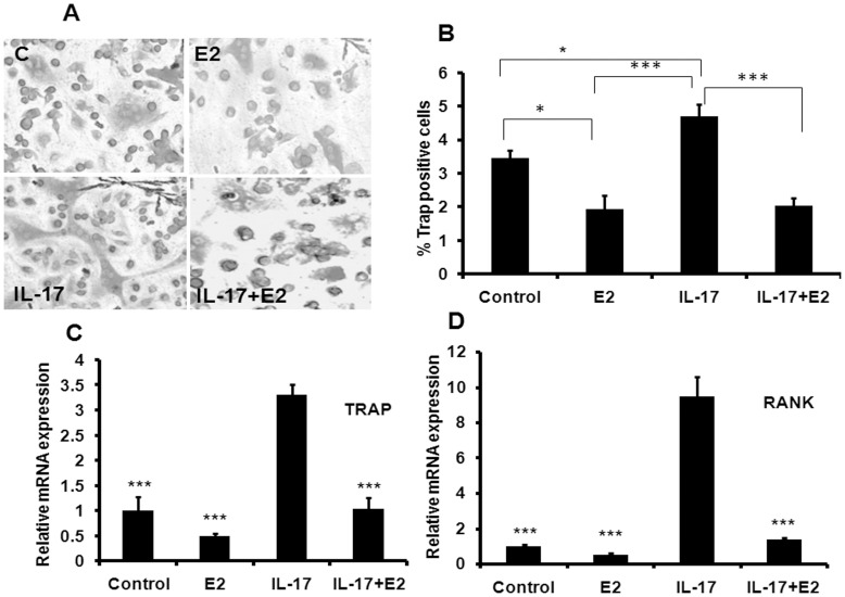
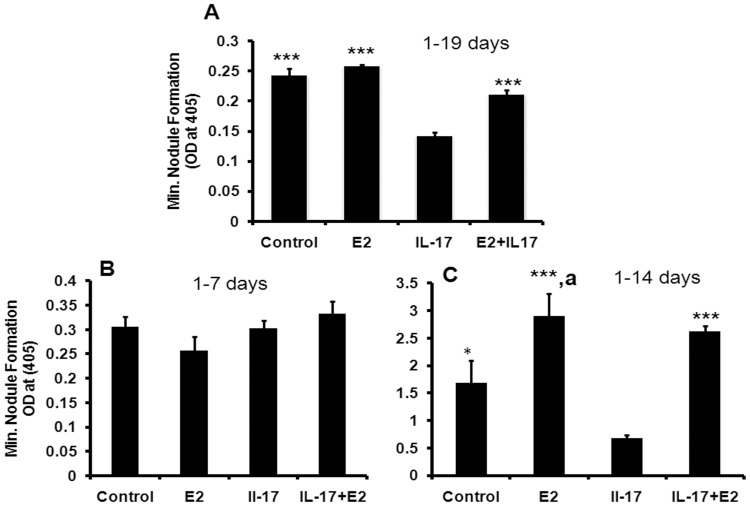
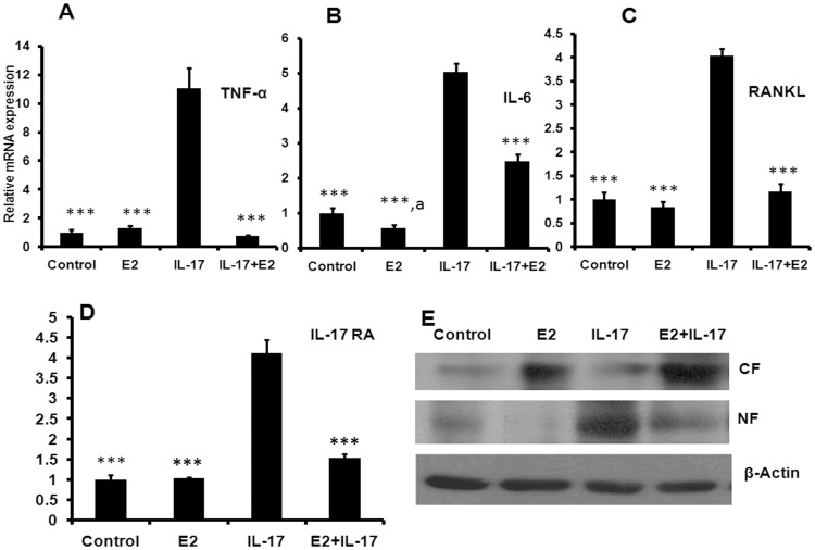
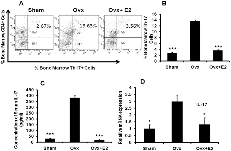
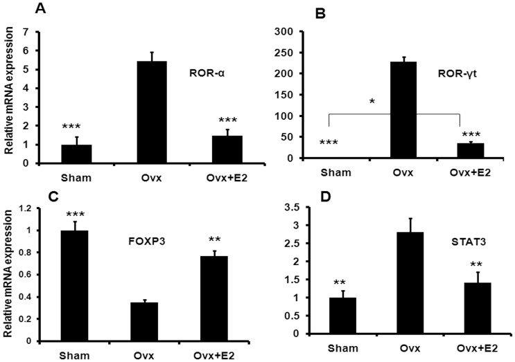
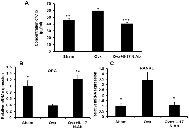
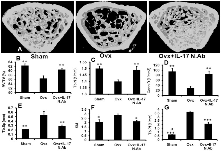
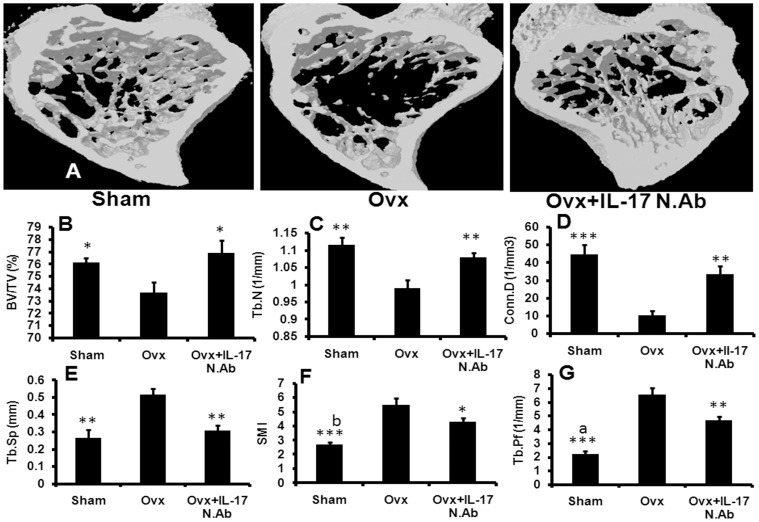
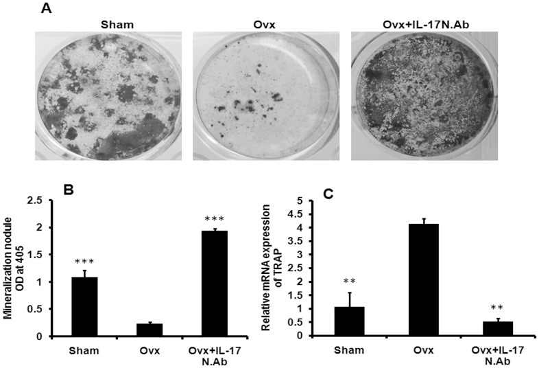
Th17 cells produce IL-17, and the latter promotes bone loss in collagen-induced arthritis in mice. Blocking IL-17 action in mouse model of rheumatoid arthritis reduces disease symptoms. These observations suggest that Th17 cells may be involved in the pathogenesis of bone loss. However, the role of Th17 cell in estrogen (E2) deficiency-induced bone loss is still not very clear. We investigated the effect of E2 on Th17 differentiation in vivo and IL-17 mediated regulation of osteoclast and osteoblast differentiation. Additionally, effect of IL-17 functional block under E2 deficiency-induced bone loss was studied. In murine bone marrow cells, E2 suppressed IL-17 mediated osteoclast differentiation. IL-17 inhibited formation of mineralized nodules in osteoblasts and this effect was suppressed by E2. E2 treatment to mouse calvarial osteoblasts inhibited the IL-17-induced production of osteoclastogenic cytokines and NF-kB translocation. In ovariectomized mice, there was increase in the number of Th17 cells, transcription factors promoting Th17 cell differentiation and circulating IL-17 levels. These effects were reversed by E2 supplementation. Treatment of neutralizing IL-17 monoclonal antibody to Ovx mice mitigated the E2 deficiency-induced trabecular bone loss and reversed the decreased osteoprotegerin-to-receptor activator of nuclear factor kappa B ligand (RANKL) transcript levels in long bones, increased osteoclast differentiation from the bone marrow precursor cells and decreased osteoblast differentiation from the bone marrow stromal cells. Our findings indicate that E2 deficiency leads to increased differentiation of Th17 cells with attendant up regulation of STAT3, ROR-γt and ROR-α and downregulation of Foxp3 which antagonizes Th17 cell differentiation. Increased IL-17 production in turn induces bone loss by increasing pro-osteoclastogenic cytokines including TNF-α, IL-6 and RANKL from osteoblasts and functional block of IL-17 prevents bone loss. IL-17 thus plays a critical causal role in Ovx-induced bone loss and may be considered a potential therapeutic target in pathogenesis of post menopausal osteoporosis.


Conflict of interest statement

Competing Interests: The authors have declared that no competing interests exist.

Figures

Figure 1
Figure 1. Estrogen prevents IL-17 induced differentiation of osteoclasts from BMCs.
(A) Representative photomicrograph (20x magnification) show that IL-17 at 100 ng/ml concentration induced osteoclastogenesis from BMCs in presence of M-CSF (10 ng/ml) and RANKL (50 ng/ml) in five days culture. E2 treatment at 10−9M concentration inhibits IL-17 induced formation of multinucleated cells. (B) Quantitative representation of TRAP+ cells at various treatment conditions. (C) mRNA level of TRAP gene, which is a marker of functional osteoclast was determined by qPCR from the total RNA isolated from cultured cells. (D) RANK expression help in the differentiation of osteoclast precursor cells in to mature osteoclast. mRNA level of RANK gene was determined by qPCR from the total RNA isolated from cultured cells. Data represent three independent experiments and expressed as mean ± SEM with 95% confidence interval. Statistical analysis was performed by ANOVA method followed by the Newman–Keuls test of significance using Prism version 3.0 software.
Figure 2
Figure 2. Estrogen prevents the IL-17 mediated reduction in mineral nodule formation.
(A) Effect of E2 on IL-17 mediated inhibition of mineralization in 19 days cultured MCO. MCO was retrieved from 1–2 days old mice pups by sequential digestion with collagenase/dispase enzymes using standard method as given in material and methods. Quantitative measurement of mineralization nodules in all the groups were performed by using alizarin red staining. (B) Effect of E2 and IL-17 on MCO in 7 days mineralization. (C) Effect of E2 and IL-17 on MCO in 14 days mineralization. Data represent three independent experiments and expressed as mean ± SEM with 95% confidence interval. Statistical analysis was performed by ANOVA method followed by the Newman–Keuls test of significance using Prism version 3.0 software.
Figure 3
Figure 3. Estrogen prevents the IL-17 induced expression of osteoclastogenic cytokines in osteoblasts.
(A) Effect of E2 on IL-17 induced osteoclastogenic cytokine TNF-α was evaluated. (B) Effect of E2 on IL-17 induced IL-6 mRNA level. (C) Effect of E2 on IL-17 induced RANKL mRNA level. (D) Effect of E2 on IL-17 induced IL-17RA mRNA level. (E) Effect of E2 on IL-17 induced NF-kB translocation. Data represent three independent experiments and expressed as mean ± SEM with 95% confidence interval. Statistical analysis was performed by ANOVA method followed by the Newman–Keuls test of significance using Prism version 3.0 software.
Figure 4
Figure 4. Ovx induces the production of IL-17 secreting Th17 cells, circulating IL-17 and transcript levels of IL-17.
(A) Bone marrow cells were stimulated with 20 ng/ml PMA +250 ng/ml ionomycin in the presence of 2 µM monensin; then IL-17-producing cells were detected by FACS. Ovx induces the proliferation of Th17 cells compared to the sham control group (P<0.01). Oral administration of E2 at 0.01 mg/kg body wt reduces the number of Th-17 cells. (B) Bar diagram represents the number of Th-17 cells as percentage of total cells acquired in flow cytometer. Ten mice per group were taken for the study. (C) Serum level of circulating IL-17 was determined by specific ELISA as described in Materials and Methods. Each group represents results from a pool of eight mice. (D) Relative mRNA expression of IL-17 in isolated (detailed method of isolation was given in material and method section) CD4+ cells was measured in all the groups. CD4+ cells of five animals were pooled in each group for RNA isolation and to run real-time PCR assay. Data represent three independent experiments and expressed as mean ± SEM with 95% confidence interval. Statistical analysis was performed by ANOVA method followed by the Newman–Keuls test of significance using Prism version 3.0 software.
Figure 5
Figure 5. Ovx induces the transcript levels of factors regulating Th17 differentiation.
Relative mRNA expression of ROR-α (A), ROR-γt (B), Foxp3 (C) and STAT 3 (D) in isolated CD4+ cells was measured in all the groups. CD4+ cells of five animals were pooled in each group for RNA isolation and to run real-time PCR assay. Data represent three independent experiments and expressed as mean ± SEM with 95% confidence interval. Statistical analysis was performed by ANOVA method followed by the Newman–Keuls test of significance using Prism version 3.0 software.
Figure 6
Figure 6. IL-17 neutralization reduces serum CTx level in Ovx mice and decreases RANKL expression in femur trabecular bone.
(A) After treatment animals were sacrificed and serum samples were collected from all the groups. C-terminal telopeptides of type I collagen (CTx) level was measured in all the groups by ELISA. ELISA was conducted in six replicates in each group. (B) mRNA level of OPG in the trabecular region of femur bone was studied in all the groups (method was explained in the material and method section). (C) mRNA level of RANKL in the trabecular region of femur bone was studied in all the groups. RNA from four femur bones was pooled for the study of OPG and RANKL in each group. Data represent three independent experiments and expressed as mean ± SEM with 95% confidence interval. Statistical analysis was performed by ANOVA method followed by the Newman–Keuls test of significance using Prism version 3.0 software.
Figure 7
Figure 7. IL-17 neutralization blocks Ovx induced bone loss and improves trabecular microarchitecture in femur.
(A) After treatment animals were sacrificed and long bones were collected in 70% isopropanol. (A) BV/TV, (B) Tb.N., (C) Conn.D, (D) Tb.Sp, (E) SMI and (F) Tb.Pf. Blocking of IL-17 with NIL-17mAb restores Ovx induced alterations in femur trabecular region. Six mice were taken in each group for the study. Data expressed as mean ± SEM with 95% confidence interval. Statistical analysis was performed by One way-ANOVA nonparametric method followed by the Newman–Keuls test of significance using Prism version 3.0 software. *** P<0.001, ** P<0.01 and * P<0.05 compared with Ovx animals; a P<0.001 compared between sham and Ovx + IL-17 N.Ab.
Figure 8
Figure 8. IL-17 neutralization blocks Ovx induced bone loss and improves trabecular microarchitecture in tibia.
(A) After treatment animals were sacrificed and long bones were collected in 70% isopropanol. (A) BV/TV, (B) Tb.N., (C) Conn.D, (D) Tb.Sp, (E) SMI and (F) Tb.Pf. Blocking of IL-17 with NIL-17mAb restores Ovx induced alterations in tibial trabecular region. Six mice were taken in each group for the study. Data expressed as mean ± SEM with 95% confidence interval. Statistical analysis was performed by One way-ANOVA nonparametric method followed by the Newman–Keuls test of significance using Prism version 3.0 software. *** P<0.001, ** P<0.01 and * P<0.05 compared with Ovx animals; a P<0.001 and b P<0.01 compared between sham and Ovx + IL-17 N.Ab.
Figure 9
Figure 9. NIL-17 mAb treatment to Ovx mice promotes mineral nodule formation and inhibits osteoclastogenesis in bone marrow microenvironment.
(A) Representative images show mineralization nodules formed in the BMCs harvested from the long bones of all groups of mice. (B) Quantitative measurement of mineralization nodules in all the groups were performed by using alizarin red staining. (C) Ex-vivo osteoclastogenesis was performed in BMCs of all the groups. After five days total RNA was isolated and real-time PCR was performed to assess the expression of TRAP gene. Data represent three independent experiments and expressed as mean ± SEM with 95% confidence interval. Statistical analysis was performed by ANOVA method followed by the Newman–Keuls test of significance using Prism version 3.0 software.

Similar articles

Cited by

References

    1. Clowes JA, Riggs BL, Khosla S (2005) The role of the immune system in the pathophysiology of osteoporosis. Immunol Rev 208: 207–227. - PubMed
    1. Ginaldi L, Di Benedetto MC, De Martinis M (2005) Osteoporosis, inflammation and ageing. Immun Ageing 2: 14. - PMC - PubMed
    1. Arron JR, Choi Y (2000) Bone versus immune system. Nature 408: 535–536. - PubMed
    1. Oostlander AE, Everts V, Schoenmaker T, Bravenboer N, van Vliet SJ, et al. (2012) T cell-mediated increased osteoclast formation from peripheral blood as a mechanism for Crohn’s disease-associated bone loss. J Cell Biochem 113: 260–268. - PubMed
    1. Manolagas SC (2000) Birth and death of bone cells: basic regulatory mechanisms and implications for the pathogenesis and treatment of osteoporosis. Endocr Rev 21: 115–137. - PubMed

Publication types