. 2012;7(9):e44580.
doi: 10.1371/journal.pone.0044580. Epub 2012 Sep 6.

Transforming growth factor-beta promotes rhinovirus replication in bronchial epithelial cells by suppressing the innate immune response

Affiliations

Transforming growth factor-beta promotes rhinovirus replication in bronchial epithelial cells by suppressing the innate immune response

Nicole Bedke et al. PLoS One. 2012.

Abstract

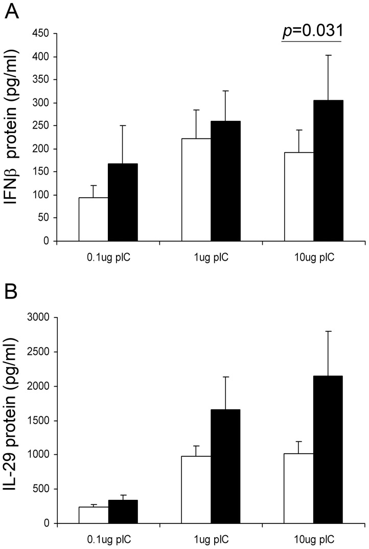
Rhinovirus (RV) infection is a major cause of asthma exacerbations which may be due to a deficient innate immune response in the bronchial epithelium. We hypothesized that the pleiotropic cytokine, TGF-β, influences interferon (IFN) production by primary bronchial epithelial cells (PBECs) following RV infection. Exogenous TGF-β(2) increased RV replication and decreased IFN protein secretion in response to RV or double-stranded RNA (dsRNA). Conversely, neutralizing TGF-β antibodies decreased RV replication and increased IFN expression in response to RV or dsRNA. Endogenous TGF-β(2) levels were higher in conditioned media of PBECs from asthmatic donors and the suppressive effect of anti-TGF-β on RV replication was significantly greater in these cells. Basal SMAD-2 activation was reduced when asthmatic PBECs were treated with anti-TGF-β and this was accompanied by suppression of SOCS-1 and SOCS-3 expression. Our results suggest that endogenous TGF-β contributes to a suppressed IFN response to RV infection possibly via SOCS-1 and SOCS-3.


Conflict of interest statement

Competing Interests: DED and STH are co-founders of Synairgen Research Ltd. and hold shares in Synairgen plc. Synairgen is currently developing inhaled IFN-β for treatment of virus-induced asthma exacerbations. This does not alter the authors’ adherence to all the PLoS ONE policies on sharing data and materials.

Figures

Figure 1
Figure 1. The effect of exogenous TGF-β2 on RV replication.
PBECs from 3 non-asthmatic volunteers were pre-incubated with 0, 1, 10, and 25 ng/ml of TGF- β2 for 24 h, followed by infection with RV1B at 5000 TCID50 units/105 cells. Cells were then further incubated for 48 h in the presence or absence of TGF-β2, as indicated. Viral replication at 24 h was measured as vRNA by RT-qPCR (A) and at 48 h by release of infectious virions into culture supernatants by TCID50 assays (B). The graph (C) shows data for infectious virus release from PBECs from 10 non-asthmatic donors treated without or with 10 ng/ml TGF-β2, followed by infection with RV1B at 5000 TCID50 units/105 cells for 48 hours. Statistical comparison was made using a Wilcoxon rank sum test. The # mark in C indicates where 2 data points overlap (1.8e6→3.1e6 TCID50 units/ml).
Figure 2
Figure 2. Exogenous TGF-β2 suppresses IFN-β release from virally infected (A) or poly IC exposed (B) PBEC cultures from non-asthmatic donors.
PBEC cultures were infected with RV1B (5000 TCID50 units/105 cells) (n = 10) or treated with poly IC (n = 5) in the presence or absence of TGF-β2 which was used at 1 (black bars in B) or 10 ng/ml (panel A and grey bars in B). Culture supernatants were harvested 48 hours p.i (A) or 8 h post stimulation (B) and IFN-β protein levels were measured by ELISA. In B, the data are expressed as a % of control cultures treated with poly IC in the absence of TGF-β2 (median (IQR) IFN-β release  = 346 (1135) and 369 (1390) pg/ml for cells treated with 1 or 10 µg/ml Poly IC, respectively. The data were analyzed using Wilcoxon’s rank sum test (A) or using a paired t-test for normally distributed data (B).
Figure 3
Figure 3. Exogenous TGF-β2 suppresses IFN-λ1/IL-29 release from virally infected (A) (n = 10) or poly IC (n = 4) exposed (B) PBEC cultures from non-asthmatic donors.
IFNλ1/IL-29 protein levels were measured by ELISA from RV-infected or poly IC exposed PBECs treated with TGF-β2 as described in Figure 2. Median (IQR) IFN-λ1 release  = 3896 (2766) and 4932 (4941) pg/ml for cells treated with 1 or 10 µg/ml Poly IC, respectively.
Figure 4
Figure 4. The effect of neutralizing endogenous TGF-β on RV replication.
PBECs from 8 asthmatic donors or 6 non-asthmatic control subjects were pretreated for 24 h in the presence of a neutralizing anti pan TGF-β antibody or isotype control antibody before infection with RV1B (1000 units/105 cells) for 48 h. The fold-decrease in viral replication by the neutralizing antibody was plotted as a ratio of the TCID50/ml of antibody-treated versus isotype controls. The figure shows median and interquartile range, with individual data points superimposed. Data were analysed using a Mann Whitney U test.
Figure 5
Figure 5. Suppression of viral replication in PBECs from asthmatic donors by neutralization of endogenous TGF-β.
PBECs from asthmatic donors were pretreated for 24 h in the presence of a neutralizing anti pan TGF-β antibody or isotype control antibody before infection with RV1B (1000 units/105 cells) for 48 h. In A, viral titre was determined as TCID50/ml using culture supernatants obtained 48 h p.i. In B, IFN-β protein was measured at 48 h and was expressed as a ratio of the viral load measured as TCID50 units. The data were analyzed using Wilcoxon’s rank sum test.
Figure 6
Figure 6. The effect of anti-TGF-β antibodies on release of IFN-β and IFNλ1/IL-29 protein in response to poly IC.
PBECs from 6 asthmatic donors were treated with 0.1–10 µg/ml poly IC in the presence of neutralizing anti-TGF-β antibodies (black bars) or an IgG isotype control (open bars) and incubated for 24 h. Supernatants were removed and IFN-β (A) or IFN-λ1/IL-29 protein levels (B) were measured by ELISA. Graphs show (mean±SEM) IFN produced in pg/ml the presence of the control or anti TGF-β antibodies.
Figure 7
Figure 7. The effect of RV infection on TGF-β isoform expression.
TGF-β1, β2, β3 mRNA levels were measured in PBECs from 4 asthmatic and 4 non-asthmatic donors following infection with RV; TGF-β mRNA expression was measured relative to GAPDH/UBC using the ΔΔCt method (A). Total TGF-β2 protein levels were measured in conditioned media from PBEC cultures of 15 non-asthmatic and 23 asthmatic donors which were harvested at 48 h. Latent TGF-β2 was activated by acid-treatment and total TGF-β2 measured by ELISA. Statistical significance was tested using Mann Whitney U test (B). PBEC culture supernatants of 2 non-asthmatic and 4 asthmatic donors were tested in a transformed mink lung cell luciferase bioassay in order to measure active TGF-β in the presence (black bars) or absence (grey bars) of rhinovirus at 5000 TCID50 units/105 cells. Superimposed is a standard curve obtained using 250 or 500 pg of active TGFβ (C). Data in panels A and B are given as box and whisker plots showing median, interquartile range and 95% confidence intervals; individual data points are superimposed.
Figure 8
Figure 8. The effect of TGF-β neutralization on basal SMAD2 activation.
PBECs from asthmatic donors were treated with RV and anti TGF-β antibody, as indicated, as described in Figure 5. Cell lysates were harvested at 1, 4, and 6 hours post-virus infection and Smad-2 phosphorylation was analysed by Western blotting. A representative Western blot is shown in (A) and densitometric quantification of the experiment repeated using PBECs from 3 different asthmatic subjects is shown in (B).
Figure 9
Figure 9. Neutralizing endogenous TGF-β suppresses RV1B mediated SOCS-1 and SOCS-3 gene expression in asthmatic PBECs.
Samples were treated as described in Figure 5. SOCS-1 (A) and SOCS-3 (B) gene expression were measured in 7 asthmatic subjects at 48 h p.i. by RT-qPCR and normalized to housekeeping genes. Results were plotted as relative fold-induction using the ΔΔCt method. The Wilcoxon rank sum test was used to analyse statistical significance.

References

    1. Holgate ST, Polosa R (2006) The mechanisms, diagnosis, and management of severe asthma in adults. Lancet 368: 780–793. - PubMed
    1. Cohn L, Elias JA, Chupp GL (2004) Asthma: mechanisms of disease persistence and progression. Annu Rev Immunol 22: 789–815. - PubMed
    1. Johnston SL, Pattemore PK, Sanderson G, Smith S, Lampe F, et al. (1995) Community study of role of viral infections in exacerbations of asthma in 9–11 year old children. BMJ 310: 1225–1229. - PMC - PubMed
    1. Wark PA, Johnston SL, Bucchieri F, Powell R, Puddicombe S, et al. (2005) Asthmatic bronchial epithelial cells have a deficient innate immune response to infection with rhinovirus. J Exp Med 201: 937–947. - PMC - PubMed
    1. Contoli M, Message SD, Laza-Stanca V, Edwards MR, Wark PA, et al. (2006) Role of deficient type III interferon-lambda production in asthma exacerbations. Nat Med 12: 1023–1026. - PubMed

Publication types

MeSH terms

Substances