. 2012;7(9):e44605.
doi: 10.1371/journal.pone.0044605. Epub 2012 Sep 7.

An exported heat shock protein 40 associates with pathogenesis-related knobs in Plasmodium falciparum infected erythrocytes

Affiliations

An exported heat shock protein 40 associates with pathogenesis-related knobs in Plasmodium falciparum infected erythrocytes

Pragyan Acharya et al. PLoS One. 2012.

Abstract

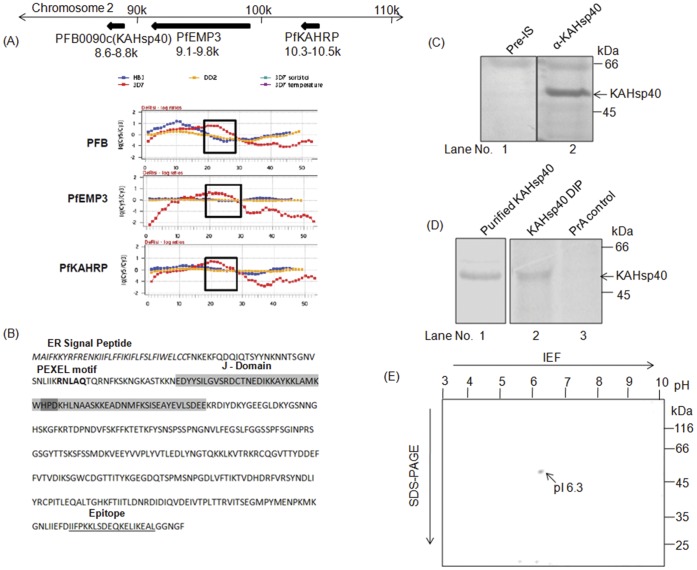
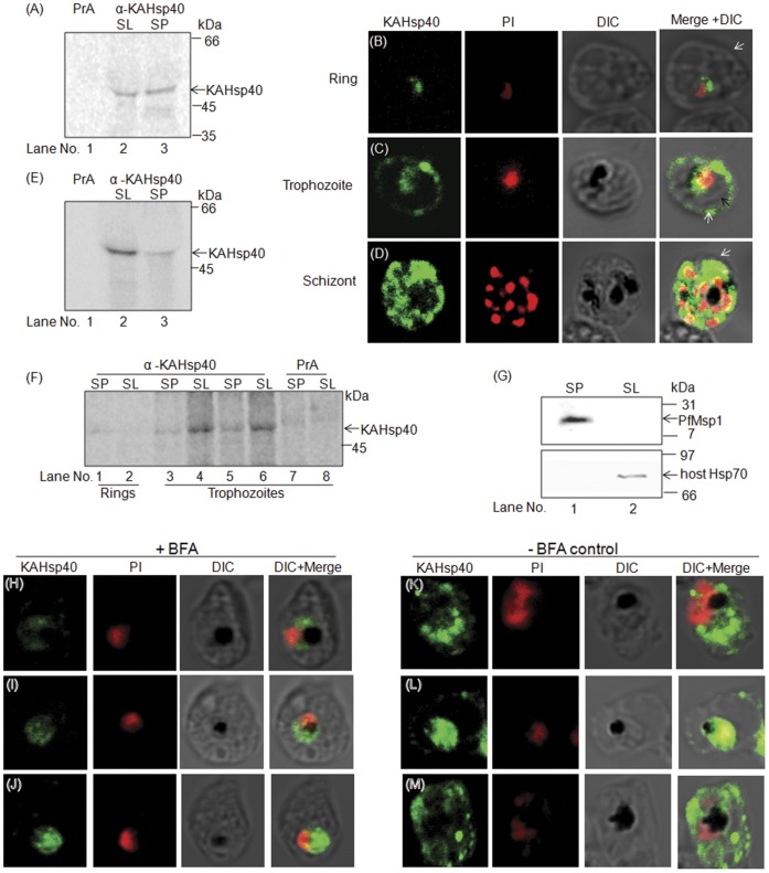
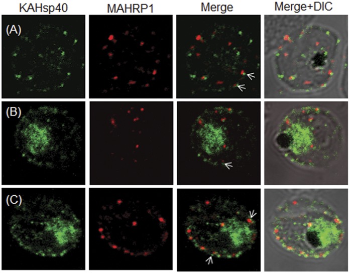
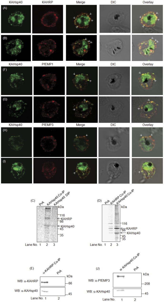
Cell surface structures termed knobs are one of the most important pathogenesis related protein complexes deployed by the malaria parasite Plasmodium falciparum at the surface of the infected erythrocyte. Despite their relevance to the disease, their structure, mechanisms of traffic and their process of assembly remain poorly understood. In this study, we have explored the possible role of a parasite-encoded Hsp40 class of chaperone, namely PFB0090c/PF3D7_0201800 (KAHsp40) in protein trafficking in the infected erythrocyte. We found the gene coding for PF3D7_0201800 to be located in a chromosomal cluster together with knob components KAHRP and PfEMP3. Like the knob components, KAHsp40 too showed the presence of PEXEL motif required for transport to the erythrocyte compartment. Indeed, sub-cellular fractionation and immunofluorescence analysis (IFA) showed KAHsp40 to be exported in the erythrocyte cytoplasm in a stage dependent manner localizing as punctuate spots in the erythrocyte periphery, distinctly from Maurer's cleft, in structures which could be the reminiscent of knobs. Double IFA analysis revealed co-localization of PF3D7_0201800 with the markers of knobs (KAHRP, PfEMP1 and PfEMP3) and components of the PEXEL translocon (Hsp101, PTEX150). KAHsp40 was also found to be in a complex with KAHRP, PfEMP3 and Hsp101 as confirmed by co-immunoprecipitation assay. Our results suggest potential involvement of a parasite encoded Hsp40 in chaperoning knob assembly in the erythrocyte compartment.


Conflict of interest statement

Competing Interests: The authors have declared that no competing interests exist.

Figures

Figure 1
Figure 1. Characteristic features of KAHsp40.
(A) Upper panel indicates sub-telomeric chromosomal cluster on chromosome 2 of Plasmodium falciparum which consists of KAHsp40, KAHRP and PfEMP3. Lower panel shows the transcription profiles of these genes from different strains of Plasmodium falciparum (Adapted from PlasmoDB.org) (B) KAHsp40 sequence consists of an N-terminal ER signal peptide, a PEXEL motif followed by a J-domain that contains a conserved HPD motif. The C-terminal epitope that was used for generating antibody has been underlined. (C) Immunoblot with α-KAHsp40 recognizes a specific band of MW at about 48 kDa in total parasite lysate whereas the pre-immune serum (Pre-IS) does not. (D) The form of KAHsp40 immunoprecipitated from radiolabeled parasite lysate corresponds to the processed form of the protein as its mobility is same as the recombinant protein on SDS-PAGE. Lane 1 is the coomassie stained purified KAHsp40 protein whereas Lanes 2 and 3 is an autoradiogram of IP with α-KAHSp40 antisera and protein-A (PrA) control respectively. (E) Two-dimensional gel of KAHsp40 immunoprecipitated from denatured metabolically labeled infected RBC lysate. DIP- Denaturing Immunoprecipitation.
Figure 2
Figure 2. KAHsp40 is exported into the infected RBC cytosol.
(A) DIP of fractionated infected RBC with α-KAHsp40 antiserum. SL: Saponin Lysate; SP: Saponin Pellet. This reveals that KAHsp40 is present in both the SP as well as SL. IFA of (B) ring stage parasites (C) trophozoite stage parasites (D) schizont stage parasites reveals the presence of KAHsp40 in discrete foci in the erythrocyte compartment. Black arrow indicates the parasite boundary and white arrow indicates the erythrocyte membrane. (E) Pulse-labeling of the parasite for 15 minutes followed by denaturing IP (DIP) for KAHsp40 reveals that it is exported out to the SL within 15 minutes of synthesis in trophozoite stages. (F) Stage specific denaturing IP of KAHsp40 reveals export of KAHsp40 occurs in trophozoite stages. Lanes 3–4 and 5–6 represent 24 h and 36 h post invasion respectively. (G) Western blot for hHsp70 and PfMsp1 to show that compartment integrity is maintained during saponin based fractionation. (H-J) IFA reveals that the signal for KAHsp40 was restricted to the parasite in BFA treated cultures. (K-M) KAHsp40 was exported to the erythrocyte compartment in mock treated cultures. DIP- Denaturing Immunoprecipitation.
Figure 3
Figure 3. KAHsp40 does not associate with Maurer’s cleft.
(A–C) IFA reveals that both KAHsp40 and MAHRP1 are present in discrete foci in the infected erythrocyte, however, they do not co-localize with each other in spite of signals being in close proximity (highlighted by white arrows). The images shown have been taken at the trophozoite stage.
Figure 4
Figure 4. KAHsp40 associates with knobs on the infected erythrocyte membrane.
(A-B) IFA reveals that KAHsp40 and KAHRP co-localize in the erythrocyte periphery near the erythrocyte plasma membrane. White arrows indicate the discrete foci in which they co-localize. (C) Co-IP of KAHsp40 from total infected erythrocytes reveals two interactors, of about 66 kDa and 150 kDa. (D) Reciprocal co-IP immunoprecipitates analyzed on a 5–15% gradient gel reveals that 66 kDa interactor of KAHsp40 is KAHRP. (E) Western blot of KAHRP co-IP immunoprecipitate shows the presence of KAHsp40. (F-G) IFA reveals that KAHsp40 and PfEMP1 co-localize with each other on the erythrocyte plasma membrane indicating the close association of KAHsp40 with knobs. (H-I) IFA reveals that KAHsp40 and PfEMP3 co-localize with each other on the erythrocyte plasma membrane indicating the close association of KAHsp40 with knobs. (J) Western blot of KAHsp40 co-IP immunoprecipitate shows the presence of PfEMP3. The images shown have been taken at the trophozoite stage.
Figure 5
Figure 5. KAHsp40 associates with the PEXEL translocon on the PVM.
(A, B) IFA analysis reveals that KAHsp40 and Hsp101 co-localize with each other on the PVM. (C, D) IFA analysis reveals that KAHsp40 and PTEX150 co-localize with each other on the PVM. White arrows indicate the discrete foci in which they co-localize. (E) Western blot of KAHsp40 co-IP immunoprecipitate shows the presence of Hsp101.

References

    1. Marti M, Good RT, Rug M, Knuepfer E, Cowman AF (2004) Targeting malaria virulence and remodelling proteins to the host erythrocyte. Science 306: 1930–1933. - PubMed
    1. Hiller NL, Bhattacharjee S, van Ooij C, Liolios K, Harrison T, et al. (2004) A host-targeting signal in virulence proteins reveals a secretome in malarial infection. Science 306: 1934–1937. - PubMed
    1. Lanzer M, Wickert H, Krohne G, Vincensini L, Braun Breton C (2006) Maurer’s clefts: a novel multi-functional organelle in the cytoplasm of Plasmodium falciparum- infected erythrocytes. Int J Parasitol 36 23–36. - PubMed
    1. Tilley L, Sougrat R, Lithgow T, Hanssen E (2008) The twists and turns of Maurer’s cleft trafficking in P. falciparum- infected erythrocytes. Traffic 9 187–197. - PubMed
    1. Su XZ, Heatwole VM, Wertheimer SP, Guinet F, Herrfeldt JA, et al. (1995) The large diverse gene family var encodes proteins involved in cytoadherence and antigenic variation of Plasmodium falciparum-infected erythrocytes. Cell 82 89–100. - PubMed

Publication types

MeSH terms

Substances

LinkOut - more resources