. 2012;7(9):e44835.
doi: 10.1371/journal.pone.0044835. Epub 2012 Sep 7.

Differential requirements in endocytic trafficking for penetration of dengue virus

Affiliations

Differential requirements in endocytic trafficking for penetration of dengue virus

Eliana G Acosta et al. PLoS One. 2012.

Abstract

The entry of DENV into the host cell appears to be a very complex process which has been started to be studied in detail. In this report, the route of functional intracellular trafficking after endocytic uptake of dengue virus serotype 1 (DENV-1) strain HW, DENV-2 strain NGC and DENV-2 strain 16681 into Vero cells was studied by using a susceptibility to ammonium chloride assay, dominant negative mutants of several members of the family of cellular Rab GTPases that participate in regulation of transport through endosome vesicles and immunofluorescence colocalization. Together, the results presented demonstrate that in spite of the different internalization route among viral serotypes in Vero cells and regardless of the viral strain, DENV particles are first transported to early endosomes in a Rab5-dependent manner. Then a Rab7-dependent pathway guides DENV-2 16681 to late endosomes, whereas a yet unknown sorting event controls the transport of DENV-2 NGC, and most probably DENV-1 HW, to the perinuclear recycling compartments where fusion membrane would take place releasing nucleocapsid into the cytoplasm. Besides the demonstration of a different intracellular trafficking for two DENV-2 strains that shared the initial clathrin-independent internalization route, these studies proved for the first time the involvement of the slow recycling pathway for DENV-2 productive infection.


Conflict of interest statement

Competing Interests: The authors have declared that no competing interests exist.

Figures

Figure 1
Figure 1. Effect of blockade of clathrin-mediated endocytosis on DENV-1 and DENV-2 strains.
A. Cells were treated with 50 µM chlorpromazine and then infected with reference strains and clinical isolates of DENV-1 or DENV-2. Virus yields were quantified by PFU at 48 h p.i. and results are expressed as % of inhibition of virus multiplication with respect to a control of infected cells without drug treatment. Each value is the mean±SD of three independent experiments. B. Cells were treated with 50 µM chlorpromazine or left untreated and incubated with TRITC-labelled transferrin. C. Cells transiently transfected with the constructs GFP-DIIIΔ2 or GFP-EH29 were infected with DENV-2 strain 16681. After 24 h cells were fixed and viral antigen expression was visualized by immunofluorescence staining using mouse anti-E glycoprotein antibody and TRITC-labelled anti-mouse IgG.
Figure 2
Figure 2. Time course of DENV penetration.
A. 100–200 PFU/well of DENV-1 HW, DENV-2 NGC, DENV-2 16681 or JUNV were bound to cells at 4°C and then allowed to internalize after rapid warming to 37°C. Ammonium chloride was added at different time points to inhibit penetration and infection. Cells were further incubated at 37°C for 3 h, then extracellular virus was inactivated and cultures were overlaid with plaquing medium. The infection levels observed were normalized to the level in control cells without ammonium chloride. Each point is the mean±SD of three independent experiments. B. Cells were treated or not with 50 mM ammonium chloride and then stained with acridine orange. C. DENV-2 NGC was adsorbed to Vero cells during 1 h at 4°C in the presence or absence of 50 mM ammonium chloride and then cultures were shifted to 37°C. At different time intervals cultures were fixed and processed to reveal C and E protein by immunofluorescence using mouse anti-C and mouse anti-E antibodies followed of FITC-labelled anti-mouse IgG, respectively.
Figure 3
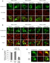
Figure 3. Transport of DENV particles to early endosomes.
A. Cells transiently transfected with the GFP-tagged versions of Rab5 wt and S34N, and the plasmid pGFP-C1 were infected with DENV-1 HW, DENV-2 NGC or DENV-2 16681 (MOI:10 PFU/cell). After 1 h of infection, cells were fixed and processed to visualize GFP transgene expression and internalized viral particles by immunofluorescence staining using mouse anti-E glycoprotein antibody and TRITC-labelled anti-mouse IgG. B. Cells transfected as in A) were infected with DENV-1 HW, DENV-2 NGC or 16681 (MOI:1 PFU/cell). After 24 h of infection cultures were fixed and immunofluorescence staining was performed as in A. C. For quantification of samples shown in B, 250 transfected cells with similar levels of GFP expression were screened and cells positive for viral antigen were scored. D. Cells transfected as in A were then incubated with TRITC-labelled transferrin during 30 min. Then, cells were fixed and fluorescence was visualized.
Figure 4
Figure 4. Transport of DENV particles to late endosomes: Rab7 dependence.
A. Cells transiently transfected with the GFP-tagged versions of Rab7 wt and DN T22N and the plasmid pGFP-C1 were infected with DENV-1 HW, DENV-2 NGC or 16681. After 24 h of infection cultures were fixed and processed to visualize GFP transgene expression and viral antigen by immunofluorescence staining using mouse anti-E glycoprotein antibody and TRITC-labelled anti-mouse IgG. B. For quantification of samples shown in A, 250 transfected cells with similar levels of GFP expression were screened and cells positive for viral antigen were scored. C. Cells transiently transfected as in A were infected with JUNV. At 24 h p.i. cells were fixed and infection was assessed by immunofluorescence using mouse anti-JUNV NP antibody and TRITC-labelled anti-mouse IgG.
Figure 5
Figure 5. Transport of DENV particles to late endosomes: effect of wortmannin.
Vero cells were infected with DENV-2 NGC or 16681 in the presence of wortmannin 100 nM and after 10 or 60 min at 37°C cultures were fixed. Rab5 was revealed using a rabbit polyclonal antibody followed by FITC-conjugated secondary antibodies, while C protein was revealed using a mouse monoclonal antibody followed by incubation with TRITC-conjugated secondary antibodies. Enlarged details of single and merged channels of the boxed areas are shown. The degree of colocalization was estimated by calculating the Mander’s overlap coefficient from 20 cells using the application imageJ. Values are indicated in each picture.
Figure 6
Figure 6. Transport of DENV particles to recycling endosomes.
Cells transiently transfected with the GFP-tagged versions of Rab22 wt and Q64L (A), Rab11 wt and S25N (B) and the plasmid pGFP-C1 were infected with DENV-1 HW, DENV-2 NGC or 16681. After 24 h of infection cultures were fixed and processed to visualize GFP transgene expression and viral antigen by immunofluorescence staining using mouse anti-E glycoprotein antibody and TRITC-labelled anti-mouse IgG. C. For quantification of samples shown in A and B, 250 transfected cells with similar levels of GFP expression were screened and cells positive for viral antigen were scored. D. Cells expressing GFP-Rab7 wt or Green Lantern-Rab11 wt were infected at 24 h post-transfection with DENV-2 NGC or 16681 during 60 min at 4°C and then shifted at 37°C. After 15 min cells were fixed and processed to visualize GFP transgene expression and viral antigen by immunofluorescence staining using mouse anti-C glycoprotein antibody and TRITC-labelled anti-mouse IgG. Cells were visualized with a confocal microscope.
Figure 7
Figure 7. Model of infectious intracellular transport of DENV-1 HW, DENV-2 NGC and DENV-2 16681 in Vero cells.
DENV-1 HW is internalized through the classical clathrin-mediated endocytic pathway, while DENV-2 NGC and DENV-2 16681 use a non-classical clathrin- and caveolin- independent route. These viruses are then transported in a Rab5-dependent manner to early endosomes. There, DENV-2 16681 is incorporated into late endosomes in a Rab7-dependent fashion and DENV-2 NGC and probably DENV-1 HW into recycling endosomes in a Rab22-dependent process. Viral fusion would take place in the corresponding organelles between 14–16 min p.i. releasing the viral nucleocapsids to the cytoplasm.

Similar articles

Cited by

References

    1. Guzmán MG, Halstead SB, Artsob H, Buchy P, Farrar J, et al. (2010) Dengue: a continuing global threat. Nat Rev Microbiol 8: S7–S16. - PMC - PubMed
    1. Kyle JL, Harris E (2008) Global spread and persistence of dengue. Annu Rev Microbiol 62: 71–92. - PubMed
    1. Altmeyer R (2004) Virus attachment and entry offer numerous targets for antiviral therapy. Curr Pharm Des 10: 3701–3712. - PubMed
    1. Perera R, Khalik M, Kuhn RJ (2008) Closing the door on flaviviruses: Entry as a target for antiviral drug design. Antiviral Res 80: 11–22. - PMC - PubMed
    1. Krishnan MN, Sukumaran B, Pal U, Agaise H, Murray JL, et al. (2007) Rab 5 is required for the cellular entry of dengue and West Nile viruses. J Virol 81: 4881–4885. - PMC - PubMed

Publication types

Substances