

En Face OCT Imaging for the Diagnosis of Outer Retinal Tubulations in Age-Related Macular Degeneration
- PMID: 22970349
- PMCID: PMC3437289
- DOI: 10.1155/2012/542417
En Face OCT Imaging for the Diagnosis of Outer Retinal Tubulations in Age-Related Macular Degeneration
Abstract
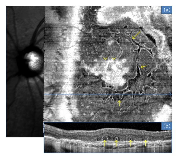
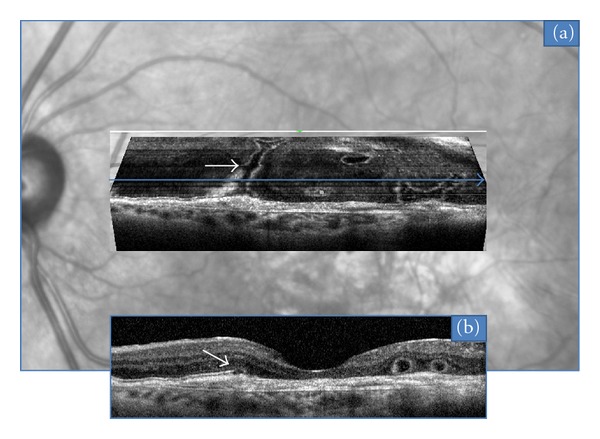
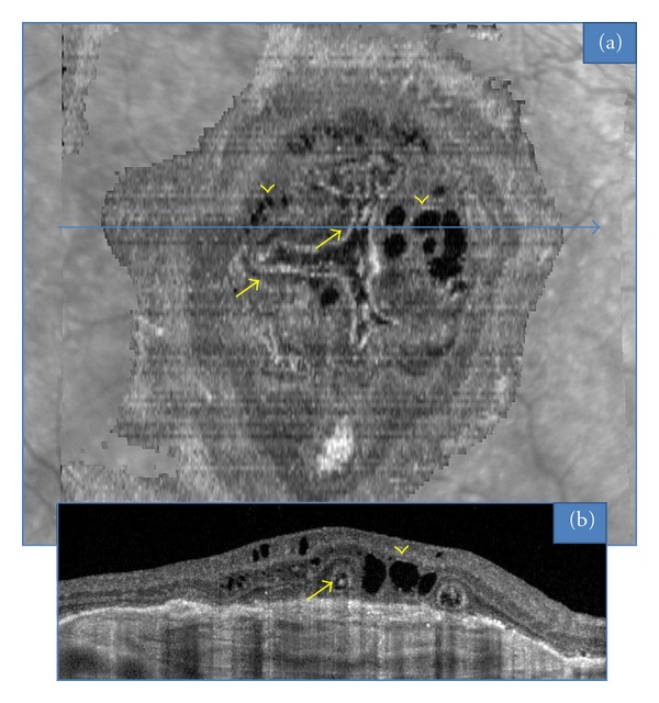
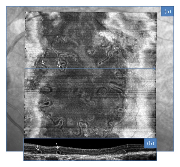
Purpose. "En face" is an emerging imaging technique derived from spectral domain optical coherence tomography (OCT). It produces frontal sections of retinal layers, also called "C-scan OCT." Outer retinal tubulations (ORTs) in age-related macular degeneration (AMD) are a recent finding evidenced by spectral-domain OCT. The aim of this study is to characterize the morphology of ORT according to the form of AMD, using "en-face" spectral domain OCT. Methods. "En face" OCT imaging was prospectively performed in 26 consecutive eyes with AMD that also had ORT. Results. There were 15 neovascular, 8 atrophic, and 3 eyes with a mixed (fibrotic and atrophic) form of AMD. Among the neovascular group, the most frequent tubulation pattern on "en-face" OCT was a branching network emanating from a fibrovascular scar; we term this pattern as "pseudodendritic." It did not require treatment when observed as an isolated finding. In all cases of atrophic AMD, the tubular network was located at the edge of the geographic atrophy area, and formed a "perilesional" pattern. Six atrophic cases showed tubular invaginations inside this area. Conclusion. "En face" OCT is a valuable technique in the diagnosis and followup of macular disease. It revealed the main characteristic patterns of ORT associated with neovascular and atrophic AMD.
Figures
References
-
- Zweifel SA, Engelbert M, Laud K, Margolis R, Spaide RF, Freund KB. Outer retinal tubulation a novel optical coherence tomography finding. Archives of Ophthalmology. 2009;127(12):1596–1602. - PubMed
-
- Podoleanu AG, Dobre GM, Webb DJ, Jackson DA. Simultaneous en-face imaging of two layers in the human retina by low-coherence reflectometry. Optics Letters. 1997;22(13):1039–1041. - PubMed
-
- Wolff B, Maftouhi MQE, Mateo-Montoya A, Sahel JA, Mauget-Faÿsse M. Outer retinal cysts in age-related macular degeneration. Acta Ophthalmologica. 2011;89(6):e496–e499. - PubMed
-
- Maftouhi MQE, Wolff B, Mauget-Faÿsse M. Outer retinal cysts in exudative age-related macular degeneration: a spectral domain OCT study. Journal Francais d’Ophtalmologie. 2010;33(9):605–609. - PubMed
-
- Yannuzi LA. The Retinal Atlas. chapter 2. New York, NY, USA: Elsevier; 2010. Hereditary chorioretinal dystrophy; pp. 158–160.
LinkOut - more resources
Full Text Sources
Other Literature Sources
Research Materials
